CENTRO DE CIÊNCIAS
DEPARTAMENTO DE BIOQUÍMICA E BIOLOGIA MOLECULAR PROGRAMA DE PÓS-GRADUAÇÃO EM BIOQUÍMICA
FABRÍCIO EULÁLIO LEITE CARVALHO
CHLOROPLAST REDOX METABOLISM AND NPQ REGULATION IN ARABIDOPSIS AND RICE EXPOSED TO EXCESSIVE LIGHT – NEW INSIGHTS
INTO PSII PHOTOPROTECTION MECHANISMS
CHLOROPLAST REDOX METABOLISM AND NPQ REGULATION IN ARABIDOPSIS AND RICE EXPOSED TO EXCESSIVE LIGHT – NEW INSIGHTS
INTO PSII PHOTOPROTECTION MECHANISMS
Tese apresentada ao programa de pós-graduação em Bioquímica da Universidade Federal do Ceará, como requisito parcial à obtenção do título de Doutor em Bioquímica. Área de concentração: Bioquímica vegetal.
Orientador: Prof. Dr. Joaquim Albenísio Gomes da Silveira (UFC-BRA)
Co-orientador: Prof. Dr. Alexander V. Ruban (QMUL-UK)
Gerada automaticamente pelo módulo Catalog, mediante os dados fornecidos pelo(a) autor(a)
C323c Carvalho, Fabrício Eulálio Leite.
Chloroplast redox metabolism and npq regulation in arabidopsis and rice exposed to excessive light : new insights into psii photoprotection mechanisms / Fabrício Eulálio Leite Carvalho. – 2016.
171 f. : il. color.
Tese (doutorado) – Universidade Federal do Ceará, Centro de Ciências, Programa de Pós-Graduação em Bioquímica , Fortaleza, 2016.
Orientação: Prof. Dr. Joaquim Albenísio Gomes da Silveira. Coorientação: Prof. Dr. Alexander V. Ruban.
Ao Prof. Dr. Joaquim Albenísio Gomes da Silveira, por orientar-me durante toda minha jornada científica, desde a graduação até a conquista do doutorado. Por tantos sábios conselhos e por todas as grandes oportunidades que sempre me concedeu. Obrigado.
To Prof. Dr. Alexander V. Ruban for promptly receive me in your excellent group, thank you for open a new world of science to me. The way I see the world will never be the same. Thank you.
Ao Prof. Dra. Marcia Margis-Pinheiro, por receber-me em Porto Alegre diversas vezes e tantos experimentos envolvendo as plantas transformadas de arroz. Por sempre manter uma porta aberta para mim e por tantas valiosas oportunidades. Obrigado.
A todos os membros convidados da banca avaliadora, por sua colaboração e disponibilidade.
À minha amiga Ana Karla M. Lobo por tantas vezes me ajudar com os experimentos e não me deixar esquecer os prazos e as provas. Obrigado.
Ao meu amigo Prof. Dr. Marcio O. Martins, por sempre me ajudar em todos os experimentos, correções e todo o trabalho de bancada. Por suas valiosas dicas com IRGA. Obrigado.
Ao meu amigo Prof. Dr. Milton C. Lima Neto, por sempre me ajudar a planejar os experimentos e por todo seu apoio na bancada. Obrigado por seus ensinamentos de fluorimetria. To my friend Dr. Erica Belgio, for help so much in London, for discuss my data and teach me so many times. Thank you.
To my friend Max Ware, for promptly help in QMUL arriving, for help in the experiments discussion and manuscript writing. Cheers mate!
To my friend Dr. Vasco Giovagnetti for help me in the lab work and keep so good working environment. Grazie amico!
To dear Dr. Petra Ungerer, for help me so many times in lab work and being always so kind. Thank you.
A todos os meus companheiros de LABPLANT, especialmente João Víctor, Lara, Juliana e Adilton, sempre acessíveis e que tanto colaboraram por toda a jornada. Obrigado amigos.
To all my friends in QMUL, especially Emma, Sunny, Jun, Amy, Yonglan, Ruth and Chris, for always make me fell in home. Thank you.
laboratório e durante toda a jornada. Muito obrigado.
Ao Prof. Dr. André L. Silva por sempre manter uma porta aberta e tantas vezes colaborar com meu trabalho. Obrigado por tantas oportunidades.
À Prof. Dra. Aurenívia Bonifácio por toda colaboração e auxílio, especialmente durante os meus primeiros anos de iniciação científica.
A todos os professores do DBBM que colaboraram muito para minha formação acadêmica especialmente ao Prof. Dr. José Tadeu Abreu de Oliveira e Prof. Dr. Francisco de Assis Paiva Campos. Muito obrigado.
A todos os colegas do DBBM que sempre me ajudaram nos momentos de necessidade. Obrigado.
A todo o pessoal da limpeza e administração que sempre fazem o melhor de seus esforços para que possamos trabalhar em um ambiente agradável e funcional.
À minha querida noiva e futura esposa Luana por sempre ser tão paciente e compreensiva com meus esforços em prol desse objetivo que foi o título de doutor. Obrigado por sempre me apoiar e confortar-me nas horas em que tudo parecia impossível e desesperador. Obrigado por tanta paciência e serenidade mesmo com os longos meses de distância em outro país. Divido essa conquista com você, amor da minha vida.
Aos meus amados pais Aloizio e Betânia que sempre me apoiaram por toda vida. Foi deles que recebi as primeiras lições do mundo e as primeiras palavras de conforto. Graças a vocês pude perceber ainda muito jovem o quanto o mundo era grandioso e como era preciso aproveitar ao máximo o que ele tinha a oferecer. Testemunhei muitas horas de dificuldades e sacrifícios, mas, acima de tudo, serenidade e amor para sempre me oferecer o melhor que podiam. Vocês me deram a vida e o mundo! Muito obrigado.
À Deus e Nossa Senhora de Fátima, que sempre iluminaram meu caminho e me trouxeram saúde e coragem para trabalhar.
Plantas são organismos sésseis e por sua natureza desenvolveram diversos mecanismos ligados a maximização da utilização dos recursos primordiais disponíveis para sua sobrevivência nas mais diversas condições ambientais. Em condições ambientais ótimas de crescimento, a intensidade de energia absorvida a partir da luz incidente equivale a demanda energética exercida pela atividade metabólica circunstancial da planta. Entretanto, na quase totalidade dos casos, tais condições não existem na natureza. O excesso de luz é caracterizado por uma condição ambiental na qual o organismo vegetal é submetido a uma quantidade de energia luminosa mais alta do que a capacidade de utilização nas reações bioquímicas que compõem o metabolismo celular necessário para o crescimento normal do indivíduo. Sendo assim, o excesso de luz pode ser consequência de altas intensidades de luz incidente ou devido a restrições nas reações de consumo de poder redutor, especialmente assimilação de CO2.
Portanto, plantas submetidas a estresse salino por exemplo, o qual está muitas vezes associado a uma restrição de natureza estomática, podem apresentar estresse de excesso de luz, mesmo sob intensidades moderadas de luz incidente. A energia excedente pode aumentar a probabilidade da ocorrência de reações colaterais tais como a formação das espécies reativas de oxigênio (EROS). Essas moléculas de alta reatividade podem gerar diversos efeitos negativos para o funcionamento celular, incluindo desnaturação de proteínas, peroxidação lipídica de membranas e fotoinibição das atividades do fotossistemas II (PSII). A ocorrência de fotoinibição é extremamente negativa para o crescimento e desenvolvimento vegetal, afetando a produção de alimentos global. Entretanto, pressões evolutivas causadas pela seleção natural conduziram ao aparecimento de diversos mecanismos de proteção fotooxidativa. Entre as respostas de fotoproteção acionadas em vegetais, duas merecem especial destaque: a dissipação de energia na forma de calor (componente qE do quenching não-fotoquímico - NPQ) e a modulação da expressão de genes ligados a rotas de síntese de antioxidantes enzimáticos e não enzimáticos. Em uma perspectiva sistêmica, entretanto, nenhuma dessas respostas ocorre de maneira isolada em uma célula vegetal. Contrariamente, esses mecanismos interagem de maneira contínua e interdependente, partilhando substratos e rotas de sinalização, e, deste modo, conduzindo à aclimatação da planta às condições de excesso de luz.
Posteriormente, buscou-se um maior entendimento acerca dos mecanismos inerentes ao parâmetro pNPQ. Utilizou-se uma abordagem simplificada consistindo em apenas três fases ontogenéticas de plantas de Arabidopsis no intuito de investigar os processos de conhecido envolvimento com a formação do qE, ΔpH, relação violoxantina/zeaxantina e quantidade da
proteína PsbS. Esses dados estão relacionados no capítulo 3 do presente trabalho e evidenciam que: a) plantas senescentes e juvenis apresentam menor quantidade de PsbS e formação de ΔpH
respectivamente em comparação a plantas maduras, b) as plantas maduras apresentam maior quantidade de pigmentos xantofilas que, em combinação com maiores níveis de clorofila reportados anteriormente, podem explicar uma maior eficiência e potencial na formação de qE, o qual estaria intimamente ligado ao pNPQ.
Finalmente, o presente trabalho de doutorado buscou a aplicação dos conhecimentos gerados acerca das novas técnicas de detecção da fotoinibição e pNPQ em uma planta modelo de interesse agronômico, arroz (Oryza sativa). Objetivando-se investigar a relação das EROS com os processos de fotoproteção associados ao pNPQ, plantas de arroz silenciadas por meio de RNAi para a expressão das APX de tilacóides (APX8) foram empregadas. Os resultados obtidos nesses estudos se encontram no capítulo 4 da presente tese e evidenciam que: a) em luz moderada, plantas silenciadas (apx8) apresentam maior susceptibilidade a fotoinibição associada a menor acúmulo de biomassa e menor produtividade, b) essas características das plantas apx8 provavelmente estiveram ligadas a menor eficiência de formação de pNPQ e qE, c) as diferenças relacionadas ao pNPQ provavelmente estiveram associadas com menor conteúdo de clorofilas e maior acúmulo de EROS nas plantas apx8, evidenciando novamente a importância desses fatores com a capacidade de fotoproteção em plantas e d) durante a aclimatação ao excesso de luz, a ausência da APX8 é compensada por outras peroxidases, levando a redução nos níveis de EROS e provavelmente recuperação dos níveis de clorofila, que levam a uma recuperação do fenótipo evidenciado em luz moderada.
Conclui-se com a presente tese que o uso das metodologias relacionadas a determinação do qPd para estudos de integridade dos RCII foi eficaz para duas espécies distintas, Arabidopsis thaliana e Oryza sativa. Adicionalmente, os resultados indicam que a eficiência de formação do pNPQ parece ser um fator determinante para a capacidade de fotoproteção, que por sua vez é dependente do conteúdo de pigmentos fotossintéticos, especialmente clorofila e carotenoides, assim como eficiência do metabolismo antioxidativo. Essas relações são primeiramente descritas no presente trabalho de doutorado e apresentam potencial de aplicação futura em estudos envolvendo obtenção de plantas mais tolerantes ao excesso de luz. Tais condições de excesso de luz são recorrentes em diversas regiões do mundo, mas especialmente importantes em regiões do semiárido do nordeste do Brasil, onde se encontram em combinação com outros fatores de estresse abiótico, tais como seca e alta salinidade do solo. Espera-se com essas descobertas, contribuir para o conhecimento científico da área de bioquímica vegetal e, posteriormente, possibilitar sua aplicação em âmbito do desenvolvimento agrícola regional.
Plants are sessile organisms, which evolved from aquatic environment to land. This crucial change in habitat opened a new word of resources, but also generated numerous adaptive challenges. Among these defies, sustain photosynthesis under an excessive light environment consist in one of the most important adverse factors to be overcame by plants. Excessive light is defined as the light absorbed by plants, which exceeds their photosynthetic capacity. Under such conditions, the over-accumulation of oxygen reactive species (ROS) could lead to impairment on photosystem two reaction centers (RCII), generating photoinhibition and affecting negatively crop productivity. In this sense, the development of photoprotective mechanisms was essential for plants surviving. Among such mechanisms, non-photochemical quenching (NPQ) and antioxidative metabolism have been intensively studied. However, the real importance of such photoprotective mechanisms and the possible integrative processes which connect NPQ and redox metabolism still not totally understood. The present study addressed to comprehend the integrative mechanisms underlying NPQ and antioxidative processes in two different plants species: one classic plant model (Arabidopsis thaliana) and one important crop model (Oryza sativa). In order to reach such objectives, first was employed new fluorimetric-based methodologies related to photoinhibition quantification, involving photochemical quenching measured in the dark subsequently illumination (qPd) and the fraction of NPQ formed which was related to photoprotection (pNPQ). The use of this methodology was well succeed with A.Thaliana plants under an important physiological problem: light tolerance during plant ontogenesis. The importance of ROS accumulation during less tolerant developmental phases (juvenile and senescent) and chlorophyll concentrations were discussed. Subsequently, the mechanisms underlying pNPQ formation were investigated in the same model, which revealed the importance of combined adjustments in PsbS protein expression, delta pH formation and zeaxanthin/violaxanthin pigment contents for phototolerance during A. thaliana ontogenesis. The importance of total xanthophyll pool in pNPQ generation was evaluated. Finally, the current study attempted to employ the methodology involving qPd in a crop model (rice) knockdown for thylakoidal ascorbate peroxidase isoform (APX8 proteins; apx8 = transformed plants). The data revealed new importance for APX8, which were more sensitive to light, specially under moderate growth light (400 µmol m-2 s-1) compared with non-transformed (NT). Transformed apx8 plants
accumulated less chlorophyll, over-accumulated H2O2 and presented impairment on thermal
component of NPQ (qE) formation. Under high light intensities growth conditions (4 days), apx8 plants activated compensatory mechanisms which were probably effective as photoprotective mechanism.
CHAPTER I
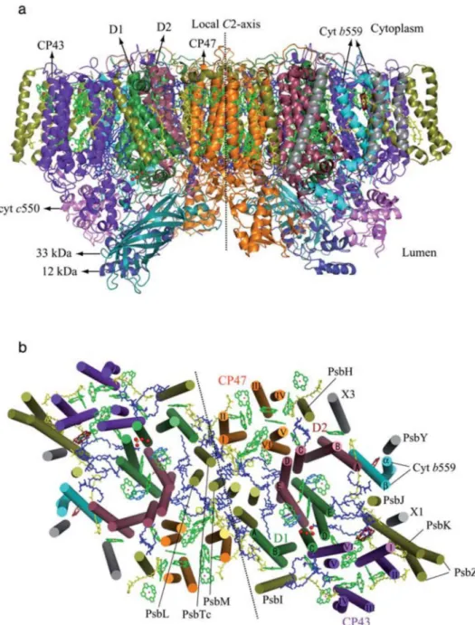
Figure 1. Crystal structure of PSII. 23
Figure 2. Light response curves for photosynthesis – Excessive light. 24 Figure 3. Schematic representation of ROS effects on photoinhibition. 26 Figure 4. Mechanisms involved in minimizing photoinhibition of PSII. 27 Figure 5. Schematic diagram of a plant/algal cell sensing and signalling
molecules that are involved in responses to excess light. 31 Figure 6. Scheme of H2O2 production in chloroplasts and a possible signal
transduction pathway for the down-regulation of lhcb genes by H2O2. 32
Figure 7. Scheme depicting photosynthetic and feedback control of
photosynthesis in higher plants. 33
Figure 8. Structural model of NPQ-related reorganization of thylakoid grana
membranes. 35
Figure 9. Topological model of A. thaliana PsbS and working mechanism. 36 Figure 10. Production of ROS by acceptor- and donor-side mechanism in PSII. 40 Figure 11. The redox metabolic process in the different plant cell organelles 42 Figure 12. Simplified photosynthetic model containing the several components
Figure 1. Visual aspect of Arabidopsis thaliana at juvenile, adult, reproductive
and senescent phases. 59
Figure 2. Photosynthetic dynamics of Arabidopsis thaliana plants at juvenile
phase. 61
Figure 3. Photosynthetic dynamics of Arabidopsis thaliana plants at adult
phase. 62
Figure 4. Photosynthetic dynamics of Arabidopsis thaliana plants at
reproductive phase. 62
Figure 5. Photosynthetic dynamics of Arabidopsis thaliana plants at senescent
phase. 63
Figure 6. Light tolerance and protective NPQ (pNPQ) dynamics in
Arabidopsis thaliana plants at 1 to 13 weeks old. 63 Figure 7. Pigment contents measured in leaves from Arabidopsis thaliana
plants aged between 1–2 and 14 weeks old. 64
Figure 8. Relationship between chlorophyll a parameters and light intensity related to photoinhibition in 50% of leaf populations of Arabidopsis
thaliana plants aged between 1 and 13 weeks. 65 Figure 9. Light tolerance and protective NPQ (pNPQ) dynamics of inner,
intermediate and external leaves from 8-week-old Arabidopsis
thaliana plants. 65
Figure 10. Age-dependent reactive oxygen species (ROS) accumulation
indicators in leaves of Arabidopsis thaliana. 66 Table S1. Physiological markers of the four ontogenetic phases studied
(juvenile, adult, reproductive and senescent). 72 Figure S1. Photosynthetic dynamics of Arabidopsis thaliana plants aged 3
weeks. 73
Figure S2. Photosynthetic dynamics of Arabidopsis thaliana plants aged 5
weeks. 74
Figure S3. Photosynthetic dynamics of Arabidopsis thaliana plants aged 9
weeks. 75
Figure S4. Photosynthetic dynamics of Arabidopsis thaliana plants aged 10
weeks. 76
Figure S5. Photosynthetic dynamics of Arabidopsis thaliana plants aged 12
weeks. 77
Figure S6. Anthocyanin visible absorption spectra profile measured in leaves of
7-week-old Arabidopsis thaliana plants. 78
Figure S7. Electron transport rate versus light intensity curves measured in
Arabidopsis thaliana plants from 1 to 13 weeks old. 79 Figure S8. Photosynthetic dynamics of intermediate leaves in Arabidopsis
thaliana aged 8 weeks. 80
Figure S9. Photosynthetic dynamics of inner leaves in Arabidopsis thaliana
CHAPTER III
Figure 1. Visual aspect of Arabidopsis thaliana at juvenile, mature and
senescent phases. 105
Figure 2. Western blotting of PSBS protein from leaves of Arabidopsis thaliana plants at juvenile, mature and senescent phases of
ontogenesis. 106
Figure 3. ΔpH gradient formation measured in chloroplasts isolated from
leaves of Arabidopsis thaliana plants at juvenile, mature and
senescent phases of ontogenesis. 107
Figure 4. Carotenoids profile quantified by HPLC in extracts from leaves of Arabidopsis thaliana plants at juvenile, mature and senescent phases
of ontogenesis. 108
Figure 5. Photosynthetic super-complexes ratios measured in stacked thylakoids enriched fraction, obtained from leaves of Arabidopsis thaliana plants at juvenile, mature and senescent phases of
ontogenesis.
109
Figure 6. 77 °K and fluorescence emission spectra measured on thylakoids extracts from Arabidopsis thaliana plants at juvenile, mature and
senescent phases of ontogenesis 110
Table 1. Peaks obtained by FPLC separation spectra at 670 nm from plants at
juvenile, mature and senescent phases of ontogenesis. 111 Table 2. Comparison between PSII and PSI 77 K fluorescence emission peaks
measured on thylakoids extracts from juvenile, mature and senescent
Figure 1. Phenotypic characterization of apx8 silenced rice. Plants were
cultivated under greenhouse conditions 142
Figure 2. Relationship between PSII yield (closed circles), qPd (open circles) and NPQ parameters measured on intact leaves from measured in leaves from non-transformed (NT) and thylakoidal ascorbate peroxidase 8 silenced (apx8) rice plants.
143
Figure 3. Relationship between NPQ, actinic light intensities and qP measured in leaves from non-transformed (NT) and thylakoidal ascorbate
peroxidase 8 silenced (apx8) rice plants. 144 Figure 4. Relationship between the most efficient pNPQ value and light
intensity and non-photochemical quenching measured in leaves from non-transformed (NT) and thylakoidal ascorbate peroxidase 8
silenced (apx8) rice plants.
145
Figure 5. Photosystem II electron transport rate (ETRII) measured in leaves from non-transformed (NT) and thylakoidal ascorbate peroxidase 8
silenced (apx8) rice plants. 146
Figure 6. Non-photochemical quenching (NPQ) measured in leaf segments from non-transformed (NT) and thylakoidal APX silenced (apx8)
rice. 147
Figure 7. Redox indicators, chlorophyll content and NPQ in NT and apx8
plants exposed to high light. 148
Figure 8. Total APX activity measured in leaves from non-transformed (NT)
and thylakoidal ascorbate peroxidase 8 silenced (apx8) rice plants. 149 Figure 9. Photosynthetic CO2 assimilation parameters measured in leaves from
non-transformed (NT) and thylakoidal ascorbate peroxidase 8
silenced (apx8) rice plants. 150
Figure 10. Actual quantum yield of PSII and ascorbate content measured in leaves from non-transformed (NT) and thylakoidal ascorbate
peroxidase 8 silenced (apx8) rice plants. 151 Figure S1. Phenotypic characterization of apx8 silenced rice lines. 152 Figure S2. Maximum photosynthetic quantum efficiency (Fv/Fm) measured in
leaves from non-transformed (NT) and thylakoidal ascorbate
peroxidase 8 silenced (apx8) rice plants. 153 Figure S3. Maximum potential PSII quantum yield (Fv/Fm) and
non-photochemical quenching (NPQ) measured in leaf segments from
non-transformed (NT) rice plants. 154
Table S1. State transition-related parameters determined on leaves from non-transformed (NT) and thylakoidal ascorbate peroxidase 8 silenced
(apx8) rice plants. 155
Figure S4. Photosynthesis-CO2 fitted curves (A-Ci) measured in leaves from
non-transformed (NT) and thylakoidal ascorbate peroxidase 8
silenced (apx8) rice plants. 156
Figure S5. Photosynthetic CO2 assimilation (Pn) measured in leaves from NT
and apx8 rice plants exposed to methyl viologen and continuous
2-DE: two dimensional electrophoresis PETC: photosynthetic electron transport apx8: tAPX silenced rice plants pNPQ: protective NPQ
ASC: ascorbate PQ: plastoquinone
CAT: catalase PQH2: double reduced plastoquinone
chl: chlorophyll PRX: peroxiredoxins
Ci: internal CO2 concentration PsbS: PSII subunit S protein
D1: PsbA encoded gene product PSI: photosystem I DHA: dehydroascorbate PSII: photosystem II
E: foliar transpiration Q A: PSII strongly bound quinone
ETR: taxa de transporte de elétrons do PSII Q B: PSII weakly bound quinone
FPLC: fast protein liquid chromatography qP: photochemical quenching (Puddle model) Fv/Fm: maximum potential PSII quantum
efficiency qPd: photochemical quenching measured in dark after illumination period GO: glycolate oxidase RCII: PSII reaction center
gs: stomatal conductance RNAi: interference RNA GSH: reduced glutathione ROS: reactive oxygen species GSSG: oxidized glutathione Rubisco: Ribulose 1,5 bisphosphate carboxylase/oxygenase H2O2: hydrogen peroxide sAPX: stromal ascorbate peroxidase
HPLC: high pressure liquid chromatography SOD: superoxide dismutase
lhcb: major antennas b proteins genes tAPX: thylakoidal ascorbate peroxidase LHII: PSII light harvesting complex VDE: violaxanthin de-epoxidase
MDHA: monodehydroascorbate Viol: violaxanthin NPQ: non-photochemical quenching Zea: zeaxanthin
CHAPTER I - How plants support photosynthesis under excess light? An integrated view of photoprotective mechanisms. 16
1. Introduction 18
2. Photosynthesis and excess light 19
2.1. Photosynthesis, a general overview 19
2.2. PSII-LHC super-complexes, the main core of higher plants photochemical
activity 21
2.3. Excessive light and photoinhibition 24
3. Photoprotective mechanisms 28
3.1. Leaf position and light incidence 28
3.2. Modulation of LHC-PSII genes/proteins and chlorophyll content under
excessive light 30
3.3. NPQ – dissipating excess energy as heat 33
3.4. Antioxidative metabolism and photoprotection 38
3.4.1. Does chloroplastic APX really play crucial role in plant responses to
excessive light? 43
4. Remarks and further perspectives 44
5. References 46
CHAPTER II - Quantifying the dynamics of light tolerance in Arabidopsis
plants during ontogenesis 56
1. Introduction 57
2. Materials and methods 59
3. Results 61
4. Discussion 65
5. References 68
CHAPTER III - Mechanisms underlying phototolerance dynamics during
Arabidopsis ontogenesis 83
1. Introduction 86
2. Materials and methods 89
3. Results 93
4. Discussion 95
1. Introduction 117
2. Materials and methods 120
3. Results 125
4. Discussion 130
5. References 135
CONCLUSION 158
Plants exposed to excessive light needed to develop several mechanisms of photoprotection as a response of evolution pressures. Therefore, in order to reach new insights into PSII photoprotection mechanisms, the current thesis had the following objectives:
1. Perform a deep and integrative review in order to understand the current state-of-the-art in terms of photoprotection of PSII reaction centers;
2. Employ new methodologies related to PSII photoinhibition monitoring and NPQ study and, subsequently, associate these results with oxidative stress;
3. Compare the results obtained with the use of these new methodologies with classical methodologies regarding qE-related processes.
Review
How plants support photosynthesis under excess light? An integrated view of photoprotective mechanisms.
Fabricio E. L. Carvalho
Fabricio E.L. Carvalho 1. Introduction
Plants are sessile organisms, which evolved from aquatic environment to land at least 475 million years ago (Wellman et al., 2003). This crucial change in habitat opened to plants a new word of resources, but also generated numerous adaptive challenges (Goss and Lepetit, 2014). Among these defies, sustain photosynthesis under an excessive light environment consist in one of the most important adverse factors to be overcame by plants (Ruban, 2015). Indeed, plants are exposed to a wide variation in light intensity incidence, which are main provoked by occasional shadowing (cloud movements, canopy competition, etc.) and seasonal variations (Raven, 1984). Nevertheless, for several plants species excessive light is a very usual condition. Excessive light is defined as the light absorbed by plants, which exceeds their photosynthetic capacity (Li et al., 2009a). This concept is very distinct from high light, which in turns, is conditional, relative and restricted to a comparison between different light regimes, and, therefore, is not suitable to define a stressful condition. Indeed, excessive light can occur even under relatively low values of photosynthetic photon flux density (PPFD), according to studied species and circumstantial metabolic demand (Silveira and Carvalho, 2016). Excessive light conditions could generate high levels of reduced power in thylakoid membranes, which can lead to photosystem II (PSII) reaction centre (RCII) photodamage (Demmig-Adams et al., 2012). In addition, excessive reduced power favours over-production of reactive oxygen species (ROS), which are closely related to membrane damage (Shigeoka and Maruta, 2014), photoinhibition (Nishiyama et al., 2011) and even plant death (Foyer and Noctor, 2003).
successfully activate defence mechanisms and sustain homeostasis, the organism becomes acclimated and, if the trait is stabilized throughout generations, the species is considered adapted (Souza and Lüttge, 2015). The current review is focused on main mechanisms related to photoprotection and photosynthetic homeostasis maintaining under excessive light conditions. Moreover, since none of these defence mechanisms act independently in a complex context such as plant cell, it will be given a special focus on integrative routes involving such mechanisms.
2. Photosynthesis and excess light
2.1. Photosynthesis, a general overview
Photosynthesis is the primordial metabolic process associated to life sustaining in planet Earth (Raven, 1984). By this process, photons are harvested by photosynthetic organisms and utilized to generate reducing power (reduced ferredoxin, NADPH and ATP). Subsequently, this energy is employed by different metabolic processes of plant cell, according to circumstantial demand (Blankenship, 2013). Among these processes, light-dependent CO2 assimilation is
believed to be the most important energetic sink in green tissues (Foyer et al., 2012; Silveira and Carvalho, 2016). However, in nature, light and CO2 amounts greatly vary according to
oscillations in environmental conditions. Thus, to succeed for millions of years, land plants needed to rise a very complex system related to photosynthesis optimization including both photochemical and CO2 assimilation phases (Ruban, 2015).
Fabricio E.L. Carvalho
1) Quantum energy from light photons are promptly harvested by antenna complexes;
2) This energy is employed to H2O splitting and oxygen evolution at PSII reaction
centre (RC);
3) Electrons obtained from water splitting are carried through an electron transport chain producing a proton gradient between lumen (acid) and stroma (basic) into chloroplasts. This gradient is a key event related to several other processes such as ATP synthesis.
4) At PSI, electrons are used in NADP+ reduction, producing NADPH (Hill and
Bendall, 1960).
The NADPH and ATP generated in photochemical activity of thylakoid membranes can be utilized as energy source for several different metabolic processes, such as CO2 assimilation
in Calvin cycle reactions. Indeed, CO2 assimilation is believed to be the most important electron
sink for PSII. Carbohydrates generated as consequence of Calvin cycle activity, such as sucrose, can be transported to different plant organs, which is crucial to sustain plant growth and development. Calvin cycle pathway involves the following three main events:
1) Carboxylation phase, which is the most primordial reaction related to CO2
assimilation (considering C3 plants) and is catalysed by the most abundant protein in Earth planet, ribulose-1-5-bisphosphate carboxylase/oxygenase enzyme (Rubisco), which can use both CO2 and O2 as substrate;
by photochemical activity in thylakoid membranes is therefore utilized in this photosynthetic phase;
3) Regeneration phase, which encompasses regeneration of ribulose-1-5-bisphosphate, restarting the cycle (Calvin and Benson, 1948). This phase involves several enzymes such as sedoheptulose-bisphosphatase and fructose-bisphosphate aldolase, which are activated by light.
2.2. PSII-LHC super-complexes, the main core of higher plants photochemical activity
Photosynthesis is a very complex metabolic process, which involves multiple interconnected sub-processes. Evidently, to none of these sub-processes can be considered as more relevant in terms of promoting understanding of higher plants responses, which can lead to excessive light resistance (Silveira and Carvalho, 2016). Nevertheless, in order to maintain current study focus on integrated photoprotective mechanisms, which allow plants support photosynthesis under excess light, a deeper comprehension into PSII-LHC super-complexes structure is required. The PSII super-complex is a protein-pigment system present in thylakoid membranes of higher plants, at stacked grana sites, which are categorized in two distinct sub-complexes:
a) Light harvesting complex (LHC) PSII antennas, which is related to light energy capture and energy partition;
b) PSII core, which encompasses PSII reaction centers (RC) intrinsic proteins. These proteins are involved in oxygen evolving complex (OEC), where H2O splitting is
associated with O2 evolution (Bricker et al., 2012; van Amerongen and Croce, 2013;
Fabricio E.L. Carvalho
c) PSII extrinsic proteins, which are associated with OEC activity enhancement, despite been not indispensable for O2 evolution Bricker et al., 2012).
Recent structural studies have reported PSII-antenna as organized into four (two strongly binding and two moderately binding) major antenna complexes (LHCII) (van Amerongen and Croce, 2013). LHCII, in turns, consist of one trimeric unit: Lhcb1, Lhcb2 and Lhcb3 proteins (Horton and Ruban, 2005; van Amerongen and Croce, 2013) and three monomeric proteins containing two independent monomers each (minor antennas), referred as CP29 (Lhcb4), CP26 (Lhcb5) and CP24 (Lhcb6) proteins (van Amerongen and Croce, 2013). It is well known that antenna proteins are capable to bind several pigment molecules, such as chlorophyll a and b, lutein (Lut), violaxanthin (Vx)/zeaxanthin (Zx) and neoxanthin (Nx) (Peter and Thornber, 1991).
Fabricio E.L. Carvalho
2.3. Excessive light and photoinhibition
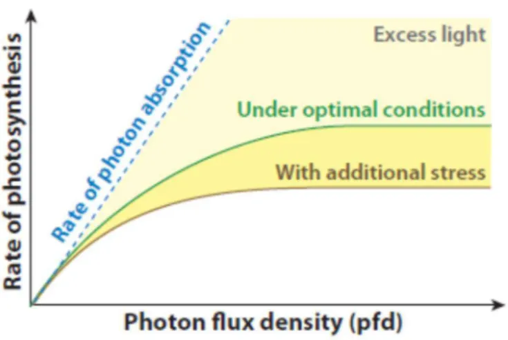
The majority of plants in nature are not exposed to optimal environmental conditions for growth (Goh et al., 2012). Indeed, for the most conditions high incident light (high input) or impairment of CO2 assimilation (low output) and in other important metabolic processes can
lead to photosynthetic unbalance, which is a serious threat for plants surviving (Ruban, 2015). This condition is defined as excess light stress (Fig. 2). Excess light stress may be particularly a relevant condition for tropical and equatorial regions, where radiation can reach very high levels between periods of cloud cover. Here, the average annual solar irradiation can exceed 1800 kWh/m2, typical of dry seasons in the tropics (Murchie et al., 2015). Regions such as
Central Luzon in Philippines and Central and East Java in Indonesia can receive accumulative annual solar irradiation of more than 2200 kWh/m2 (Murchie et al., 2015). Moreover,
combination of high light irradiance and drought, or salt stress, both usual conditions in semi-arid regions could severely aggravate excessive light stress in plants (Ferreira-Silva et al., 2011; Silva et al., 2015; Silveira and Carvalho, 2016).
Plants exposed to excess light accumulates more reducing power than circumstantial energetic demand (Li et al., 2009a; Takahashi and Badger, 2011; Goh et al., 2012). Under these conditions, several side-reactions associated to photosynthetic electron transport chain become more probable, such as reduction of O2 at PSII (Zulfugarov et al., 2014) and PSI (Asada, 1999),
Figure 2. Light response curves for photosynthesis compared with the rate of light absorption. Figure copied from Li et al., (2009a) - Annual Review of Plant Biology.
Photoinhibition is the reduction of photosynthetic rate in response to conditions of prolonged or pronounced excess light absorption. Under excessive light conditions, double reduction of plastoquinone (PQ) pool ceases the electron transport, which inhibits PSII activity. In addition, charge recombination reactions occurring in the inhibited PSII are more likely to lead to triplet state of the primary donor (P680), favouring production of oxygen singlet (Aro et al., 1993). Photoinhibition of PSII occurs in consequence of the produced singlet oxygen as well as by weakly coupled chlorophyll molecules (Goh et al., 2012). It has been therefore considered that, even in a healthy leaf, oxygen evolving complex (OEC) does not always function in all PSII centres, and these RC are prone to a rapid and irreversible photoinhibition (Goh et al., 2012).
At PSI level, Mehler-reaction is capable to also reduces O2 generating superoxide ions
O2- and in a subsequent reaction catalysed by superoxide dismutase (SOD), O2- is rapidly
converted in H2O2 (Mehler, 1951). Hydrogen peroxide is capable to easily transpose organelles
membranes and defund to virtually all cellular compartments (Mubarakshina et al., 2010). Moreover, H2O2 is believed to be associated with several signalling routes involved in plant
Fabricio E.L. Carvalho
opposition, when H2O2 is over-accumulated in plant cells, this molecule is able to interfere in
elongation factor G, affecting D1 de novo synthesis and generating impairment in RC repair mechanisms (Nishiyama et al., 2011). Figure 3 summarises ROS-dependent photoinhibitory mechanisms involving elongation factor G and PSII RC repair mechanisms.
Figure 3. A schematic representation of the interactive regulation of photosynthesis and protein synthesis. In light, reducing equivalents that are generated by the photosynthetic transport of electrons (e−) are transmitted to EF-G via thioredoxin-dependent (Trx-dependent) redox pathways. The resultant reduction of EF-G activates the translational machinery, leading to induction of the synthesis of the D1 protein and other proteins. The repair of PSII, in turn, requires the synthesis de novo of proteins and, in particular, that of the D1 protein. High levels of ROS, produced by the photosynthetic machinery, interrupt the redox signals by maintaining EF-G in an oxidized state and, consequently, they suppress protein synthesis that is required for the repair of PSII. Copied from Nishiyama et al., (2011) - Physiologia Plantarum.
NPQ; b) increase of energy output, which involves stimulation of CO2 assimilation, higher
photorespiratory activity, triggering of antioxidant defences, induction of NO3+ assimilatory
metabolism and activation of other reductant metabolic processes (Takahashi and Badger, 2011; Silveira and Carvalho, 2016). In figure 4, is possible to summarise the main mechanisms related to photoprotection in plants (Takahashi and Badger, 2011).
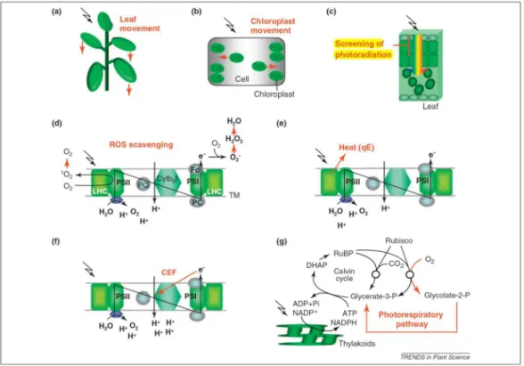
Figure 4. Examples of leaf and chloroplast mechanisms involved in minimizing photoinhibition of PSII. (a) Leaf movement. Leaves move to minimize the absorption of excessive light. (b) Chloroplast movement. Chloroplasts change their position to minimize the absorption of light. (c) Screening of photoradiation, for example, UV screening by phenolic compounds in epidermis cells. (d) ROS scavenging. O2 produced at PSII is scavenged by
membrane-bound a-tocopherol and carotenoids. O2- and H2O2 produced at PSI are scavenged
enzymatically and non-enzymatically by ascorbate. (e) Thermal energy dissipation of absorbed light energy (qE). qE dissipates light energy absorbed by photosynthetic pigments as heat at minor light-harvesting proteins. (f) CEF around PSI. CEF includes both the NAD(P)H dehydrogenase complex-dependent and PGR5-dependent pathways and helps to generates ΔpH
Fabricio E.L. Carvalho 3. Photoprotective mechanisms
In order to survive under different light conditions as previously mentioned, natural selection pressures have induced the development of several different strategies to optimization of light capture and energy use in plants (Ruban, 2015). Some of these strategies are related to maximization of light capture, which include emergence of antenna complex structures, increases in proportion of antennas/RC ratios and optimization of light quality use by processes such as PSII/PSI state transitions (Puthiyaveetil et al., 2012). These mechanisms are generally associated to shadowing and low light conditions and will not be focused on current review. In contrast, under excessive light, a very usual situation, several other mechanisms related to minimization of over-accumulated energy are described in literature (Takahashi and Badger, 2011; Goh et al., 2012). These mechanisms are now focus of the next section.
3.1. Leaf position and light incidence
Plant canopy are complex three-dimensional objects with a great variability in leaf size, shape, area, angle, curvature, twisting, and clumping (Burgess et al., 2015). These properties are especially important for studies involving gramineae plants, such as rice and wheat (Kriedeman et al., 1964; Murchie et al., 1999; Fourcaud et al., 2008). In these plant species, young leaves can exhibit 90° on light incidence angle and, under such conditions, cos θ equals to zero. Thus, differences in leaves positioning can represent a great strategy of photoprotection for several plant species, especially at physiological phases where leaves are particularly less light-tolerant (Carvalho et al., 2015). Interestingly, several plant species evolved complex physiological mechanisms associated to leaf movements, which, in turns can actively modulates θ angle of light incidence, and protect photosystems from photoinhibition under excessive energy (Takahashi and Badger, 2011).
Heliotropism, the leaf movement related to sunlight angle of incidence, consists in two distinct movements: diaheliotropism (leaf blade becomes oriented perpendicular to the angle of light incidence) and paraheliotropism (leaf blade becomes oriented perpendicular to light incidence angle). Especially, paraheliotropism is associated with minimizing energy capture, consisting in a photoprotective mechanism (Kao and Forseth, 1992; Pastenes et al., 2005). Paraheliotropism movements can reduces photoinhibition under excessive light conditions by two main strategies:
a) Reducing light input into antennas super-complexes by alterations in θ angle; b) Reducing the probability of ROS generation, because of lower energy
accumulated in thylakoid membranes, and therefore avoiding detrimental effects of ROS in PSII repair mechanisms (Takahashi and Badger, 2011).
Fabricio E.L. Carvalho
conditions (Takahashi and Badger, 2011). In A. thaliana plants, two proteins have been characterized as photoreceptors phototropin 1 (PHOT1) and phototropin 2 (PHOT2) and as essential to chloroplast movement (Sakai et al., 2001; Kasahara et al., 2002; Oikawa et al., 2003). The mechanistic basis of leaf chloroplast movements are actin filaments and chloroplast outer envelope protein CHUP1 is believed to be very important for this photoprotective mechanism (Takahashi and Badger, 2011).
3.2. Modulation of LHC-PSII genes/proteins and chlorophyll content under excessive light
Plants have evolved several different mechanisms to avoid excessive input of light energy into photosynthetic electron carriers. These mechanisms include active modulation in the organization and/or abundance of protein complexes in thylakoid membranes (Timperio et al., 2012), decreased levels of major chlorophyll a/b-binding light-harvesting complexes in relation to of reaction centers (Borisova-Mubarakshina et al., 2015), higher chl a/b ratios (Bailey et al., 2001; Ballottari et al., 2007), and modulation of PSII/PSI ratios (Puthiyaveetil et al., 2012; Borisova-Mubarakshina et al., 2015). Nevertheless, Zivcak et al., 2014 working with barley leaves concluded that decrease of chl a/b ratios in low light conditions strongly depends on plant species (Zivcak et al., 2014). Moreover, the authors evoked the theory of PSII connectivity to explain obtained data. According to this theory, PSII RC’s are interconnected and excitation energy should be distributed among then under illumination (Kramer et al., 2004). If it is true, the sigmoidal shape of fluorescence induction could affect the efficiency of utilization of absorbed light for trapping electrons in RC and hence, it should reflect on the entire fluorescence kinetics (Zivcak et al., 2014).
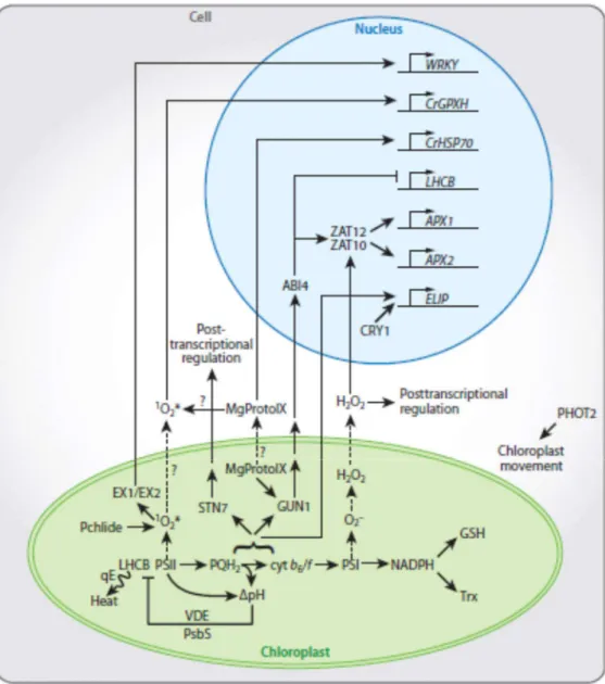
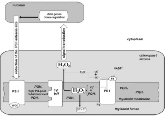
regulation of various intermediates of tetrapyrrole synthesis, such as 5-aminolevulinic acid - ALA (Li et al., 2009a). Moreover, increased Mg-Proto levels in plants exposed to photooxidative stress have been reported in the literature (Strand et al., 2003; Strand, 2004). In addition, ROS levels are believed to be related to accumulation of Mg-ProtoMe (Aarti et al., 2006). Figure 5 shows some of the most important routes of chloroplast signalling responses related to excessive light.
Fabricio E.L. Carvalho
Regulation of LHCB genes expression have recently been related to H2O2-dependent
signalling pathways (Borisova-Mubarakshina et al., 2015). Indeed, H2O2 roles in signalling
mechanisms are widely reported in literature (Desikan et al., 2001; Ślesak et al., 2007; Karpiński et al., 2013). Accordingly, results reported by Borisova-Mubarakshina et al., (2015) support the hypothesis that H2O2 is a molecular signal, which contributes to regulation of PSII
antenna size under excessive light, a response directly related to long-term high light acclimatory performance. In summary, the authors proposed H2O2 molecule released by
PQ-pool (Zulfugarov et al., 2014), as one of the main candidates by which redox state of PQ-PQ-pool provides antennas size regulatory effects (Figure 6).
Figure 6. A tentative scheme of H2O2 production in chloroplasts and a possible signal
transduction pathway for the down-regulation of lhcb genes by H2O2 originated in the PQ pool,
involved in the reduction of PSII antenna size under HL conditions. White arrows, superoxide radical and hydrogen peroxide production in chloroplast; Fd, ferredoxin; PSII, photosystem II; PSI photosystem I; PQ, PQ·−, PQH
2, plastoquinone, plastosemiquinone, plastohydroquinone
respectively; Pc, plastocyanin; SOD, superoxide dismutase; WOC, water-oxidizing complex. The PQH2/PQ ratio rises under long-term HL conditions; PQH2 spreads along the entire
3.3. NPQ – dissipating excess energy as heat
Natural selection pressures have conducted the development of several molecular mechanisms in plants related to excessive light tolerance (Goss and Lepetit, 2014). These molecular mechanisms are classified into two distinct groups of responses: long-term acclimation and short-term regulatory responses (Ruban et al., 2012). Among short-term acclimatory responses, an additional control loop in the form of a proton effect upon the PSII energy conversion event have been considered a crucial mechanism for plant defence against excessive light (Briantais et al., 1979; Genty et al., 1989). This process can be monitored by chlorophyll a fluorescence yield declining under conditions of photochemical excitation energy accumulation and is called non-photochemical quenching (NPQ) - (Govindjee et al., 1966; Murata and Sugahara, 1969; Wraight and Crofts 1970; Ruban, 2016). The figure 7 displays a synthetic model for NPQ activation in higher plants.
Figure 7. Scheme depicting photosynthetic and feedback control of photosynthesis in higher plants. PSII absorbs light and produces electrons (E.T.) which are transported through the thylakoid membrane to produce NADPH and drive formation of ΔpH for ATP synthesis. Build
up ΔpH exerts control over photosynthesis via inhibition of electron transport (photosynthetic
Fabricio E.L. Carvalho
NPQ is a complex mechanism composed of at least four major processes known to date: state transition (qT), photoinhibition (qI), heat dissipation (qE) and zeaxanthin-dependent (qZ) quenching (Nilkens et al., 2010; Ruban, 2016). These processes have individual features and probably reflect very distinct metabolic routes. Redistribution of light energy capture between PSII and PSI is believed to be related with qT (Benson et al., 2015). This process involves a partial detachment and migration of LHCII between PSII and PSI according to light conditions and is believed to be very important in algae (Baker, 2008; Puthiyaveetil et al., 2012). In higher plants, nevertheless, qT protective roles under excessive light have been questioned and qT functioning have been more associated to low light conditions (Baker, 2008).
The qI component is a slowly reversible process, which probably reflects damage to RCII and photoinhibition (Ruban et al., 2012). Photoinhibition by itself process can be considered as a protective mechanism in terms of whole plant surviving, since closed RCII confers protection compared to open ones (Matsubara and Chow, 2004). However, the strict threshold, which photoinhibition can be considered as a defence mechanism or, contrastingly represents an excessive light stress symptom is a hard to define. Indeed, classification of stress symptoms against defence responses is not a simple task (Silveira and Carvalho, 2016).
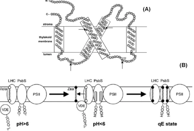
Two components are believed to act as direct protective mechanisms for avoiding damages to RCII, and therefore, photoprotection under excessive light. The fastest component of NPQ, and also the more frequently studied in plants, is named qE, and its photoprotective role is probably related to dissipation of excess energy as heat (Ruban et al., 1993). More recently described, qZ is probably related to synthesis and epoxidation of zeaxanthin and probably display roles in photoprotection. The lifetime of qE and qZ formation/relaxation present order of 5 and 15 minutes respectively, therefore consisting in the faster NPQ components (Nilkens et al., 2010; Ruban, 2016).
because of photosynthetic electron transport in thylakoid membranes is the primordial event triggering qE (Krause, 1974; Ruban et al., 1992). Subsequently, zeaxanthin binding to light harvesting complexes is related to a new state of aggregation, which allows this system to work as a thermal dissipater of energy (Demmig-Adams and Adams, 1992; Ruban et al., 1992; Farber et al., 1997; Ruban et al., 1999). Finally, an important transmembrane chl a-b binding protein with 22 kDa was discovered to display a crucial role in qE activation (Funk et al., 1995; Li et al., 2002; Niyogi et al., 2005). This protein called PsbS, presents a particular role of increase sensitivity of thylakoid membranes to pH changes, allowing qE formation under physiological
ΔpH values (Johnson and Ruban, 2011). Figure 8 and 9 summarises the mechanisms underlying
qE formation in higher plants.
Figure 8. Structural model of NPQ-related reorganization of thylakoid grana membranes. In the dark and low light, LHCII is fairly distributed in the grana, forming large C2S2M2
super-complexes with PSII and minor antenna proteins. In excess light, ΔpH triggers a conformational
Fabricio E.L. Carvalho
Nevertheless, the precise site of qE formation still have been theme of intense discussion in the last decades (Ruban et al., 1992; Ruban et al., 2007; Ruban et al., 2012; Chmeliov et al., 2015). Presently, there are three more discussed theories for explaining the site of qE formation, which all of them predicting for LHC antennas involvement:
1. Energy transfer by interactions between chlorophyll molecules bound to antenna protein complexes (Horton et al., 1996);
2. Quenching interactions between lutein bound at the L1 site and chlorophylls a612–a611–a610 of LHC antennas (Ruban et al., 2007);
3. Xanthophyll–chlorophyll quenching interactions between zeaxanthin bound at the L2 site and chlorophylls A5 and B5 of LHC complexes (Ahn et al., 2008). In order to determine the precise region of LHC antennas related to qE formation, several studies involving genetic transformation approaches have been performed. Absence of CP26 subunit did not affected qE, while CP29 and CP24 deficiencies promoted 30% and 50% respectively of qE impairment (Andersson et al., 2001; Kovács et al., 2006). Intriguingly, double deletion for CP29 and CP24 combined with 50% reduction in CP26 levels did not affected qE (de Bianchi et al., 2008). In parallel, deficiency in Lhcb1 and Lhcb2 (major antennas), lead to 35% decrease in qE (Andersson et al., 2003). Accordingly, reports on plants containing only CP26 exhibited only a partial reduction in qE (Jahns and Krause, 1994). Therefore, it is plausible to suggest no individual LHCII complex acting as the sole site of qE and, in principle, the quenching could occur in any of the four types of LHCII (Ruban et al., 2012).
Fabricio E.L. Carvalho
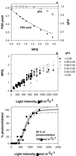
actual photoprotection of PSII has not been demonstrated yet (Jahns and Holzwarth, 2012). Therefore, in order to understand the physiological importance of different NPQ processes and their photoprotection effects, future work will have to explore this relationship in detail. A new fluorimetric methodology for photoinhibition tracking, based on qPd parameter quantification have been introduced recently (Ruban and Murchie, 2012).
The qPd approach presents a high potential to investigations involving NPQ and photoprotection since it allows to estimate the precise NPQ fraction associated with photoprotection, pNPQ (Ruban and Belgio, 2014; Ware et al., 2014; Carvalho et al., 2015; Giovagnetti and Ruban, 2015). This methodology has been successfully employed so far and positive interactions have been demonstrated between pNPQ parameter and PsbS protein expression (Ware et al., 2014), oxygen evolution complex activity (Giovagnetti and Ruban, 2015) and ontogenesis (Carvalho et al., 2015). Moreover, evidences have connected pNPQ and photoprotection to hydrogen peroxide and oxidative stress, however further studies are needed in that direction to clarify this question (Carvalho et al., 2015).
3.4. Antioxidative metabolism and photoprotection
Plants exposed to excessive light, generally, display an over-accumulation of reduction power (Li et al., 2009a). Usually, this excess of reducing power is produced because of unbalance between NADPH and ATP production in photosynthetic electron transport chain (PETC) and energetic demand of general cell metabolism (Foyer et al., 2012). Under such conditions, formation of reactive oxygen species (ROS) is extremely favoured. The two most important sites of ROS generation in green leaf cells are peroxisomes and chloroplasts, followed by mitochondria (Foyer and Noctor, 2003). In peroxisomes, ROS are produced especially by activity of glycolate oxidase (GO), which scavenges glycolate, producing H2O2 (Corpas, 2015).
phosphor-glycolate produced by Rubisco oxygenation activity, also called photorespiration (Voss et al., 2013).
The most of cellular H2O2 produced is related to photorespiratory pathway in
peroxisomes (Corpas, 2015). Indeed, peroxisomes of illuminated leaves are believed to generate up to 10 µmol m-2 s-1 of H
2O2 (Foyer and Noctor, 2003). Several studies have reported
H2O2 as important signalling molecules involved in abiotic stress responses (Karpinski et al.,
1997; Desikan et al., 2001; Ślesak et al., 2007; Li et al., 2009a; Suzuki et al., 2013). However, H2O2 over-accumulation is involved in several harmful processes, such as protein carbonylation
and lipid peroxidation, which are related to oxidative stress (Davletova et al., 2005). In peroxisomes, catalase (CAT) is a very important enzyme associated to H2O2 scavenging,
independent of electron donors and presenting a high kM to hydrogen peroxide (Mhamdi et al., 2010).
Chloroplasts are the second most important site of H2O2 generation in illuminated leaf
cells, reaching ratios up to 4 µmol m-2 s-1 (Foyer and Noctor, 2003). In chloroplast H2O2 is
generated by different routes from that evidenced in peroxisomes, which involves other ROS such as radical ion superoxide (O2-) - (Mubarakshina and Ivanov, 2010). Indeed, two distinct
theories have been used to explain H2O2 generation in chloroplasts (Asada, 1999; Mubarakshina
and Ivanov, 2010; Zulfugarov et al., 2014). Earlier, H2O2 production was credited to PSI site,
as a consequence of Mehler reactions (Mehler, 1951). In the PETC, electrons are carried to PSI Fe-S clusters and then to ferredoxin (Hill and Bendall, 1960). As O2 is a very abundant molecule
in chloroplasts, this molecule could take place of ferredoxin at PSI and act as electron receptor in the Hill chain (Mehler, 1951; Asada, 1999). The product of such reaction, O2-, is very
reactive and harmful, and rapidly scavenged by superoxide dismutase activity (SOD), generating H2O2 (Asada, 1999).
However, a second theory supports H2O2 as being generated at PSII, instead PSI
Fabricio E.L. Carvalho
on electron acceptor side of PSI is more negative as compared with PSII, therefore O2 reduction
in PSI should occurs with high efficiency (Asada, 1999). However, evidences of possible involvement of PSII acceptor side in molecular oxygen reduction to superoxide have been recently reported (Pospísil, 2009; Pospíšil, 2014; Zulfugarov et al., 2014). This reaction should occur when semiquinone (QA-) reduces O
2 generating superoxide, and subsequently, H2O2
(Fig. 10).
Figure 10. Production of ROS by acceptor- and donor-side mechanism in PSII. On the electron acceptor side of PSII, one-electron reduction of molecular oxygen by the reducing side of PSII results in the formation of O2-, which subsequently either dismutate it into free H2O2. In the
presence of free metals (Fe2+, Mn2+), the reduction of H
2O2 results in the formation of HO via
Fenton-type reaction. On the electron donor side of PSII, two-electron oxidation of H2O by the modified water-splitting Mn complex results in the formation of H2O2. Hydrogen peroxide has
been proposed to be either oxidized to O2- by tyrosine radical TyrZ or reduced to HO by free
metals the most likely being Fe2+, Mn2+released from damaged PSII. Reproduced from Pospíšil
(2009) - Biochimica et Biophysica Acta.
In chloroplasts, CAT is absent, and therefore, ascorbate peroxidase (APX) was classically believed to be the most important enzyme involved in H2O2 scavenging (Nakano
almost all cell compartments (Teixeira et al., 2004). APX are encoded by multi-genic families and isoforms are classified according to different subcellular localization (Teixeira et al., 2004). In Arabidopsis thaliana L., there are at least six genes encoding APX isoforms: two cytosolic isoforms (At1g07890 and At3g09640); two peroxisomal isoforms (At4g35000 and At4g35970); a thylakoid-bound isoform (At1g77490) and a stromal/mitochondrial isoform (At4g08390) (Teixeira et al., 2006). In contrast, rice present 8 different APX isoforms: two cytosolic isoforms (OsAPX1 and OsAPX2), two peroxisomal isoforms (OsAPX3 and OsAPX4), one mitochondrial/chloroplastic isoform (OsAPX5), one mitochondrial (OsAPX6) and two chloroplastic, one stromal (OsAPX7) and one thylakoidal isoform (OsAPX8) - (Bonifacio et al., 2011).
Regardless cell compartment, APX activity is dependent of ascorbate (ASC) as electron donor for H2O2 scavenging reaction (Nakano and Asada, 1981). Ascorbate is the most abundant
soluble antioxidant in chloroplasts, reaching concentrations up to 300 mM in these organelles (Smirnoff, 2000). ASC is synthetized in mitochondria and must be imported to chloroplasts in order to act as photoprotective molecule (Smirnoff, 2000). After ASC reach chloroplasts, this molecule can act in several mechanisms of photoprotection, such as:
1. Electron donor for APX activity; 2. Direct scavenging of other ROS;
3. Regeneration of α-tocopheryl radicals produced when α-tocopherol reduces lipid peroxyl radicals;
4. Alternative electron donor to PSII in absence of OEC function; 5. Substrate of violaxanthin de-epoxidase (VDE), crucial for qE.
Fabricio E.L. Carvalho
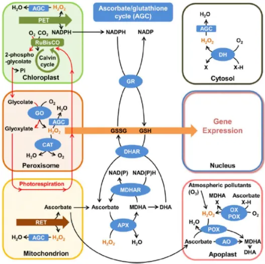
rapidly recovered by MDHA reductase or DHA reductase enzymes (DHAR) - (Foyer and Noctor, 2011). This last enzyme employs reducing power from reduced glutathione (GSH) for its activity, which, in turns, is regenerated from its oxidized form (GSSG) by activity of glutathione reductase (GR), using NADPH as electron donor (Foyer and Noctor, 2011). This cyclic process is referred as ascorbate-glutathione cycle and is considered a very important mechanism for ROS scavenging in chloroplasts under excessive light (Munné-bosch et al., 2013). Figure 11 summarises redox metabolic process in different plant cell organelles.
Figure 11. Schematic representation of subcellular hydrogen peroxide (H2O2) metabolism and
3.4.1. Does chloroplastic APX really play crucial role in plant responses to excessive light?
Plants exposed to excessive light display a strong induction of different isoforms of APX, especially cytosolic (Karpinski et al., 1997) and chloroplastic (Klein et al., 2012). Moreover, plants genetically transformed to over-express APX isoforms display increased tolerance to several abiotic stress conditions, for instance high salinity (Badawi et al., 2004; Li et al., 2009b; Singh et al., 2014). Based on these data, the answer to the question that intitules this section should be obvious. However, several studies employing the use of knocked out plants in order to assess the importance of these enzymes to abiotic stress responses have reported contrasting results (Danna et al., 2003; Giacomelli et al., 2007; Kangasjärvi et al., 2008; Maruta et al., 2010). Surprisingly, the majority of these works have reported no differences in transformed plants deficient in chloroplastic APX, in comparison to NT, under optimal growth conditions, and, sometimes even under abiotic stress (Danna et al., 2003; Giacomelli et al., 2007; Kangasjärvi et al., 2008; Maruta et al., 2010).
Fabricio E.L. Carvalho 4. Remarks and further perspectives
During ~500 million years of evolution, land plants must develop several strategies to cope with oscillations in light intensity, sustain its photosynthetic activity and survive. Under the majority of environmental conditions, light provides more energy than the whole plant metabolism can use and, therefore, plants are commonly subjected to excessive light conditions. In the current review some important mechanisms of photoprotection are discussed, employing an integrative view, which encompass fast adjustments processes such as NPQ and ROS scavenging in chloroplasts and long-term acclimatory mechanisms, involving regulation of expression of several genes/proteins. An especial focus is given to chloroplastic APX, believed to be one of the most important peroxidases in chloroplasts.
As evidenced here, several reports concerning chloroplastic APX deficiency are not able to explain its importance. Complexity of systemic responses and gene/protein networks in leaf cells should have contributed in great part for this difficulty. Certainly, understanding the importance of other peroxidases such as peroxiredoxins (PRX) should be essential for further interpretation of such controversial results involving APX proteins. Moreover, in order to achieve a deeper knowledge regarding plant cell metabolism and, therefore, take more robust conclusion about the importance of each mechanism of photoprotection related to sustain photosynthesis under excessive light, new integrative approaches are needed.
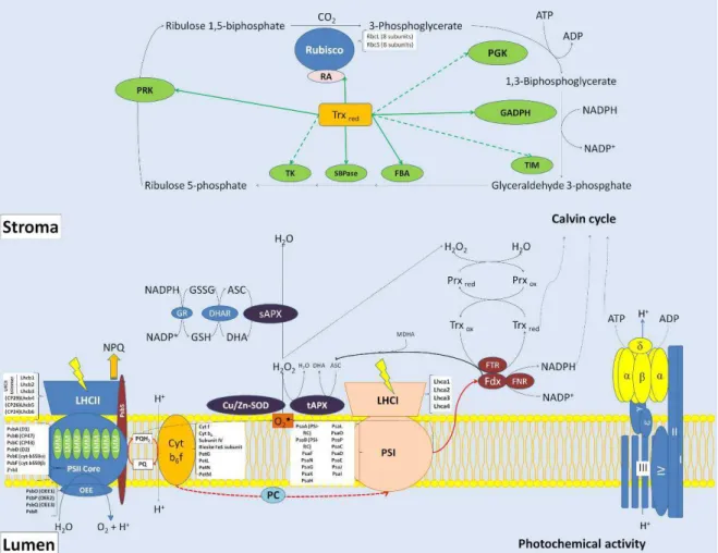
Figure 12. Simplified photosynthetic model containing the several components of photochemical (PSII and PSI) and Calvin cycle phases. The scheme shows the main photosynthesis proteins, including those related to redox reactions, which are commonly characterized by proteomics approaches. Abbreviations: ASC, ascorbate; Cu/Zn-SOD, Cu/Zn superoxide dismutase; Cyt b6f, cytochrome b6f; DHA, dehydroascorbate; DHAR, dehydroascorbate reductase; FBA, fructose biphosphate aldolase; FDX, ferredoxin; FNR, ferredoxin NADP(H) oxireductase; FTR, ferredoxin-thioredoxin reductase; GADPH, glyceraldehyde phosphate dehydrogenase; GR, glutathione reductase; GSH, reduced glutathione; GSSG, oxidized glutathione; LHCI, light harvest complex antenna of PSI; LHCII, light harvest complex antenna of PSII; LMM, PSII low molecular mass proteins; MDHA, monodehydroascorbate; NPQ, non-photochemical quenching; OEE, oxygen evolving enhancer proteins; PC, plastocyanin; PGK, phosphoglyceratekinase; PQ, oxidized plastoquinone; PQH2,
reduced plastoquinone; PRK, phosphoribulokinase; PRXox, oxidized peroxiredoxin; PRXred,
reduced peroxiredoxin; PSI, photosystem I; PSII, photosystem II; RA, rubisco activase; Rubisco, ribulose 1,5-biphosphate carboxylase/oxygenase; sAPX, stromal ascorbate peroxidase; SBPase, sedoheptulose biphosphate phosphatase; tAPX, thylakoidal ascorbate peroxidase; TIM, triose phosphate isomerase; TK, transketolase; TRXox, oxidized thioredoxin;
TRXred, reduced thioredoxin. Reproduced from Silveira and Carvalho (2016) - Journal of
Fabricio E.L. Carvalho References
Aarti PD, Tanaka R, Tanaka A (2006) Effects of oxidative stress on chlorophyll biosynthesis in cucumber (Cucumis sativus) cotyledons. Physiol Plant 128: 186–197
Ahn TK, Avenson TJ, Ballottari M, Cheng Y-C, Niyogi KK, Bassi R, Fleming GR (2008) Architecture of a charge-transfer state regulating light harvesting in a plant antenna protein. Science 320: 794–797
van Amerongen H, Croce R (2013) Light harvesting in photosystem II. Photosynth Res 116: 251–263
Andersson J, Walters RG, Horton P, Jansson S (2001) Antisense inhibition of the photosynthetic antenna proteins CP29 and CP26: implications for the mechanism of protective energy dissipation. Plant Cell 13: 1193–1204
Andersson J, Wentworth M, Walters RG, Howard CA, Ruban AV, Horton P, Jansson S (2003) Absence of the Lhcb1 and Lhcb2 proteins of the light-harvesting complex of photosystem II - Effects on photosynthesis, grana stacking and fitness. Plant J 35: 350– 361
Aro EM, McCaffery S, Anderson JM (1993) Photoinhibition and d1 protein degradation in peas acclimated to different growth irradiances. Plant Physiol 103: 835–843
Asada K (1999) The water-water cycle in chloroplasts: scavenging of active oxygens and dissipation of excess photons. Annu Rev Plant Physiol Plant Mol Biol 50: 601–639 Baccar R, Fournier C, Dornbusch T, Andrieu B, Gouache D, Robert C (2011) Modelling
the effect of wheat canopy architecture as affected by sowing density on Septoria tritici epidemics using a coupled epidemicvirtual plant model. Ann Bot 108: 1179–1194
Badawi GH, Kawano N, Yamauchi Y, Shimada E, Sasaki R, Kubo A, Tanaka K (2004) Over-expression of ascorbate peroxidase in tobacco chloroplasts enhances the tolerance to salt stress and water deficit. Physiol Plant 121: 231–238
Bailey S, Walters RG, Jansson S, Horton P (2001) Acclimation of Arabidopsis thaliana to the light environment: the existence of separate low light and high light responses. Planta 213: 794–801
Baker NR (2008) Chlorophyll fluorescence: a probe of photosynthesis in vivo. Annu Rev Plant Biol 59: 89–113
Benson SL, Maheswaran P, Ware MA, Hunter CN, Horton P, Jansson S, Ruban AV, Johnson MP (2015) An intact light harvesting complex I antenna system is required for complete state transitions in Arabidopsis. Nat Plants 1: 15176
de Bianchi S, Betterle N, Kouril R, Cazzaniga S, Boekema E, Bassi R, Dall’Osto L (2011) Arabidopsis mutants deleted in the light-harvesting protein Lhcb4 have a disrupted photosystem II macrostructure and are defective in photoprotection. Plant Cell 23: 2659– 2679
de Bianchi S, Dall’Osto L, Tognon G, Morosinotto T, Bassi R (2008) Minor antenna proteins CP24 and CP26 affect the interactions between photosystem II subunits and the electron transport rate in grana membranes of Arabidopsis. Plant Cell 20: 1012–1028
Blankenship RE (2013) Molecular mechanisms of photosynthesis. John Wiley & Sons
Bonifacio A, Martins MO, Ribeiro CW, Fontenele AV, Carvalho FEL, Margis-Pinheiro M, Silveira JAG (2011) Role of peroxidases in the compensation of cytosolic ascorbate peroxidase knockdown in rice plants under abiotic stress. Plant, Cell Environ 34: 1705– 1722
Borisova-Mubarakshina MM, Ivanov BN, Vetoshkina DV, Lubimov VY, Fedorchuk TP, Naydov IA, Kozuleva M a, Rudenko NN, Dall’Osto L, Cazzaniga S, et al (2015) Long-term acclimatory response to excess excitation energy: evidence for a role of hydrogen peroxide in the regulation of photosystem II antenna size. J Exp Bot 66: 7151–7164 Briantais JM, Vernotte C, Picaud M, Krause GH (1979) A quantitative study of the slow
decline of chlorophyll a fluorescence in isolated chloroplasts. BBA - Bioenerg 548: 128– 138
Bricker TM, Roose JL, Fagerlund RD, Frankel LK, Eaton-Rye JJ (2012) The extrinsic proteins of Photosystem II. Biochim Biophys Acta - Bioenerg 1817: 121–142
Burgess AJ, Retkute R, Pound MP, Foulkes J, Preston SP, Jensen OE, Pridmore TP, Murchie EH (2015) High-resolution three-dimensional structural data quantify the impact of photoinhibition on long-term carbon gain in wheat canopies in the field. Plant Physiol 169: 1192–1204
Calvin M, Benson AA (1948) The path of carbon in photosynthesis. US Atomic Energy Commission, Technical Information Division
Carvalho FEL, Ware MA, Ruban AV (2015) Quantifying the dynamics of light tolerance in Arabidopsis plants during ontogenesis. Plant Cell Environ 38: 2603–2617