123
Rev Bras Med Esporte – Vol. 17, No 2 – Mar/Abr, 2011 ABSTRACT
Therapeutic ultrasound is seen today as one of the most useful resources in the practice of clinical medicine and physical exercise is consolidated as an effective and efficient therapeutics in several cases; however, they are still little investigated when associated. Therefore, the present work has as the aim to analyze the influences of ultrasound and physical exercise on serum and muscle triglycerides concentrations in experimental diabetes rats. Adult Wistar rats were used and divided in eight groups: Sedentary Diabetics (SD), Trained Diabetics (TD), Sedentary Diabetic and Ultrasound (SDUs), Trained Diabetic and Ultrasound (TDUs), Sedentary Control (SC), Trained Control (TC), Sedentary and Ultrasound Controls (SUCs), Trained Control and Ultrasound (TCUs). The train-ing protocol was composed of swimmtrain-ing exercise five days a week, 30 daily minutes and with maximum load of 8% of body mass during three weeks. The ultrasound therapy was performed five days a week, for two weeks, with intensity of 0.2W/cm² and frequency of 1.0MHz. No significant differences were observed in the serum triglycerides or in the tibialis anterior and gastrocnemius muscles. Concerning the soleus muscle, the diabetic groups showed lower concentrations com-pared to the control groups and TD, and TDUs groups showed lower concentrations comcom-pared to SD and SDSU, with the trained groups presenting the lowest concentrations. The pulsed ultrasound in the intensity investigated did not influence serum triglycerides or IMTG. However, exercise was effective in reducing soleus muscle triglycerides.
Keywords: physical exercise, ultrasound therapy, triglycerides.
INTRODUCTION
One of the physical resources mostly used nowadays in the sports clinical practice is the thera-peutic ultrasound. Due to its extensive use, ultrasonic energy has been investigated as an excellent supporting aid for many biological processes. It is a therapeutic agent able to reach tissues at depth of 3 to 5cm1, producing many effects such as increase of local metabolism, increase of circulation,
extensibility of the conjunctive tissue and tissue regeneration (1-7), increase of enzymatic activity as
well as alteration of the contractile activity of the muscle (4,5). Ultrasound can be used in
combina-tion with other therapies, especially physical exercise, boosting hence its benefits (2).
The intramuscular triglycerides (IMTG) have been target of many investigations having as focus the correlation of exercise and diabetes mellitus. It is common sense among researchers that the unwanted increase of IMTG is a factor which contributes to insulin resistance (8-16). It was found out
that rats induced to experimental diabetes present IMTG buildup. Besides the IMTG excess, decrease in the fat oxidation rate has been reported in diabetic and obese humans (8,11,14,16).
The majority of the IMTGs are stored within the muscle fibers, near the myofibrils and constitute in important storage of intramuscular fuel (12,17), with the energy provided by the triglycerides is 60
times higher than the glycogen in the muscle (16).
Many techniques have been used to revert the deleterious effects produced by the exacerbated buildup of IMTG; however, until the present moment the only non-pharmacological method known which directly reduces the IMTG is the muscle activity (15). Search in the database did not confirm
the use of ultrasound therapy as isolate or combined method to alter the IMTG concentrations. Due to the ultrasound effects already confirmed, the present study has the aim to analyze the ul-trasound and exertion exercise influence on the serum and muscular triglycerides concentrations in rats submitted to experimental diabetes.
The Influence of Ultrasound and Physical
Trainning on Serum and Muscle Tryglicerides
in Experimental Diabetic Rats
Rodrigo Augusto Dalia¹ Marcelo Renato Guerino² Nivaldo Antonio Parizotto² Maria Alice Rostom de Mello1 Eliete Luicano¹
1. Physical Education Department, UNESP, Rio Claro, SP.
2. Physiotheraphy Department UFSCar – SP.
Mailing Address:
Avenida 39, 158, entre Ruas 2 e 3 – 13501-180 – Rio Claro, SP, Brasil E-mail: [email protected]
EXERCISE AND SPORTS SCIENCES
124 Rev Bras Med Esporte – Vol. 17, No 2 – Mar/Abr, 2011 MATERIALS AND METHODS
Animals
Wistar rats, with approximate age of 120 days were used for the experiment. The animals were obtained from the Central Animal Facility of UNESP, Botucatu Campus, and kept during the entire experiment in the Animal facility of the Laboratory of Biodynamics of the Physical Education Department of the Bio-sciences Institute of UNESP, Rio Claro Campus. The animals were kept in collective cages with a maximum of five animals, being placed in a room with controlled temperature of 25°C and light/ dark cycle of 12/12h, with water ad libitum and fed with balanced
solid chow for rodents (Labina Purina
®
). All experimental proce-dures are according to the guidelines from the Brazilian College of Animal Experimentation (COBEA).Experimental diabetes
The animals selected to the diabetic group were kept at fast for a period of 12 hours. After this period, the animals were anes-thetized in a CO2 chamber and received an aloxane solution in
citrate buffer 0.01M, pH 4.5 in the 35mg for each kg of body mass ratio. Administration was intravenous in the penis dorsal vein. After induction, the animals were treated with glucose added to water (125g of sugar for each 500ml of water) during 24 hours, with the aim to avoid death by severe hypoglycemia(18). Experimental
diabetes was confirmed by glycemia check by glucose oxidase method (19). The animals which presented glycemia higher or equal
to 250mg/dl were included in the diabetic group(18) ,while the
ani-mals which presented values lower than 250mg/dl were discarded from the experiment.
Experimental groups
Four distinct experimental diabetic groups were formed as described: Sedentary Diabetics (SD): the rats from this group were induced to experimental diabetes and kept with not any kind of physical activity; Trained Diabetics (TD): they were induced to experimental diabetes and performed the physical exercise protocol; Sedentary Diabetic and Ultrasound (SDUs): the rats from this group were induced to experimental diabetes, kept with not any kind of physical activity, but performed the ultrasound therapy; Trained Diabetic and Ultrasound (TDUs): they were in-duced to experimental diabetes, performed the physical training protocol and also the ultrasound therapy. In the same way, four control groups were formed, which received the same physical training and ultrasound therapy treatment: Sedentary Control (SC); Trained Control (TC); Sedentary Control and Ultrasound (SCUs); Trained Control and Ultrasound (TCUs). The difference in the number of animals observed between groups was due to the loss of some animals occurred during the experiment period, which is common in these cases.
Physical training protocol
The protocol was constituted of three weeks of a swimming program in a collective tank with water at temperature between 30°C ± 2°C. The training was performed five times per week, one session a Day, in which on the first week of training load equivalent to 5% of body mass attached to the animals’ trunk was used, with duration of 15 minutes on the first day, 20 minutes on the second day and 30 minutes on the following days until
the end of the week, being it considered the adaptation period to the water environment of the animal. At the beginning of the second week, the load was increased to 8% of body mass, repeating the same duration of the previous week and on the third week the load was kept at 8% of body mass and period of 30 minutes during the entire week(20).
Ultrasound therapy protocol
The equipment used in this study was the (Sonacel Plus, Bioset
®
), which was calibrated before the beginning of the ex-periment by the manufacturer himself, presenting the following physical parameters: pulsed wave in the repetition frequency of 100Hz, pulsed wave 1:2 or 50%, frequency of 1.0MHz and intensity of 0.2W/cm², considering that the ERA was exclusively designed for the procedure. The probe presented effective ra-diation area (ERA) of 0.5cm², adapted to this experiment by the manufacturer, during 10 minutes. The ultrasound treatment consisted in 10 sessions, five days a week, one daily session, for a period of two weeks. The rats selected for the ultrasound therapy were trichotomized and their entire right hinder legs were sterilized with alcohol 97% to facilitate the wave transmis-sion. In order to improve the connection between the ultra-sound probe and the muscle regions a hydrosuluble gel was used. The ultrasound was directly applied on the entire right hinder leg, including the anterior and posterior parts of the paw.Biochemical evaluations
24 hours after the end of the experimental period, all animals were euthanized by decapitation. The blood was collected, imme-diately centrifuged and the serum separated and used for glyce-mia and triglycerides (TG) evaluation. The glucose and triglycerides amount in the serum was determined by colorimetric enzymatic method using commercial kits (LaborLab
®
)(19). Subsequently, thetibialis anterior, gastrocnemius and soleus muscles of the right hinder leg were collected and separately weighted on a high preci-sion analytical scale for evaluation of the amount of intramuscular triglycerides (IMTG). The samples were placed in Eppendorf tubes of 1.5ml, containing 0.5ml of TritonX-100 at 0.1%. The samples were triturated in Polytron
®
for 10 seconds in a pulsatile way; after this procedure the samples were centrifuged at 4,000rpm for 10 min-utes(21). The supernatant was extracted for TG determination byphotospectrometry with use of a commercial kit (LaborLab
®
).Statistical analysis
The data obtained in each group were described as mean ± standard deviation and were compared between each other by analysis of variance (ANOVA) and application of Tukey test. In all cases the significance level was set at 5%. The software used was the Statistica 7.
RESULTS
125
Rev Bras Med Esporte – Vol. 17, No 2 – Mar/Abr, 2011
Figure 1. Triglycerides of the tibialis anterior muscle of the groups at the end of the experiment. Results expressed in mean ± standard deviation, n = 8 animals per group.
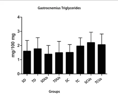
Figure 2. Triglycerides of the gastrocnemius muscle of the groups at the end of the experiment. Results expressed in mean ± standard deviation, n = 8 animals per group.
Figure 3. Triglycerides of the soleus muscle of the groups at the and of the experiment. Results expressed in mean ± standard deviation, n = 8 animals per group. a ≠ of SC, TC, SCUs and TCUs, b ≠ of TD and TDUs, ANOVA p < 0.05.
The results concerning the triglycerides of the tibialis anterior and gstrocnemius muscles were described in figures 1 and 2, respectively. None statistically significnat difference was found between groups in both cases.
Concerning the soleus muscle, the triglycerides concentra-tions found in the musculature were expressed in figure 3. The concentrations observed in the diabetic groups SD, TD, SDUs and TDUs were significantly lower than the in the control groups SC, TC, SCUs and TCUs. Another important and significant differ-ence was observed between the trained and sedentary diabetic groups, regardless of the ultrasound therapy; the TD and TDUs groups presented significantly lower values compared to the SD and SDUs groups.
DISCUSSION
The ultrasound is very much applied in sports medicine prac-tice, being the musculoskeletal system one of its main targets. However, little is known about its influence on some muscular energetic substrates, such as the IMTGs. The is no record in the literature on this kind of analysis prior this study here.
In the present investigation, the main finding was that thera-peutic ultrasound in its pulsed modality, did not influence on the intramuscular triglycerides concentrations (IMTG), regardless of physical training or type of muscle fiber. According to Guo and Jensen when using electrical stimulation, they observed in mon-keys that such physical parameter during five hours did not alter the IMTG concentrations in gastrocnemius or soleus muscles(22),
being it the only investigation in the literature which used a thera-peutic physical device to investigate its influence on the IMTGs.
The relatively low intensity as well as reduced time of therapy can explain the absence of effects on the IMTG concentrations both in diabetic rats and control rats. Dissipation of total energy used due to adaptation of the application probe may also have occurred. One of the main ultrasound effects on the IMTG would be increase in metabolism, also due to increase of local tissue temperature, which is dependent on the time of therapy; that is to say, the longer the time of therapy, the greater the increase of local tissue temperature(6).
Such effects can be produced by the pulsed modality; how-ever, the best effectiveness of the thermal effects is produced by the continuous mode, which can also increase the permeability of the biological membranes and alterations in the membrane Table 1. Glucose and TG serum concentrations of the groups at the end of the experimental period.
Groups Glucose (mg/dL) TG (mg/dL)
SD 312.92 ± 30.40* 134.84 ± 34.48
TD 249.00 ± 44.83*# 129.44 ± 36.19
SDUs 287.61 ± 44.40* 169.35 ± 53.76
TDUs 227.55 ± 64.86*# 128.10 ± 45.33
SC 113.23 ± 15.33 123.67 ± 47.19
TC 123.33 ± 12.08 174.24 ± 17.47
SCUs 121.70 ± 14.79 130.49 ± 30.40
TCUs 115.91 ± 30.15 143.35 ± 43.32
Results expressed in mean ± standard deviation, n = 8 animals per groups. * ≠ of SC, TC, SCUs and TCUs, # ≠ of SD and SDUs, ANOVA p < 0.05.
Tibialis Anterior Triglycerides
Gastrocnemius Triglycerides
Soleus Triglycerides
SD TD SDU
s TDU
s
SC TC SCUs TCUs
Groups
SD TD SDU
s TDU
s
SC TC SCU
s TCUs
Groups
SD TD SDU
s TDU
s
SC TC SCU
s TCUs
126 Rev Bras Med Esporte – Vol. 17, No 2 – Mar/Abr, 2011 potential(23). Therefore, the results obtained in this study were not
conclusive concerning the modality selection and physical param-eters so that alterations on the IMTGs may occur; hence, further investigation is still needed.
Physical exercise is well-consolidated as having effect similar to the insulin activity in the glycemic homeostasis control(24). In our
study, physical training decreased glycemia of the diabetic animals, which corroborates many investigations in the literature(25). The
serum TG concentrations have not been altered, contrasting with previous reports in the literature which report decrease of these concentrations with physical training, probably due to the fact that the training proposed in this study is of short duration.
The data concerning the IMTG shown in figure 1 are in agree-ment with the literature, which shows that use of both IMTG and glycerol is dependent on the type of muscle fiber (13,14,16,22,26,27). Since
the tibialis anterior is classified as a muscle with predominance of type II fiber, with approximately 90%, it would be more dependent on the glycidic metabolism(28). According to some researchers, the
type I fibers manifest increase of oxidative capacity (11,13), which
jus-tifies the IMTG reduction only in the soleus muscle, which contains only type I fibers in its composition.
The treatment association by ultrasound to training seems to have contributed to the increase in the substrate concentration; however, the ultrasound alone did not reestablish the low levels induced by the disease. The gastrocnemius is a mixed muscle (28),
but with predominance of type II fibers, in which the capacity of fat uptake and oxidation, such as IMTG, is very low.
The IMTG concentrations found in the soleus muscle were similar to the ones found in other studies(13,22), that is to say, all
the diabetic groups presented triglycerides decrease. The soleus muscle presents predominance of type I odidative fibers and
con-tains higher amount of IMTG and greater oxidative capacity(13,22).
The main difference found in the IMTG concentrations in the soleus muscle was due to the physiological status of the diabetic animal. Higher use of IMTG as energetic substrate, at rest or exercise status, was observed in diabetic rats, compared to the control rats(9,11).
This higher use is due to the lack of insulin, which has its oxidation regulating role, both of fatty acids and IMTG, at exercise and rest
(9,12); therefore, the amounts and use of IMTG have a strong
correla-tion with insulin sensitivity and concentracorrela-tion(10,11,15,29).
The hormone-sensitive lipase enzyme (HSL) is one of the main IMTG hydrolysis regulators(16,17,30) and insulin plays a regulating role
of this enzyme. Low insulin concentrations, as in the case of ex-perimental diabetic, may stimulate the HSL, promoting increase of IMTG hydrolysis in diabetic individuals.
It is also acknowledged that IMTG oxidation is higher during ex-ercise compared to at rest (12); which was corroborated in our study
by the significantly higher difference of the sedentary diabetic groups compared to the animals from the trained diabetic groups.
CONCLUSIONS
Considering the found results, it can be concluded that the application of pulsed ultrasound at the used intensity and protocol, did not alter the IMTG concentrations of the soleus, gastrocnemius and tibialis anterior muscles assessed. On the other hand, experi-mental diabetes considerably reduced the IMTG intramuscular concentrations. Physical training boosted the IMTG decrease in the soleus muscle, probably due to predominance of oxidative fibers.
All authors have declared there is not any potential conflict of interests concerning this article.
REFERENCES
1. Berna-Serna JD, Sanchez-Garre J, Madrigal M, zuazu I, Berna-Mestre JD. Ultrasound therapy in rectus sheath hematoma. Phys Ther. 2005;85:352-7.
2. Van der Windt DA, van der Heijden GJ, van den Berg SG, ter Riet G, de Winter AF, Bouter LM. Ultrasound therapy for musculoskeletal disorders: a systematic review. Pain. 1999;81:257-71.
3. Rose S, Draper DO, Schulthies SS, Durrant E. The Stretching Window Part Two: Rate of Thermal Decay in Deep Muscle Following 1-MHz Ultrasound. J Athl Train. 1996;31:139-43.
4. Chan AK, Myrer JW, Measom GJ, Draper DO. Temperature Changes in Human Patellar Tendon in Response to Therapeutic Ultrasound. J Athl Train. 1998;33:130-5.
5. Baker KG, Robertson VJ, Duck FA. A review of therapeutic ultrasound: biophysical effects. Phys Ther. 2001;81:1351-8.
6. De Deyne PG, Kirsch-Volders M. In vitro effects of therapeutic ultrasound on the nucleus of human fibroblasts. Phys Ther. 1995;75:629-34.
7. Johns LD. Nonthermal Effects of Therapeutic Ultrasound: The Frequency Resonance Hypothesis. J Athl Train. 2002;37:293-9.
8. Bonen A, Parolin ML, Steinberg GR, Calles-Escandon J, Tandon NN, Glatz JF, et al. Triacylglycerol accu-mulation in human obesity and type 2 diabetes is associated with increased rates of skeletal muscle fatty acid transport and increased sarcolemmal FAT/CD36. FASEB J. 2004;18:1144-6.
9. Bruce CR, Lee JS, Kiens B, Hawley JA. Postexercise muscle triacylglycerol and glycogen metabolism in obese insulin-resistant zucker rats. Obes Res. 2004;12:1158-65.
10. Sinha R, Dufour S, Petersen KF, LeBon V, Enoksson S, Ma Yz, et al. Assessment of skeletal muscle tri-glyceride content by (1)H nuclear magnetic resonance spectroscopy in lean and obese adolescents: relationships to insulin sensitivity, total body fat, and central adiposity. Diabetes. 2002;51:1022-7. 11. Kelley DE. Skeletal muscle triglycerides: an aspect of regional adiposity and insulin resistance. Diabetes
Care. 2001;967:135-45.
12. Sacchetti M, Saltin B, Olsen DB, van Hall G. High triacylglycerol turnover rate in human skeletal muscle. J Physiol. 2004;561:883-91.
13. Schrauwen-Hinderling VB, Hesselink MK, Schrauwen P, Kooi ME. Intramyocellular lipid content in human skeletal muscle. Obesity. 2006;14:357-67.
14. Tucker Mz, Turcotte LP. Aging is associated with elevated muscle triglyceride content and in-creased insulin-stimulated fatty acid uptake. Am J Physiol Endocrinol Metab. 2003;285:E827-35. 15. Stannard SR, Johnson NA. Insulin resistance and elevated triglyceride in muscle: more important for
survival than “thrifty” genes? J Physiol. 2004;554:595-607.
16. Kiens B. Skeletal muscle lipid metabolism in exercise and insulin resistance. Physiol Rev. 2006;86:205-43.
17. Watt MJ, Heigenhauser GJ, Dyck DJ, Spriet LL. Intramuscular triacylglycerol, glycogen and acetyl group metabolism during 4 h of moderate exercise in man. J Physiol. 2002;541:969-78.
18. Luciano E, Lima FB. Metabolismo de ratos diabéticos treinados submetidos ao jejum e ao exercício agudo / Metabolism of diabetic trained rats during fasting and acute exercise. Revista de Ciência Biomédica. 1997;18:47-60.
19. Henry RJ, Cannon DC, Wilkeman J. Clinical chemistry, principles and techniques. 2.ed. New York: Harper and Harper Row Publishes, 1974, 1288p.
20. Gobatto CA, Mello MAR, Sibuya CY, Azevedo JRM, Santos LA, Kokobum E. Maximal lactate steady state in rats submitted to swinning exercise. Comp Biochem Physiol. 2001;130:21-7.
21. Belmonte M, Aoki M, Tavares F, Seelaender M. Rat myocellular and perimysial intramuscular triacylg-lycerol: a histological approach. Med Sci Sports Exerc. 2004;36:60-7.
22. Guo z, Jensen MD. Blood glycerol is an important precursor for intramuscular triacylglycerol synthesis. J Biol Chem. 1999;274:23702-6.
23. Robinson SE, Buono MJ. Effect of Continuous-Wave Ultrasound on blood flow in Skeletal muscle. Phys Ther. 1995;75:145-50.
24. zinman B, Ruderman N, Campaigne BN, Devlin JT, Schneider SH, American Diabetes Association Physical Activity/Exercise and Diabetes. Diabetes Care. 2004;27:S58-S62.
25. Gomes RJ, Caetano FH, De Mello MAR, Luciano E. Effects of chronic exercise on growth factors in diabetic rats. J Exerc Physiol. 2005;8:16-23.
26. Rico-Sanz J, Hajnal JV, Thomas EL, Mierisova S, Ala-Korpela M, Bell JD. Intracellular and extracellular skeletal muscle triglyceride metabolism during alternating intensity exercise in humans. J Physiol. 1998;510:615-22.
27. He J, Watkins S, Kelley DE. Skeletal muscle lipid content and oxidative enzyme activity in relation to muscle fiber type in type 2 diabetes and obesity. Diabetes. 2001;50:817-23.
28. Minamoto VB, Bunho SR, Salvini TF. Regenerated rat skeletal muscle after periodic contusions. Braz J Med Biol Res. 2001;34:1447-52.
29. Coyle EF, Jeukendrup AE, Oseto MC, Hodgkinson BJ, zderic TW. Low-fat diet alters intramuscular substrates and reduces lipolysis and fat oxidation during exercise. Am J Physiol. 2001;280:E391-8. 30. Watt MJ, Holmes AG, Pinnamaneni SK, Garnham AP, Steinberg GR, Kemp BE, et al. Regulation of