63
Agenesis of the internal carotid artery: a case report
Radiol Bras. 2008;41(1):63–16 Case Report
Agenesis of the internal carotid artery: a case report*
Agenesia da artéria carótida interna: relato de casoWilliam da Silva Neves1, Milton Yochiharu Kakudate2, Crescêncio Pereira Cêntola3, Raphael Gouveia Garzon4, Américo Poça d’Água5, Rafaelo Sanches2
The present paper reports a case of a 14-year-old-female adolescent who presented a single episode of syncope, without any other symptom. Axial and coronal T2-weighted magnetic resonance imaging demonstrated an absent right internal carotid artery flow void. A subsequent magnetic resonance angiography utilizing the time-of-flight technique showed absence of the right internal carotid artery. This finding was confirmed by magnetic resonance angiography of the cervical vessels, and axial computed tomography angiography showed agenesis of the right carotid canal. The literature reports such finding in association with other anomalies such as transsphenoidal encephaloceles and circle of Willis aneurysms. These associations were not ob-served in the present case. The patient remained asymptomatic.
Keywords: Internal carotid artery; Agenesis; Syncope.
Relata-se, aqui, caso de uma adolescente de 14 anos de idade que apresentou episódio isolado de síncope, sem outros sintomas. No exame de ressonância magnética observou-se, nos cortes nos planos axial e coro-nal ponderados em T2, ausência do flow void da artéria carótida interna direita na sua porção intracaver-nosa. Realizou-se, então, angiorressonância magnética técnica time-of-flight, que mostrou ausência da arté-ria carótida interna direita, o que foi comprovado com a angiorressonância magnética de vasos cervicais e com angiotomografia computadorizada, que mostrou, nos cortes axiais, agenesia do canal carotídeo direito. Tal achado é relatado na literatura, em associação com outras anomalias, como encefaloceles transesfenoi-dais e aneurismas do polígono de Willis. No presente caso, não foram observadas tais associações. A paciente permaneceu assintomática.
Unitermos: Artéria carótida interna; Agenesia; Síncope.
Abstract
Resumo
* Study developed in the Centro Regional de Radiologia Inter-vencionista Vascular do Hospital Beneficência Portuguesa de São José do Rio Preto, and Instituto de Radiodiagnóstico Rio Preto (Ultra-X), São José do Rio Preto, SP, Brazil.
1. MD, formerly Resident in Radiology at Instituto de Radio-diagnóstico Rio Preto (Ultra-X), São José do Rio Preto, SP, Bra-zil.
2. MDs, Radiologists, Preceptors at Instituto de Radiodiagnós-tico Rio Preto (Ultra-X), São José do Rio Preto, SP, Brazil.
3. Professor, Division of Interventional Radiology, Instituto de Radiodiagnóstico Rio Preto (Ultra-X), São José do Rio Preto, SP, Brazil.
4. MD, Unit of Interventional Radiology, Instituto de Radiodiag-nóstico Rio Preto (Ultra-X), São José do Rio Preto, SP, Brazil.
5. MD, Neurologist at Hospital Beneficência Portuguesa de São José do Rio Preto, Collaborator for Faculdade de Medicina de São José do Rio Preto, São José do Rio Preto, SP, Brazil.
Mailing address: Dr. William S. Neves. Rua Javari, 4060, Vila Marin. Votuporanga, SP, Brazil, 15500-000. E-mail: wsneves@ hotmail.com
Received September 14, 2006. Accepted after revision March 23, 2007.
model to be established after the alterations along the other five phases as follows: the third phase, involving the vertebral arter-ies; the fourth phase, involving the anterior cerebral artery; the fifth phase, ophthalmic arteries; the sixth phase, the circle of Willis; and the seventh or fetal phase(4).
CASE REPORT
A female, caucasian, 14-year-old pa-tient, coming from the interior of the state of São Paulo, who, 15 days ago, had under-gone an episode of syncope witnessed by her relatives, where transitory signs of som-nolence and dysarthria were observed. The patient was immediately referred to a neu-rologist.
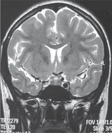
Both during the clinical examination and magnetic resonance imaging (MRI) scanning, the patient presented asymptom-atic, with no new episode of syncope or other complaints being reported. The sus-picion of ICA agenesis was raised by MRI Neves WS, Kakudate MY, Cêntola CP, Garzon RG, Poça d’Água A, Sanches R. Agenesia da artéria carótida interna: relato de caso. Radiol Bras. 2008;41(1):63–66.
Padget(2), and McLone & Naidich(3) two branches of the primitive internal carotid artery (ICA) develop early in the embryo-genesis, originating from the third aortic arch.
In its primitive form, the ICS reaches the cephalic region up to the level of the Rathke’s pouch where two primary divi-sions will occur. Only one cranial branch will extend anteriorly to supply the devel-oping forebrain. In summary, the anterior choroidal, middle cerebral, anterior cere-bral and primitive olfactory arteries will develop from this vessel. Posteriorly, an-other branch will give rise to the posterior choroidal, diencephalic and mesencepha-lic arteries. As these branches advance cau-dally, anastomosis will be made with the developing longitudinal neural arteries sup-plied by the trigeminal artery connections to the primitive ICA.
Ultimately, this pattern describes the first and second developmental phases and results in the adult cerebral circulation INTRODUCTION
The embryonal development of the ce-rebral arterial circulation occurs in seven phases. Certain alterations in this process may lead to agenesis or hypoplasia of the carotid vessels. According to Streeter(1),
Radiol Bras. Jan/Fev 2008;41(1)
64
Neves WS et al.
Radiol Bras. 2008;41(1):63–66 in the absence of the typical flow voidin
the ICA region, on the spin-echo sequences (Figure 1).
Considering the suspicion of agenesis or hypoplasia of the right ICA, the patient was submitted to non-contrast enhanced computed tomography (CT) and MRI an-giography of the brain and cervical vessels, for diagnostic confirmation.
The CT study was aimed at differenti-ating a congenital from an acquired agen-esis of the bony carotid canal, but the ab-sence of the right carotid canal was ob-served.
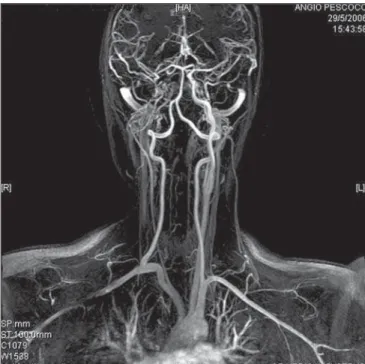
The MRI angiography of the brain and cervical vessels was performed in an at-tempt to depict a global picture of the ce-rebral arterial network, and the absence of the right ICA from the aortic arch (Figures 2, 3 and 4).
Additionally, the patient was submitted to multislice CT, in an attempt to demon-strate some other skull bone abnormality, but only a confirmation of the other stud-ies findings was observed by means of the 3D imaging reconstruction with volume rendering, demonstrating the absence of the right carotid canal.
DISCUSSION
An extremely rare incidence of ICA agenesis has been reported, with few scarce tens of cases being worldwide reported, maybe, to some extent, because the dysgen-esis is generally asymptomatic in the great-est majority of patients. This occurs be-cause there is a sufficient cerebral circula-tion supplied by anastomosis in the circle of Willis, intracavernous and external ca-rotid artery anastomosis, besides persistent embryonal arteries. In these cases, the pa-tients are referred for medical assistance because of complications resulting from abnormalities associated with carotid artery agenesis(5–7)
.
The ICA agenesis is usually unilateral, although there are reports of asymptomatic bilateral agenesis(8). If the agenesis is de-tected by MRI angiography, it must be con-firmed by CT, in an attempt to find hypo-plasia or absence of the carotid canal. Gen-erally, the main secondary source of blood supply is the vertebrobasilar system (in cases of bilateral agenesis) or the dominant ICA (in cases of unilateral agenesis or hy-poplasia)(7).
The main findings associated with these anomalies are transsphenoidal encephalo-celes, circle of Willis aneurysms, and an extensive rete mirabilis in the cranial base. Intracranial aneurysms are found in about 25% of cases of symptomatic internal ca-rotid artery agenesis with intracranial hem-orrhagic manifestations(5,6,9,10).
Also, other less frequent findings are re-ported. There are few cases where agenesis is associated with neuropsychomotor de-velopment delay, agenesis of the corpus callosum and persistent cavum vergae, in patients with bilateral agenesis. Few cases of unilateral agenesis in association with arachnoid cyst are reported(11,12). Also, there is a report about association of megadoli-chobasilar anomaly and olivopontocere-bellar atrophy with unilateral ICA agen-esis(13), besides rare cases of hypopituitar-ism associated with unilateral artery agen-esis, although intracavernous anastomosis seem to be efficient(14,15).
The absence of clinical or neurological symptoms of ischemia in the majority of patients with unilateral or bilateral agenesis or hypoplasia of the ICA leads to the as-sumption that, most of times, perfusional
Figure 2. Non-contrast-enhanced, axial CT, bony window technique showing the absence of the right carotid canal.
65
Agenesis of the internal carotid artery: a case report
Radiol Bras. 2008;41(1):63–66
to confirm the patency or dysgenesis of the carotid canal, corresponding to the vessel agenesis or hypoplasia(16). Multidetector CT with 3D reconstruction also is useful in cases of dubious diagnosis.
CONCLUSION
In this brief case report, there is no in-tention to affirm that agenesis of the ICA should be always suspected. However, it may be concluded that a detailed and meth-odological evaluation of the carotid canals and ICA flow void in the investigation of primary or acquired stenosis (a usual find-ing in patients with neurological com-plaints) may lead to the finding of this anomaly that, although asymptomatic, may be associated with other potentially severe malformations and disorders.
The carotid canals should always be evaluated during the reading of routine cra-nial CT studies with axial slices and bone window images, considering that this is the confirmatory sign of the ICA absence, and does not imply an increase neither in the ac-quisition time nor in the cost of the study. The finding of an episode of syncope in this patient may be associated with a tran-sitory ischemic accident.
Figure 4. Cervical vessels MRI angiography confirming the right ICA absence.
deficit is not present. However, 123-iodine single-photon emission computed tomog-raphy (SPECT) can be utilized, and has already been utilized to demonstrate the absence of areas with perfusional deficit, considering that some few patients have experienced episodes of transitory is-chemic accidents of unidenfied etiology (11).
Likewise the present case, other cases of agenesis of the internal carotid artery have been incidentally found by MRI an-giography. In these cases, it is always nec-essary to confirm if the hypoflow is caused by an acquired stenosis or dysgenesis of the vessel and the bony carotid canal. So, axial CT of the skull base should be performed
Figure 3. Cerebral MRI angiography showing the right ICA absence.
66
Neves WS et al.
Radiol Bras. 2008;41(1):63–66
REFERENCES
1. Streeter GL. The developmental alterations in the vascular system of the brain of the human embryo. Philadelphia, PA: Lippincott Williams & Wilkins Contrib Embryol Carnegie Inst. 1918;8:5–38. 2. Padget DH. The development of the cranial venous
system in man, from the viewpoint of compara-tive anatomy. Philadelphia: Lippincott Williams & Wilkins Contrib Embryol Carnegie Inst. 1957; 36:81–140.
3. McLone DG, Naidich T. Embryology of the cere-bral vascular system. In: Edwards MSB, Hoffman HJ, editors. Cerebral vascular disease in children and adolescents. Baltimore, MD: Williams & Wilkins; 1989:1–16.
4. Truwit CL, Barkovich AJ. Disorders of brain de-velopment. In: Atlas SW, editor. MRI of the brain and spine. 2nd ed. Philadelphia, PA: Lippincott-Raven; 1997:1–25.
5. Blustajn J, Netchine I, Frédy D, et al. Dysgenesis of the internal carotid artery associated with transsphenoidal encephalocele: a neural crest
syndrome? AJNR Am J Neuroradiol. 1999;20: 1154–7.
6. Quint DJ, Silbergleit R, Young WC. Absence of the carotid canals at skull base CT. Radiology. 1992;182:477–81.
7. Tasar M, Yetiser S, Tasar A, et al. Congenital ab-sence or hypoplasia of the carotid artery: radioclinical issues. Am J Otolaryngol. 2004;25: 339–49.
8. Rumboldt Z, Castillo M, Solander S. Bilateral congenital absence of the internal carotid artery. Eur Radiol. 2003;13 Suppl 6:L130–2.
9. Gailloud P, Clatterbuck RE, Fasel JHD, et al. Seg-mental agenesis of the internal carotid artery dis-tal to the posterior communicating artery leading to the definition of a new embryologic segment. AJNR Am J Neuroradiol. 2004;25:1189–93. 10. Cali RL, Berg R, Rama K. Bilateral internal
ca-rotid artery agenesis: a case study and review of the literature. Surgery. 1993;113:227–33. 11. Kidooka M, Okada T, Handa J. Agenesis of the
internal carotid artery – report of a case combined
with arachnoid cyst in a child. No To Shinkei. 1992;44:371–5.
12. Owada Y, Sakuta Y. Bilateral carotid artery agen-esis with corpus callosum hypogenagen-esis – a case report. No To Shinkei. 1995;47:589–94. 13. Okabe S, Oda N, Ishii M. Agenesis of left
inter-nal carotid artery associated with megadolicho-basilar anomaly and olivopontocerebellar atrophy. Rinsho Shinkeigaku. 1989;29:1395–400. 14. Moon WJ, Porto L, Lanfermann H, et al.
Agen-esis of internal carotid artery associated with con-genital anterior hypopituitarism. Neuroradiology. 2002;44:138–42.