Ana Margarida
Gonçalves Santos
Relevância clínica do antigénio sialil-Tn no
cancro da bexiga
Clinical relevance of the sialyl-Tn antigen in
bladder cancer
Ana Margarida
Gonçalves Santos
Relevância clínica do antigénio sialil-Tn no
cancro da bexiga
Clinical relevance of the sialyl-Tn antigen in
bladder cancer
Dissertação apresentada à Universidade de Aveiro para cumprimento dos requisitos necessários à obtenção do grau de Mestre em Bioquímica, ramo de Bioquímica Clínica, realizada sob a orientação científica do Professor José Alexandre Ferreira, Investigador de Pós-Doutoramento do Centro de Investigação do Instituto de Oncologia do Porto (IPO-Porto) e do Departamento de Química da Universidade de Aveiro
Apoio financeiro da FCT ao projeto PEst-C/QUI/UI0062/2011 e projeto PEst-OE/SAL/UI0776/2011, Financiados no âmbito do Programa Operacional Temático Fatores de Competitividade (COMPETE) E Comparticipado pelo Fundo Comunitário Europeu (FEDER).
À minha mãe (in memoriam)
o júri
Presidente Professora Doutora Rita Maria Pinho Ferreira
Professora Auxiliar Convidada do Departamento de Química da Universidade de Aveiro
Doutor Carlos Alberto Palmeira de Sousa
Auxiliar da Faculdade de Ciências da Saúde da Universidade Fernando Pessoa
Doutor José Alexandre Ribeiro de Castro Ferreira
Investigador de Pós-Doutoramento do Centro de Investigação do Instituto Português de Oncologia do Porto (IPO-Porto) e do Departamento de Química da Universidade de Aveiro
agradecimentos Ao Doutor José Alexandre Ferreira, pela orientação científica única e constante, pela motivação, disponibilidade e, acima de tudo, por me incentivar sempre a confiar em mim, a ser perseverante e a acreditar num amanhã mais animador!
A todos os professores que neste longo percurso me transmitiram os conhecimentos necessários para poder terminar mais esta etapa de formação académica com sucesso.
Ao Doutor Lúcio Lara Santos, por me ter acolhido no Grupo de Patologia e Terapêutica Experimental do Instituto Português de Oncologia do Porto (IPO-Porto), e ao Doutor Ricardo Cruz, do Departamento de Urologia, por todas as indicações prestadas. Ao Doutor Luís Lima, pela simpatia com que me recebeu no grupo, bem como por todos os conhecimentos científicos transmitidos. À Sofia Pereira, pela cedência e paciência com que me reviu as lâminas de imunohistoquímica, bem como pelos conhecimentos partilhados. À Ana Tavares, e a todo o Departamento de Anatomia Patológica, pela boa-disposição e espírito de entreajuda com que me receberam, bem como por todas as dicas dadas na procura infinda de lâminas e blocos! Um grande obrigada à Elisabete Fernandes e à Daniela Oliveira por toda a ajuda laboratorial prestada e, especialmente, pela amizade que me permitiu integrar-me tão bem neste grupo. Sem dúvida que as nossas gargalhadas foram o remédio mais eficaz contra os dias menos bons!
À Marlene Esteves, à Fátima Monteiro, à Joana Ribeiro e à Andreia Peixoto pelo apoio prestado no decorrer desta dissertação, e pelos ótimos momentos de convívio e descontração que me proporcionaram. À Joana Justino, amiga e companheira de faculdade, de casa, de sonhos, de inseguranças, de vitórias. Aos restantes colegas de trabalho e de faculdade, por terem contribuído para o meu crescimento pessoal e profissional.
Aos amigos, que nunca me deixaram baixar os braços, mesmo quando o desânimo era inevitável. Muito obrigada por tudo. São a minha segunda família, sem dúvida.
Por fim, ao Xavier Nunes e à minha família o maior obrigada de todos. Pelos conselhos, pela motivação constante, pela paciência e amor incondicionais. Devo-vos tudo.
i Clinical relevance of the sialyl-Tn antigen in bladder cancer
resumo Aproximadamente 50% dos doentes com carcinomas musculo invasivos de bexiga (MIBC) desenvolvem metástases num período de 5 anos após a cirurgia, mesmo quando submetidos a regimes de quimioterapia pré- e pós-cirurgia. Assim, surge a necessidade de se desenvolverem biomarcadores específicos para identificar fenótipos celulares agressivos e terapias diretas baseadas em marcadores moleculares. Recentemente, surgiram evidências de que tumores de bexiga em estadio avançado expressam sialil-Tn (STn) na superfície celular, um antigénio associado a tumores que pode ser usado para detetar fenótipos agressivos no cancro da bexiga. Este antigénio resulta de uma paragem prematura na O-glicosilação de proteínas presentes na superfície celular, e tem demonstrado ser capaz de prevenir o reconhecimento imunológico das células cancerígenas, evitando a eliminação das células metastáticas, modular o fenótipo maligno e induzir a capacidade metastática destas mesmas células. No presente estudo, avaliou-se por imunohistoquímica a expressão de STn e Ki-67 (marcador de proliferação celular) numa série de 96 doentes com cancro de bexiga em diferentes estadios. Este estudo demonstrou uma associação entre a expressão de STn e a proliferação tumoral e invasão. A expressão de STn também foi observada em metástases ganglionares e à distância de tumores STn positivos. O antigénio STn foi maioritariamente observado em glicoproteínas de alto peso molecular (>250 kDa) e em proteínas de baixo peso molecular a 25, 15 e 10 kDa. Estas espécies de baixo peso molecular predominaram nas metástases ganglionares que também não apresentaram proteínas de alto peso molecular, sugerindo uma assinatura molecular associada com metastização. De modo geral, o antigénio STn apresenta potencial para o desenvolvimento de novas terapias contra tumores de bexiga agressivos. Estudos futuros deverão ser realizados para determinar a natureza das glicoproteínas que expressam o STn e confirmar a possibilidade de uma assinatura molecular associada com metastização.
A segunda parte do trabalho focou-se na validação de um xenógrafo de tumor de bexiga expressando STn, como modelo para teste de fármacos e identificação de biomarcadores de prognóstico. Um tumor de bexiga musculo invasivo e STn positivo e os xenotransplantes resultantes da primeira, segunda e terceira passagens foram comparados em relação a proliferação (Ki-67), diferenciação (p63 e CK20) e expressão de STn, por imunohistoquímica. Os padrões de histologia e imunohistoquímica entre o tumor primário e os xenógrafos eram idênticos, revelando um grau de similaridade entre o modelo animal e o tumor humano. Contudo, os níveis de p53 e Ki-67 aumentaram ao longo das passagens enquanto os níveis de STn diminuíram, sugerindo uma seleção dos clones mais proliferativos.
Estas observações possuem uma importância fundamental na expansão do conhecimento sobre a relevância clínica do STn no cancro da bexiga e na criação dos fundamentos para uma terapia baseada neste antigénio.
iii Clinical relevance of the sialyl-Tn antigen in bladder cancer
abstract Approximately 50% of muscle invasive bladder cancers (MIBC) develop metastasis within 5 years after surgery, despite being subjected to pre- and post-surgery chemotherapy regimes. Thus, specific biomarkers to target aggressive cell phenotypes and direct molecular-based therapy are warranted. Recently, evidences have been presented that advanced stage bladder cancers express the cell-surface tumor-associated carbohydrate antigen sialyl-Tn (STn), which may be used to target aggressive bladder cancer cells. The STn antigen results from a premature stop in the O-glycosylation of cell-surface proteins and has been found to prevent immune recognition, contributing to avoid metastatic cell elimination, modulates the malignant phenotype and enhances the metastatic ability of cancer cells. In the present study, a series of 96 patients with bladder cancer of different stages was screened for STn expression and proliferation (over-expression of Ki-67) by immunohistochemistry. This showed an association between STn expression and tumor proliferation and invasion. STn expression was also observed in lymph node and distant metastases of STn positive tumors. The STn antigen was mainly detected in high-molecular weight glycoproteins (>250 kDa) and low-high-molecular weight proteins at 25, 15 and 10 kDa. These low-molecular weight species predominated in lymph node mestastasis samples that also did not present high-molecular weight proteins, suggesting a molecular signature associated with metastasis. Altogether, the STn antigen presents potential for the development of new therapies against aggressive bladder cancer. Studies should be conducted to determine the nature of the STn-expressing glycoproteins and disclosing the possibility of a molecular signature associated with metastasis.
The second part of the work focused on the validation of a STn-expressing bladder cancer xenograft as a model to drug testing and identification of prognostic biomarkers. A STn-positive muscle-invasive bladder tumor and its first, second and third generation xenotransplants were compared in relation to proliferation (Ki-67), differentiation (p63 and CK20), aggressiveness (p53) and STn expression, by immunohistochemistry. Histological and histochemical expression patterns were similar between primary tumor and xenografts, highlighting a degree of similarity between the animal model and the human tumor. However, p53 and Ki-67 levels increased along passages while STn decreased, suggesting a selection of the most proliferative clones.
The generated information is regarded of primary importance to expand the knowledge about the clinical relevance of STn in bladder cancer and create the rationale for a STn-based therapy.
A ignorância afirma ou nega veementemente. A ciência duvida.
Index of tables ... ix
Index of figures ... xi
Abbreviations ... xiii
Chapter I | Introduction ... 15
Bladder cancer – clinicopathological classification, therapeutic challenges and the sialyl-Tn antigen ... 1
Protein O-glycosylation in eukaryotic cells ... 4
Regulation of O-glycosylation ... 8
Alterations of O-glycosylation in cancer ... 9
STn – a pan-carcinoma antigen ... 12
Abnormal O-glycosylation in bladder cancer ... 15
The STn antigen in bladder cancer ... 20
Chapter II | Aims and scopes ... 21
Aims and scopes ... 23
Chapter III | Material and methods ... 25
Overview ... 27
Chapter IV | Sialyl-Tn over-expression is associated with invasion and metastasis in bladder cancer ... 29
Abstract ... 31
Introduction ... 32
Materials and Methods ... 33
Population ... 33
Expression of STn in bladder tumours ... 33
Protein extraction and Western blot ... 34
Statistical analysis ... 34
Results ... 35
Association of STn expression with invasion and proliferation ... 35
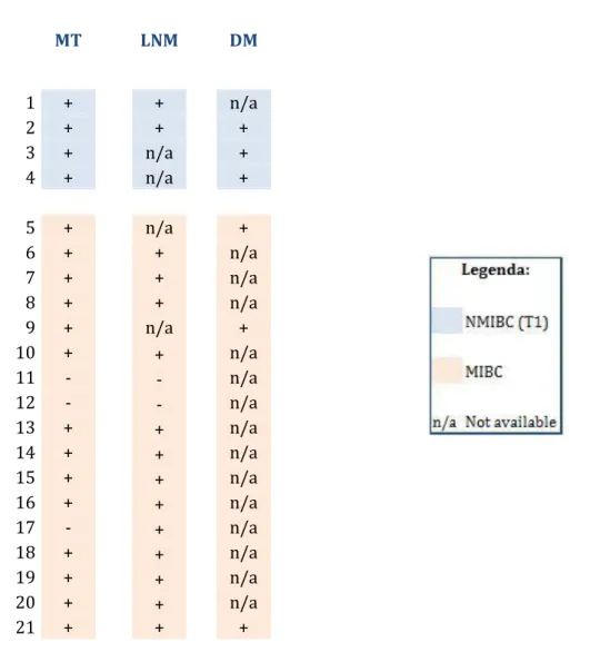
Association of STn expression with lymph node and distant metastasis ... 38
STn glycoprofiling ... 41
Discussion ... 43
Chapter V | Patient-derived sialyl-Tn positive invasive bladder cancer xenografts in nude mice: an exploratory model study ... 49
Abstract ... 51
Animals ... 54
Establishment of xenografts ... 55
Histological analysis ... 56
Immunohistochemical analysis ... 56
Protein extraction and Western blot ... 57
Results ... 58
Histological and immunohistochemical analysis of the primary tumor and xenografts ... 59
Discussion ... 65
Chapter VI | Conclusions ... 69
Conclusions ... 71
Chapter VII | Future work and perspectives ... 73
Future work and perspectives ... 75
Chapter VIII | Bibliography ... 77
ix Clinical relevance of the sialyl-Tn antigen in bladder cancer
Table 1 | Literature review on the expression of tumor-associated glycans in
healthy, pre-neoplastic and neoplastic urothelium……… 18/19
Table 2 | Comparison between the immunoexpression of tumor markers p53, p63, Ki-67, CK20 and STn in the primary tumor and the third generation xenografts (P2), that showed high and homogeneous growth rates……… 63
xi Clinical relevance of the sialyl-Tn antigen in bladder cancer
Figure 1 | Extension of the different stages of bladder cancer (NMIBC and
MIBC)………... 2
Figure 2 | Pathways of the biosynthesis of O-GalNAc glycans – synthesis of Tn and STn antigens and cores 1 to 4, which are the most common in humans………. 7
Figure 3 | Graphic overview of the expression of STn in cancer………... 14
Figure 4 | Overview on the analytical approach………... 28
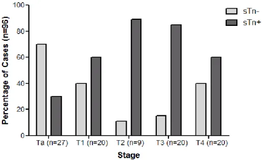
Figure 5 |Association between STn expression and tumor stage………... 36
Figure 6 | Association between STn expression and NMIBC and MIBC ……… 36
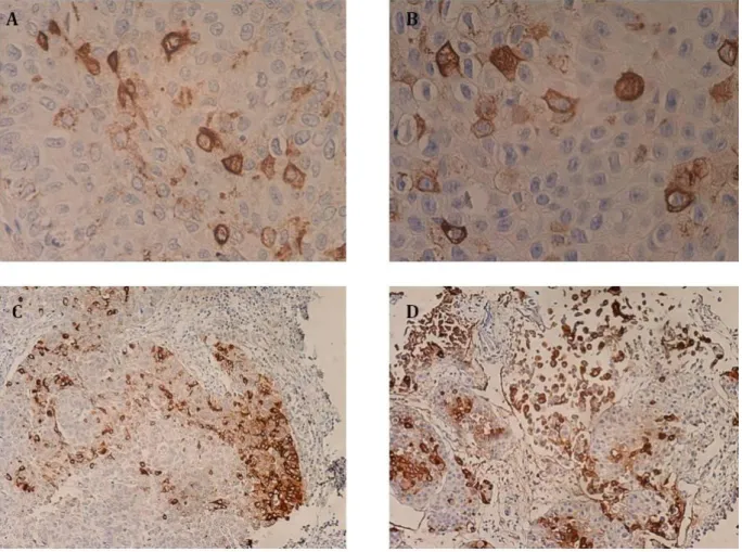
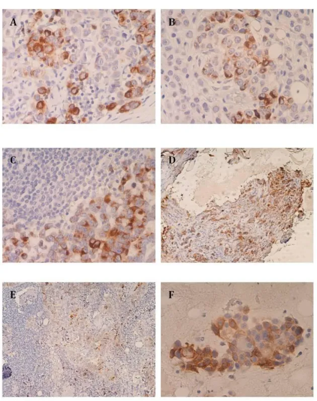
Figure 7 | Expression of STn in bladder tumors…..………... 37
Figure 8 | Expression of STn and Ki-67 in bladder tumors……… 38
Figure 9 | Expression pattern of STn in primary bladder tumors (MT, main tumor) and the correspondent lymph node (LNM) and/or distant metastasis (DM)……….… 39
Figure 10 |Expression of STn in lymph node and distant metastasis.……….…. 40
Figure 11 | STn expression pattern in protein lysates from bladder tumors (T1-T4, n=9) and ganglia (n=2)……….…... 42
Figure 12 | Ratio between low-molecular weight proteins and MUC expression in primary tumors……… 43
Figure 13 | MIBC tissue was used to establish the xenograft model……….. 55
Figure 14 | Tumor growth curve for the first (P0), second passage P1 (a and b) and third generation (P2) xenografts………...………...…... 59
Figure 15 | Histology and tumor molecular markers (p53, p63, Ki-67, CK20) immunoexpression of primary tumor (Pt) and first (P0), second (P1), third (P2) generation xenografts (original magnification x200).………..…………. 60
Figure 16 | STn immunoexpression in the primary tumor and xenografts in the first (P0), second (P1) and third generations (P2) (original magnification x200)………...………. 61
xii Clinical relevance of the sialyl-Tn antigen in bladder cancer
Figure 18 | Western blot for the proteins expressing the STn antigen in primary tumor and P2 xenografts. ………..………...………. 64
β3/4 Gal-T β3/4-galactosyltransferase
β3/4 Gn-T N-β3/4-acetylglucosaminyltransferase
BCG Bacillus Calmette-Guérin
C1Gal-T Core 1 β(1-3) galactosyltransferase
C2Gn-T Core 2 β(1-6)-N-acetylglucosaminyltransferase C3Gn-T Core 3 β(1-3) N-acetylglucosaminetransferase
CIS Carcinoma in situ
Cosmc Core 1 β(1-3) galactosyltranferase-specific molecular chaperone
ER Endoplasmatic reticulum Fuc-T Fucosyltransferase Gal Galactose GalNAc N-acetylgalactosamine GlcNAc N-acetylglucosamine IHC Immunohistochemistry
MIBC Muscle invasive bladder cancer
MoAb Monoclonal antibody
Neu5Ac N-acetyl neuraminic acid
NMIBC Non-muscle invasive bladder cancer
ppGalNAc-T UDP-GalNAc:polypeptide N-acetylgalactosaminyltransferase
Ser Serine
ST3Gal α2,3-sialyltransferase
ST6GalNAc GalNAc α2,6-sialyltransferase
STn Sialyl-Tn antigen
Thr Threonine
1 Clinical relevance of the sialyl-Tn antigen in bladder cancer
Bladder cancer – clinicopathological classification,
therapeutic challenges and the sialyl-Tn antigen
Urinary bladder cancer is the fifth most frequent neoplasia in western countries (and ninth worldwide), and has the highest treatment costs per patient of all cancers [1– 3]. High recurrence rate and long-term follow-up, as well as repeated interventions, are the major causes that turn bladder cancer the costliest to treat among all solid tumors [2, 4].
Most of diagnosed bladder cancer cases in western countries (70-80%) are superficial non-muscle invasive carcinomas (NMIBC) that do not reach the muscularis
propria [3–5]. NMIBC cases include papillary low-grade tumors confined to the mucosa
(pTa), high-grade papillary lesions, that may invade the subepithelial connective tissue but not the muscle (pT1) or high-grade flat lesions termed carcinoma in situ (CIS), as shown in
Figure 1 [6–8]. High-grade tumors present poorly differentiated cells and higher aggressiveness, while low-grade tumors exhibit well differentiated cells. The standard treatment for NMIBC is complete transurethral resection (TUR) [4, 5, 7]. Approximately 10-30% of the patients with NMIBC are at an high risk of recurrence that may be accompanied by muscle invasive disease (MIBC), correlated with poor prognosis [3, 9]. The risk of recurrence and/or progression is determined by clinicopathological features – according to the European Organization for Research and Treatment of Cancer (EORTC), this group includes high-grade papillary tumors, CIS and multifocal disease [5, 7]. To decrease the risk of recurrence/progression, these patients are submitted to schedule of intravesical instillations with attenuated strains of Mycobacterium bovis (the Bacillus
Calmette-Guérin (BCG) vaccine) [3, 4], after removal of the tumor. This treatment is known
to promote a strong immunologic response in the bladder that ultimately contributes to eliminate the residual tumor [7, 10]. Despite effective in delaying recurrence, one third of the patients either do not respond or present intolerance to BCG, 70% of the responders relapse within 5 years and approximately 15% progress to muscle invasive disease [7, 8]. Moreover, many patients develop chronic cystitis or other side-effects during and after BCG therapy [3]. Upon therapeutic failure and/or muscle invasion, the patient is generally appointed for cystectomy [4, 5]. Therefore, alternative organ-spearing therapeutic options for NMIBC patients that either do not respond or show significant intolerance to BCG immunotherapy are warranted.
2 Clinical relevance of the sialyl-Tn antigen in bladder cancer
The remaining 20-30% of the newly diagnosed cases worldwide are muscle invasive bladder cancers (MIBC), that comprise pT2-pT4 stages(Figure 1) [3, 4]. These neoplasias are treated by radical cystectomy [3, 5], and also submitted to pre-and post-cystectomy neo-adjuvant chemotherapy, to reduce the risk of metastasis. However, the current chemotherapy regime carries significant toxicity, and approximately 50% of MIBC cases develop metastasis within 5 years [5, 11].
Figure 1 |Extension of the different stages of bladder cancer (NMIBC and MIBC) (reprinted from [4]).
At the moment, there is a lack of specific biomarkers to target aggressive cell phenotypes in bladder tumors, predict MIBC response to chemotherapy or assist the design of optimal treatment schemes, which would translate in better outcomes and improved overall survival. Therefore, the identification of these biomarkers is considered of main importance, since they may be used to avoid preventive cystectomy and/or reduce the chance of poor outcome, greatly improving the management of bladder cancer [3, 11].
Recent studies have pointed out that advanced stage NMIBC and MIBC express the sialyl-Tn (STn) antigen, a cancer-associated glycan that results from a premature stop in
3 Clinical relevance of the sialyl-Tn antigen in bladder cancer
the O-glycosylation of cell-surface proteins [11–13]. This antigen is not present in the healthy urothelium, denoting a cancer-specific nature, and has been found to promote the invasive and migration potential of bladder cancer cells in vitro [11]. Given its cell-surface nature, STn offers potential to target aggressive bladder cancer cells. However, despite these preliminary evidences, the clinical relevance of these alterations, such as the association with invasion and metastasis, remains to be verified. Additionally, the proteins carrying this alteration remain unknown. Thus, the confirmation of the association of the sialyl-Tn antigen with invasion and metastasis is warranted in order to establish the rationale for a novel therapeutics. The development of animal models expressing this antigen would also greatly benefit such a goal.
4 Clinical relevance of the sialyl-Tn antigen in bladder cancer
Protein O-glycosylation in eukaryotic cells
The sialyl-Tn (STn) glycan can be found as a post-translational modification of O-glycoproteins at the cell-surface of cancer cells. Glycosylation is the most common post-translational modification of proteins and plays a pivotal role in structural and functional features of these molecules – in fact, virtually, all proteins can be glycosylated [14, 15]. Glycans are more complex and diverse in structure and in composition than proteins. Furthermore, the glycosylation of proteins is not a template driven event and frequently the same protein may assume different glycoforms, making of glycan characterization a challenging analytical task [16, 17]. Moreover, glycan structures may vary significantly in response to changes in physiological conditions, with implications in cell-cell adhesion, cell recognition, activation/modulation of immune response and the activation of several intracellular signaling pathways [17, 18].
Protein O-glycosylation is a stepwise pathway that begins in the Golgi apparatus of eukaryotic cells, in which monosaccharides are added individually and sequentially to serine (Ser) and threonine (Thr) residues of proteins by a complex set of enzymes [15, 19].
O-glycans are highly expressed in mucins, a class of heavily glycosylated proteins secreted
by mucosa and some exocrine glands [14, 20]. The high content in VNTR (Variable Number
Tandem Repeat) regions exhibited by mucins, which are rich in Ser and Thr residues,
provides the necessary backbone for a bulk of O-glycosylation [14, 20, 21]. As a result of its association with mucins, cell surface glycans are generally designated as mucin-type O-glycans; nevertheless, O-glycans can also be found in many other cell surface glycoproteins exhibiting Ser and Thr residues [14]. A certain variation of O-glycosylation seems to also occur in the nucleus and cytosol, a process termed nuclear glycosylation. This nuclear O-glycosylation is performed by a single N-acetylglucosamine (GlcNAc) residue that binds to a serine (Ser)/threonine (Thr), and appears to perform a signaling role similar to protein phosphorylation [22, 23]. In addition to mucins, a recent proteomic study has described almost 3000 glycosites in over 600 O-glycoproteins, 80% of each from the cell surface, but also from the cytoplasm and nucleous. This work greatly expanded the view of the O-glycoproteome and the array of protein function it may regulate [24].
Mucin-type O-glycans synthesis begins with the transfer of an α-acetylgalactosamine (GalNAc) from the donor-nucleotide sugar uridine diphosphate – N-acetylgalactosamine (UDP-GalNAc) to the hydroxyl group of a residue of Ser//Thr within the glycoprotein being synthesized [14, 15, 19]. This reaction forms the simplest mucin O-glycan – the Tn antigen (GalNAcα-O-Ser/Thr). This structure can be sialylated at O-6
5 Clinical relevance of the sialyl-Tn antigen in bladder cancer
position by a ST6GalNAc-I sialyltransferase, originating the sialyl-Tn (STn, Neu5Acα2-6GalNAcα-O-Ser/Thr) antigen; nevertheless, sialyl-Tn cannot function as substrate for any other glycosyltransferase, and thus the elongation of oligosaccharide chain stops (Figure 2) [14, 18, 20].
The initial step of O-glycosylation is catalyzed by a family of 20 membrane-bound enzymes denominated UDP-GalNAc:polypeptide glycosyltransferases (ppGalNAc-Ts), that have distinct but overlapping specificities [15, 19]. This family of enzymes has a C-terminal lectin domain, which make them unique among all others eukaryotic glycosyltransferases [22, 25]. This diversity, as well as the fact that O-glycosylation has no consensus sequence to occur, allows a fine tuned control of the initiation of this process in a specific cell or even in a specific protein [14, 26].
When the first step of the synthesis is concluded, a galactose (Gal) residue is transferred from an uridine diphosphate - galactosamine (UDP-Gal) donor to the GalNAc of the Tn antigen by a specific galactosyltransferase (β(1-3)-galactosyltransferase, C1Gal-T1 or T-synthase), yielding the most common core structure – core 1, also called T antigen or Thomsen-Friedenreich antigen (Galβ1-3GalNAcα-O-Ser/Thr), as shown in Figure 2 [14, 22, 25]. In 2009, Wang et al. proposed that the active state of T-synthase depends on the co-expression of an unusual molecular chaperone localized in the ER, Cosmc [27]. Apparently, this chaperone prevents the aggregation and proteasomal degradation of T-synthase, and is required for the export of the enzyme from the ER, leading to the formation of core 1 [27].
T antigen can be sialylated at O-3 position by a ST3Gal-sialyltransferase, yielding the sialyl-3-T antigen (S3T, Neu5Acα2-3Galβ1-3GalNAcα-O-Ser/Thr); this antigen can be further sialylated at O-6 position, by a ST6Gal-sialyltransferase, originating the di-sialyl-T antigen (diST, Neu5Acα2-3Galβ1-3(Neu5Acα2-6)GalNAcα-O-Ser/Thr) (Figure 2) [14, 20]. Alternatively, the GalNAc residue of the T antigen may be sialylated at the O-6 position by a ST6GalNAc-sialyltransferase, yielding the sialyl-6-T antigen (S6T, Galβ1-3(Neu5Acα2-6) GalNAcα-O-Ser/Thr), which can then originate the diST antigen. Tn and T antigens, as well as their sialylated structures, block further elongation of the O-chain, and are generally designated simple mucin-type O-glycans [20, 28].
Core 1 may function as a precursor of other core structures (from core 2 to 8), by the addition of different monosaccharides, such as galactose, acetylgalactosamine, N-acetylglucosamine and sialic acids [19, 22]. However, cores 1-4 are the most common in humans – in fact, cores 5-8 have been characterized in tissues and appear to be formed
6 Clinical relevance of the sialyl-Tn antigen in bladder cancer
through direct modification of the nascent O-GalNAc glycan, but the enzymatic machinery underlying this process remains unclear [15, 20, 25].
Core 2 is synthesized in many epithelial (as intestinal mucosa) and hematopoietic tissues by the addition of a branching GlcNAcβ(1-6)-linked residue to core 1, by core 2 β(1-6) N-acetylglucosaminyltransferases (or C2Gn-T) (Figure 2) [14, 20].
Core 3 is formed by the addition of a GlcNAc residue by a core 3 β(1-3) N-acetylglucosaminetransferase (C3Gn-T3) to Tn antigen, and the subsequent addition of a GlcNAc residue onto core 3 by a C2Gn-T yields core 4, as represented in Figure 2.Thus, the prior synthesis of core 3 is required for the formation of core 4 [19, 20, 25].
The extension of core units provides a vast array of glycan structures, and is catalyzed by N-acetylglucosaminyltransferases (β3/4 Gn-Ts) and/or β3/4-galactosyltransferases (β3/4 Gal-Ts), leading to the formation of side chains designated type-1 (Galβ1-3GlcNAc-R) and type-2 (Galβ1-4GlcNAc-R) chains [14, 19, 22]. These chains present a ubiquitous expression, and therefore are widely expressed among epithelial tissues. Type-1 and type-2 chains can be modified by the action of fucosyl and sialyltransferases, yielding ABO blood group determinants and/or Lewis blood group related antigens (Lea, SLea, Lex, SLex, Leb and Ley), which function as terminal structures by stopping chain elongation [14, 19, 22]. After O-glycosylation is concluded, the formed structures can undergo modifications, such as phosphorylation, sulfation and methylation, which constitutes an additional mean for their diversity [22].
O-GalNAc glycans are crucial structures for cells viability, once they play several and
distinct roles in the organism, depending on the structure they present – this structural variability allows them to function as signaling, recognition and adhesion molecules [16, 17, 20].
7 Clinical relevance of the sialyl-Tn antigen in bladder cancer
Figure 2 |Pathways of the biosynthesis of O-GalNAc glycans – synthesis of Tn and STn antigens and cores 1 to 4, which are the most common in humans (reprinted from [14]).
Enzymes: β(1-3)-galactosyltransferase (β3Gal-T); β(1-4)-galactosyltransferase (β4Gal-T); core 3 β(1-3) N-acetylglucosaminyltransferase (β3Gn-T); core 1 β(1-3)-galactosyltransferase (C1Gal-T1); core 2 β(1-6)-N-acetylglucosaminyltransferase (C2Gn-T); UDP-GalNAc:polypeptide
N-acetylgalactosaminyltransferase (ppGalNAc-T); α2,3-sialyltransferase (ST3Gal); GalNAc α2,6-sialyltransferase (ST6GalNAc).
8 Clinical relevance of the sialyl-Tn antigen in bladder cancer
Regulation of O-glycosylation
As O-GalNAc glycans are complex and widely diverse structures, their synthesis is tightly regulated. However, exactly mechanisms by which this regulation occurs remain to be clarified [20, 21]. The regulation of O-glycosylation seems to be dependent on the pattern of the enzymes involved in this process – substrate specificity, intracellular localization and level of relative activity are the main control factors of O-glycosylation [20, 21, 23].
Substrate specificity of glycosyltransferases and other enzymes involved in O-glycosylation allows a restriction of the number of possible O-GalNac glycans that can be formed, since these enzymes act in well-defined substrates [20, 25]. On the other hand, this specificity reduces the number of pathways by which O-glycans can be synthesized [20, 25]. For example, ST antigen can only be generated by the addition of a sialic acid residue to T antigen (core 1). The adding of a galactose residue to STn antigen does not yield the same structure, since the sialic acid present in STn antigen blocks the action of other glycosyltransferases, namely C1Gal-T1 [20].
Intracellular localization of glycosyltransferases is another important regulation factor of O-glycosylation, which is determined by the physical separation of enzymes within the Golgi compartments [25, 28]. These enzimes appear to be arranged in an assembly line in the Golgi apparatus – early acting glycosyltransferases occupy the cis-Golgi, intermediate acting enzymes are localized in the medial Golgi and terminal acting enzymes in the trans-Golgi, which allows a tight control of O-glycosylation [20, 28].
The third major control factor is the level of relative activity of glycosyltransferases, that consequently dictate the relative amounts of synthesized O-glycans – the activity of two competitive enzymes will determine the nature of the O-GalNAc glycan being synthesized, depending on which activity predominates [16, 25]. Besides this competition between glycosyltransferases, there is also the influence of other regulatory components, namely specific binding proteins required for the activation of glycosyltransferases [20]. Co-expression of Cosmc required for the activation of C1Gal-T1 is one example of this type of regulatory components [27].
9 Clinical relevance of the sialyl-Tn antigen in bladder cancer
Alterations of O-glycosylation in cancer
Abnormal protein O-glycosylation is considered a hallmark of malignant transformation [14, 20, 21]. Tumor-associated glycans can be found in the surface of cancer cells, and therefore are easily accessible to antibodies and lectins [29, 30]. Moreover, they are released to the peripheral circulation, either in secreted glycoproteins or by shedding from cell surfaces – these features allow them to be explored in various serological assays [20, 29]. Therefore, the presence of some tumor-associated glycans in serum is nowadays for post-surgical follow-up, to determine disease recurrence, progression and/or response to therapeutics [31].
Among the most common structural features associated with cancer are the altered expression of terminal structures, which includes loss of ABO determinants by secretor individuals and changes of Lewis antigenic patterns [20, 28], over-expression of mucins [32, 33], particularly MUC1, and incomplete O-glycosylation, yielding low molecular weight O-glycans [14, 20].
The first evidences of alterations in glycosylation patterns of tumor cells concerns loss of ABO blood group determinants in gastric cancer [34], and the correlation of the degree of A/B determinants deletion with invasion and metastatic spread was then described in lung [35] and head and neck [36] carcinomas. Thereafter, the relationship between expression of ABH antigens and survival of patients with non-small-cell carcinoma of the lung was also studied, and it was concluded that continued A determinant expression was correlated with a longest average survival, while A deletion was correlated with a reduced overall survival [37]. Nevertheless, correlation between B antigen expression and survival rate was not found [37]. Similar results were observed in bladder cancer patients with non-invasive and invasive carcinomas [38, 39].
Changes in Lewis antigenic patterns are related to over-sialylation of terminal structures, resulting in an over-expression of SLea and SLex antigens. The referred alterations in glycosylation stem from an aberrant expression of the genes encoding sialyl and fucosyltransferases, which increases the synthesis of these sialylated structures [14, 40]. The comprehension of the role of SLea and SLex in cancer begun in 1980s, when several studies demonstrated the expression of these antigens in cancer specimens, using monoclonal antibodies [41]. Later, in 1991, a study demonstrated the capability of SLea and SLex to function as selectin ligands for ELAM-1 (Endothelial Leukocyte Adhesion Molecule 1) in endothelials cells [42]. Subsequently, it was also discovered that this
10 Clinical relevance of the sialyl-Tn antigen in bladder cancer
mechanism allowed malignant cells to adhere to endothelium, facilitating invasion and metastasis [14]. Therefore, over-expression of SLea and SLex in different carcinomas has been demonstrated to be correlated with poor prognosis [17, 43].
Sialylated Lewis antigens can also be released to the bloodstream by malignant cells – thus, soluble forms of these antigens are also expressed in high amounts in the blood of many cancer patients [20, 21]. Studies based on the average survival of oncological patients with gastric carcinomas after surgery revealed that high levels of SLea and SLex correlated with lower survival [44, 45]. Based on these features, SLea and SLex are currently used in non-invasive assessment of tumor progression and metastatic spread [14, 17, 20].
The over-expression of mucins during neoplastic transformation, namely MUC1 mucin, has been widely documented [32, 33, 46]. MUC1 is a transmembrane glycoprotein with a large extracellular mucin-like domain, formed by 30-90 repeats of 20 homologous amino acids rich in O-glycosylation sites [47, 48]. Normally, MUC1 is expressed exclusively in the apical domain of epithelial cells; however, on carcinomas cells, the correct topology is lost and MUC1 is expressed over the entire cell surface, in abnormally large amounts [20, 33]. Since the extracellular domain of MUC1 is long, dense and relatively rigid, due to the abundance of O-glycan oligosaccharides, adhesion molecules present in cell surface, such as cadherins and integrins, become shielded. Thus, cell-cell and cell-extracellular matrix interactions decrease, creating an anti-adhesion effect [33, 49]. Consequently, MUC1 mucin has the capability to induce detach of a cell from the primary lesion, leading to invasion and metastasis. This process escapes from immune surveillance, because MUC1 inhibits the interaction between cytotoxic lymphocytes and the target cell and promotes apoptosis of lymphocytes, allowing the detach cell to survive in bloodstream or in distant organs [20, 49]. Moreover, the metastatic ability induced by MUC1 is also associated with the intracellular domain of the protein, which enters the nucleus and initiates the transcription of a set of genes responsible for tumor metastasis [50]. Over-expression of MUC1 is generally correlated with higher aggressiveness and metastatic capability of the tumor [33, 49].
Serological assays for MUC16 have also been shown the usefulness of this mucin in the prognosis of ovarian cancer, in which MUC16 is detected in 80% of the patients. Moreover, a correlation between increase/decrease of MUC16 expression and regression/progression of the disease has also been observed [14]. Similarly, serological
11 Clinical relevance of the sialyl-Tn antigen in bladder cancer
assays for MUC1 mucin in early stage breast cancer specimens have revealed that high levels of this glycoprotein are useful in the prognosis of the disease [40, 51].
The mechanisms underlying aberrant expression of mucins in malignant transformation are still poorly understood; nevertheless, it was suggested that they may arise from an up- or down-regulation of the genes encoding mucin proteins [13]. These genes may be deregulated in neoplasias originating from tissues where mucins are constitutively expressed, or these macromolecules may be ectopically expressed in cancers derived from tissues normally devoid of them [13].
Another common structural feature in cancer results from a premature stop of O-chain elongation, normally by sialylation. This event leads to de novo expression of a family of low molecular weight O-glycans, that includes Tn and T antigens and their sialylated counterparts [14, 20, 21].
High levels of ST and STn antigens have been observed in several carcinomas, namely gastric [18], colon [52], bladder [53], breast [40] and pancreatic [54], whereas low levels of expression are found in healthy tissues. Therefore, ST and STn antigens have been extensively studied in last decades and are widely assumed as pan-carcinoma biomarkers [18, 21, 55].
T antigen is also considered a pan-carcinoma antigen, once it is substantially over-expressed in several carcinomas, namely breast [40], bladder [53], colon [52], gastric [56] and prostate [57]. Nevertheless, the expression of T antigen is not limited to cancer – it is also expressed in normal tissues, although in low levels, since it undergoes further glycosylation [52, 56, 57]. In cancer tissues, the glycosylation process becomes incomplete, and the expression of T antigen increases significantly. The increment of T antigen expression is associated with a worse prognosis in cancer patients [58, 59].
The incomplete synthesis observed during malignancy can arise from diverse alterations in cancer cells. They commonly stem from altered regulation of sialyl and glycosyltransferases expression in cancer cells (up/downregulation) [14, 40, 53]. Namely, the level of expression of sialyltransferases in breast cancer has been studied as a biomarker for the follow-up of oncological patients – apparently, a higher expression of ST3Gal-III and ST6Gal-I in human breast tumors is correlated with poor prognosis [60]. At the same time, the activity of these enzimes may also be altered in cancer cells [18, 53]. Increase of sialyltransferases activity leads to preclude of O-glycosylation, due to the blocking effect of sialic acid on glycosyltransferases involved in chain extension [28]. Consequently, neoplastic cells express heavily sialylated and truncated O-GalNAc glycans,
12 Clinical relevance of the sialyl-Tn antigen in bladder cancer
namely ST and STn antigens, which have an increased ability to bind to adhesion molecules present in endothelial cells, such as selectins [18, 28, 53]. Therefore, these highly sialylated O-glycans have an enormous invasive potential and metastasis capability [18, 28].
Moreover, these alterations can also stem from a deregulation in the location of glycosyltransferases. Gill et al. (2010) [26] suggested a redistribution of ppGalNAc-Ts involved in O-glycosylation induced by the activation of Src kinase. This activation promotes a relocation of the enzymes from Golgi apparatus to the ER, allowing them to be more time in physical contact with potential substrates. Thus, occurrence of glycosylation is dramatically increased, resulting in an enhanced synthesis of truncated O-GalNAc glycans [26].
Another possible mechanism underlying the expression of cancer-associated O-glycans, suggested by Ju et al. (2011) [61], focus on a mutation on the molecular chaperone Cosmc, affecting the enzyme responsible for the synthesis of T antigen, T-synthase. According to Wang et al. (2009) [27], a functional T-synthase depends on the co-expression of a molecular chaperone, Cosmc. Ju et al. (2011) [61] also demonstrated that a mutation in the gene that encodes for Cosmc is responsible by the accumulation of cancer-associated precursors both in neoplastic lesions, including colon cancer and melanoma-derived cell lines, and in human cervical cancer specimens.
Overall, simple mucin-type O-GalNAc glycans are pan-carcinoma antigens, associated with malignant phenotypes such as increased invasive and metastatic potential – therefore, these structures correlate with poor prognosis [14, 18, 40].
STn – a pan-carcinoma antigen
STn detection among healthy tissues is heterogeneous [55, 62]. The expression of this antigen was found to be positive in normal salivary glands and in globlet and parietal gastric cells of upper digestive tract [55, 63, 64], but not in the esophagus [62]; conversely, STn can only be detected in colonic cells after removal of O-acetyl groups [65], and it was never found in normal liver and pancreas [62, 66]. In relation to the urogenital tract, STn was visible in some uterine and cervix cells, but not in the ovarian ones [55, 62]. Finally, it was also detected in some cells of normal lung [62] and breast [67] tissue. It is noteworthy that the authors report, overall, that the expression of STn antigen in normal tissues is rare
13 Clinical relevance of the sialyl-Tn antigen in bladder cancer
and/or low, when compared to cancer tissues. Moreover, these studies evidence an expression of STn restricted to secreting cells, which suggest that the spread expression of this antigen in healthy tissues relates to external fluids of the body [55].
STn antigen was also found to be over-expressed by pre-neoplastic lesions, such as gastric intestinal metaplasia, breast ductal hyperplasia and chronic ulcerative colitis [18, 55]. STn was also over-expressed in epithelial benign lesions in ovaries [68] and pancreas [69], two tissues devoid of this antigen in the healthy state. Of note, the expression of STn in pre-neoplastic pancreas occurred only in the last histological grade relative to benign tumor [69]. This suggests that STn over-expression occurs earlier in carcinogenesis in tissues that normally express this antigen, rather than in tissues normally devoid of STn [55].
In relation to cancer tissues, STn was reported to be neo- or over-expressed in more than 80% of human carcinomas, namely pancreatic [69], endometrial [70], breast [40], colon [71] and gastric [72]. Therefore, STn is considered a pan-carcinoma antigen and a good tumor marker of carcinogenesis [55]. In line with these observations, several in vitro and in situ studies have associated STn antigen with aggressive cell phenotypes [18, 40, 55]. It has been demonstrated that STn expression promotes major morphological alterations on the cell surface glycosylation profile, inducing or preventing the recognition by lectins. This process contribute to a cancer phenotype, decreasing cell-cell aggregation and increasing extracellular membrane adhesion, migration, invasion and metastasis [18, 55].
Kakeji et al. (1995) [73] have reported that an increased percentage of STn-positive cells was correlated with invasion in gastric cancer. In agreement with this study, Flucke et
al. (2001) [74] reported a decreased overall survival of the patients with more than 35%
of STn-positive cells in esophageal cancer, when compared to the low expressing group (<35% of STn-positive cells). Recently, Ozaki et al. (2012) [75] described that STn-positive gastric cancer cells have demonstrated higher intraperitoneal metastatic ability in comparison with STn-negative control in nude mice, resulting in depth of invasion and shorter survival. Moreover, Ogata et al. (1992) [76] have previously demonstrated that mucins bearing STn antigen are effective inhibitors of natural killer (NK) cells cytotoxicity. Thus, it was suggested that over-expression of STn in cancer specimens induces impairment in NK cell function of the immune system and, subsequently, that mucins expressing STn antigen allow cancer cells to escape from immune surveillance [76].
14 Clinical relevance of the sialyl-Tn antigen in bladder cancer
Given its biomarker value, STn was established as a tumor marker in serological assays (CA 72-4) [14, 55, 77]. This antigen is present in the bloodstream due to O-glycoprotein secretion or to cell shedding from tumors, which occurs only when the tumor reaches a critical mass. Therefore, the presence of STn in serum is usually detected in advanced tumors, and STn is considered a poor prognosis marker [55]. In fact, raised levels of serological STn have been associated with decreased overall survival of patients with gastric, ovarian and colorectal cancers [14, 55]. Overall, STn is considered a pan-carcinoma antigen, since it is expressed in the majority of human pan-carcinomas, and it is associated with adverse outcome and decreased overall survival of the patients, as summarized in Figure 3.
Figure 3 |Graphic overview of the expression of STn in cancer (adapted from [55]).
Top part: reports of positive (blue) or negative (white) expression in healthy, pre-malignant and cancer tissues. Bottom part: reports assessing the correlation between STn expression and clinical features of cancer, with a significant correlation found in pink and no correlation found in white. Size of circles is proportional to the number of published reports.
Obs.: The positive expression of STn in healthy adult tissue (first line) relates to a sparse or low expression, as described in the first paragraph of this section.
15 Clinical relevance of the sialyl-Tn antigen in bladder cancer
Given the cancer-associated nature of the STn antigen, a cancer vaccine named
Theratope, comprehending a synthetic STn disaccharide coupled to a immunogenic carrier
protein, keyhole limpet hemocyanin (KHL), was developed [55, 78]. Tests in animal models and phase II trials for breast and ovarian cancers have showed that the antigen is safe and produces a strong immune response against these tumors [78]. However,
Theratope failed phase III trials for metastatic breast cancers, since it did not improved
overall patient survival. In a recent review on the subject, Julien et al. (2012) [55] discussed that the failure of Theratope was related with the design of the study, namely that it disregarded the fact that only 20-30% of the patients with metastatic breast cancers expressed the STn antigen. In 2009, nonetheless, Julien et al. [79] investigated the efficacy of STn-carrying immunogens to inhibit tumor growth in a MUC1 transgenic mouse model. The authors reported that Theratope induced antibodies to STn that recognized the glycan carried in a number of glycoproteins. Moreover, a significant delay in tumor growth was observed in these mice, and the protection effect seemed to be dependent on STn being expressed by the tumor [79]. Altogether, the notion prevails that STn based therapeutics may constitute a strategy to control invasion and metastisis, and consequently poor outcomes. The fact that it is a cell-surface antigen that may be more easily accessible to antibodies and/or other ligands also offers potential in the context of guided therapeutics.
Abnormal O-glycosylation in bladder cancer
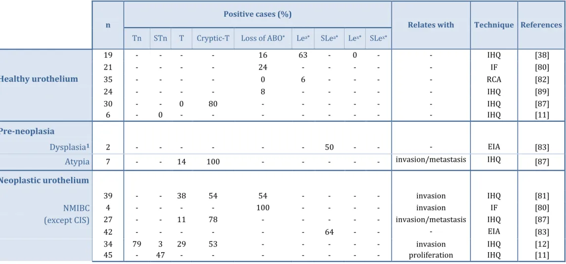
The first reports of alterations in glycosylation of bladder tumors have been presented over 40 years ago and relate to the loss of ABO blood group determinants in advanced stage carcinomas of secretor individuals (the Se – secretor – locus dictates the capability of an individual to express soluble mucins carrying blood group antigens in saliva and other tissues). Subsequent studies have shown that malignant transformations in the bladder are accompanied by changes in Lewis antigens pattern and over-expression of simple mucin type O-GalNAc glycans. Table 1 summarizes the literature on the subject in healthy, pre-neoplastic and neoplastic urothelium and its correlation with clinicopathological features. The studies focused on the expression of STn, the core antigen of this thesis, in the context of bladder cancer will be discussed further forward, in the subtopic 2.3.1..
Six studies have described the loss of ABO blood group antigens in healthy and neoplastic urothelium, as documented in Table 1. Nevertheless, this event is more
16 Clinical relevance of the sialyl-Tn antigen in bladder cancer
pronounced in the neoplastic urothelium (44-100%) when compared with the healthy urothelium (0-24%). Thorpe et al. (1983) [80] and Summers et al. (1983) [81] have correlated the loss of ABO antigens with the invasive potential of the tumor; moreover, Limas et al. (1985) [82] have also associated this event with higher grade bladder cancer.
Altered Lewis antigenic profile has been reported in pre-neoplastic and neoplastic urothelium (Table 1). Namely, several authors have studied the expression of Lea in bladder tumors. According to Cordon-Cardo et al. (1988) [38], there were no alterations in Lea expression patterns with malignant transformations in the bladder. Conversely, Limas
et al. (1985) [82] reported significantly lower expression of this antigen in healthy
urothelium (6%), when compared to invasive tumors of the bladder (35%). In agreement with this study, Juhl et al. (1986) [39] reported a high expression of Lea antigen in invasive bladder carcinomas (93%). Both these studies were performed using a boarder patient cohort (n>35) than the one used by Cordon Cardo et al. (n=19), and strongly suggest that the expression of Lea is associated with a malignant phenotype of bladder tumor [39, 82].
The sialylated form of Lea, the SLea antigen, has been observed in bladder dysplasia, CIS, non-invasive and invasive carcinomas of the bladder [83]. Nevertheless, no correlation was found with invasive or metastatic potential was not established.
Three studies referred in Table 1 have focused on the expression pattern of Lex antigen, and revealed a high percentage of bladder cancer patients expressing altered levels of Lex (78-100%). Specifically, Cordon-Cardo et al. (1988) [38] have compared the expression pattern of this antigen in healthy urothelium with invasive carcinomas and CIS. The study revealed that none of the healthy urohelium specimens expressed Lex; conversely, the antigen was observed in invasive carcinomas of the bladder (100%) and CIS (79%). Furthermore, the authors proposed that Lex expression could be a reliable indicator of malignant transformation in bladder urothelium [38].
Its sialylated form, SLex expression has also been studied in bladder urothelium, as demonstrated in Table 1. According to Numahata et al. (2002) [84], 70% of the invasive carcinomas expressed SLex expression. These findings were corroborated by Kajiwara et
al. (2005) [85] that observed altered SLex patterns in all of the invasive carcinomas specimens studied. Both studies referred altered SLex expression as a predictor of invasive potential and metastatic outcome.
Increased levels of truncated O-GalNAc glycans have also been observed in bladder cancer (Table 1). Yokoyama et al. (1988) [86] have compared the spontaneous expression
17 Clinical relevance of the sialyl-Tn antigen in bladder cancer
of T antigen (48%) with it expression after neuraminidase treatment (cryptic-T antigen (18%)), in bladder cancer specimens of various stages. The authors revealed that 70% of the T-positive cases presented recurrence, while only 11% of the cryptic-T-positive did. Similarly, Summers et al. (1983) [81] reported that those patients who did not spontaneously expressed the T antigen (54%), did not suffer invasive recurrences; conversely, the patients that expressed spontaneous T antigen (38%) suffered subsequent invasive recurrences.
Tn (79%), STn (3%), T (29%) and cryptic-T (53%) antigens were evaluated by Langkilde et al. (1992) [12] in patients with initially non-invasive carcinomas who experienced different courses of the disease. No association was found between Tn and STn antigens and tumor progression to invasion. Nevertheless, 70% of the patients that expressed T antigen and 39% of those who expressed cryptic-T antigen experienced invasive recurrence, while the other patients did not [12].
Furthermore, Limas et al. (1986) [87] demonstrated a crescent expression of T antigen through healthy (0%), pre-neoplastic (14%) and neoplastic (11-65%) urothelium. Cryptic-T antigen showed a homogeneous expression among all the specimens evaluated (78-100%). According to the authors, the spontaneous expression of T antigen is associated with aggressiveness of the tumor; moreover, in invasive carcinomas of the bladder, this expression correlates with a greater metastatic potential.
Altogether, these findings suggest that bladder cancer patients who spontaneously express T antigen present invasive recurrences more frequently than those who do not express this antigen; furthermore, T antigen expression seems to correlate with metastatic potential and aggressiveness of the tumor [12, 81, 86].
The studies presented so far suggest that both pre-neoplastic lesions and bladder tumors express altered glycosylation patterns. Some studies also point out that changes in cell glycosylation patterns are generally associated with tumor grade, invasive disease and metastasis – therefore, they are correlated with poor prognosis [81, 86]. Thus, targeting these antigens may allow determining the malignant potential of the tumor and controlling the disease. Nevertheless, there studies have been performed in small and heterogeneous patient cohorts using different antibodies. Therefore, a careful interpretation of the results should be conducted, since several antibodies have shown to have affinity for similar structurally-related glycosylated structures [88]. Different methodologies have also been used, which may contribute to biased interpretations.
18 Clinical relevance of the sialyl-Tn antigen in bladder cancer
1Evaluated indirectly in exfoliated urine cells
* May also be found as terminal structures of N-glycans and lipids
n
Positive cases (%)
Relates with Technique References Tn STn T Cryptic-T Loss of ABO* Lea* SLea* Lex* SLex*
Healthy urothelium
19 - - - - 16 63 - 0 - - IHQ [38] 21 - - - - 24 - - - IF [80] 35 - - - - 0 6 - - - - RCA [82] 24 - - - - 8 - - - IHQ [89] 30 6 - - - 0 0 - 80 - - - - - - - - - - - - - IHQ IHQ [87] [11]Pre-neoplasia
Dysplasia
1 2 - - - - - - 50 - - - EIA [83]Atypia
7 - - 14 100 - - - invasion/metastasis IHQ [87]Neoplastic urothelium
NMIBC
(except CIS)
39 - - 38 54 54 - - - - invasion IHQ [81] 4 - - - - 100 - - - - invasion IF [80] 27 - - 11 78 - - - invasion/metastasis IHQ [87] 42 - - - 64 - - - EIA [83] 34 45 79 - 3 47 29 - 53 - - - - - - - - - - - invasion proliferation IHQ IHQ [12] [11]19 Clinical relevance of the sialyl-Tn antigen in bladder cancer
2Evaluated the urinary levels
* May also be found as terminal structures of N-glycans and lipids n
Positive cases (%)
Relates with Technique References Tn STn T Cryptic-T Loss of ABO* Lea* SLea* Lex* SLex*
MIBC
44 - - - 70 invasion/metastasis IHQ [84] 33 - - - 76 - - - EIA [83] 19 - - - 53 100 - - IHQ [38] 1 - - - - 100 - - - - invasion IF [80] 48 - - - - 44 35 - - - grade/invasion RCA [82] 17 - - 65 88 - - - invasion/metastasis IHQ [87] 52 - - - 100 invasion/recurrence IHQ [85] 85 - - - 93 - - - invasion/recurrence IHQ [90] 93 19 - - - 74 - - - - 44 - - - - - - - - - - proliferation IHQ IHQ [39] [11]CIS
1 - - - 100 - - - EIA [83] 14 - - - - 50 64 79 - - IHQ [38] 5 - - 20 100 - - - invasion/metastasis IHQ [87] 62 5 - - - 20 - - - - - - - - - - 100 - - - - proliferation IHQ IHQ [91] [11]Not discriminated
83 - - 48 18 - - - grade/recurrence IF [86] 32 - - - 81 - - IHQ [92] 78 - - - 78 - - IHQ [91] Technique’s Abbreviation:IHQ – Immunohistochemistry RCA – Red Cell Adherence Test IF – Immunofluorescence EIA – Enzyme Immuno Assay – Not evaluated
20 Clinical relevance of the sialyl-Tn antigen in bladder cancer
The STn antigen in bladder cancer
Despite the cancer-associated nature of the STn antigens, few studies have been presented for bladder cancer (Table 1). The first study by Langkilde et al. (1992) [12] assessed this antigen on a series of transitional cell carcinomas (currently classified as high-grade urothelial cell carcinomas according to WHO guidelines), and in a control group, comprehending normal mucosal specimens of patients with non-malignant bladder urologic diseases. The authors reported that STn was not expressed by the control group and showed a very restricted pattern of expression in bladder tumors. Moreover, no association with recurrence and progression were observed [12].
In vitro studies performed by Bergeron et al. (1996) [13] showed that mucins MUC1,
MUC2 and MAUB (mucin antigen of the urinary bladder) isolated from bladder cancer cell lines carried the STn antigen. Nevertheless, that expression was not found in tumors.
Recently, Ferreira et al. (2013) [11] addressed the expression of STn in 6 necropsies of normal urothelium, as well as in 69 bladder cancer patients. STn antigen was absent from the healthy urothelium, showing the STn tumor-associated expression. The authors also reported that approximately 70% of high-grade (HG) bladder tumors over-expressed STn. Conversely, only less than 25% of low-grade (LG) tumors over-expressed this antigen – altogether, the obtained results suggest that the expression of STn is associated with advanced stage bladder tumors, known for an aggressive phenotype. Moreover, Ki-67 antigen, a proliferation tumor marker, was also assessed in 12 LG and 12 HG tumors from the initial series. Only 8% of LG bladder tumors over-expressed Ki-67, as opposed to 75% of HG tumors – overall, these findings suggest that the expression of STn is a characteristic of proliferating tumors [11]. In vitro studies have further demonstrated that STn expression enhanced the invasion capability of bladder cancer cells.
Despite these observations, little information is available about the biological and clinical significance associated with STn expression in bladder cancer. Namely, doubts persist about its association with invasion and metastasis, which is of prime importance to encourage the development of therapeutics targeting this antigen.
Chapter II |
Aims and
scopes
23 Clinical relevance of the sialyl-Tn antigen in bladder cancer
Aims and scopes
The main therapeutic concerns in bladder cancer currently includes the management of NMIBC at a high-risk or recurrence with progression, that do not respond or show intolerance to BCG immunotherapy, and MIBC [7, 10]. These patients are conservatively treated by partial or radical cystectomy with neo- and post-surgical chemotherapy to decrease the risk of metastasis. However, over 50% of the patients succumb within five years [5, 11]. Due to the limited therapeutic options, the identification of a specific biomarker associated with invasion and metastasis could encourage the development of new treatments.
The modification of cells glycosylation patterns is a recognized hallmark of cancer [14, 20, 21] and the STn antigen, in particular, presents potential to target malignant cells in invasive bladder tumors. It is known that STn contributes to avoid metastatic cell elimination in the blood stream by preventing immune recognition [76], modulates the malignant phenotype [18] and enhances the metastatic ability of cancer cells [75]. Furthermore, this antigen has already been suggested to be over-expressed by 70% of high-grade NMIBC and MIBC cases and therefore associated with tumor aggressiveness [11]. However, this study was conducted in a rather small and heterogeneous series (n=69) comprehending only few MIBC cases. The present work aims to determine the association of STn with invasion as well as lymph node and distant metastasis. The second part of the work focuses on the validation of a STn-expressing bladder cancer xenograft model to support drug testing. The generated information is regarded of primary importance to expand the knowledge about the clinical relevance of the STn antigen in bladder cancer and create the rationale for a STn-based therapy.
Chapter III |
Material and
methods
27 Clinical relevance of the sialyl-Tn antigen in bladder cancer
Overview
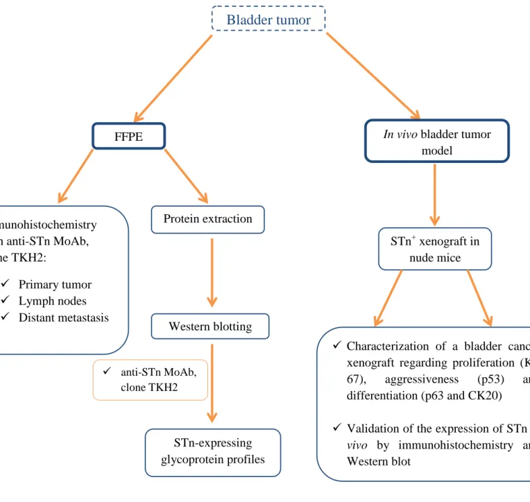
The first part of the project devotes to verify the association of the STn antigen with invasion in a series of 96 primary bladder tumors including different stages of the disease (Ta, T1, T2, T3 and T4). This series will also be characterized according to the degree of proliferation, based on Ki-67 expression. Moreover, STn expression will be evaluated in a a smaller and independent series of 21 bladder tumors isolated by radical cystectomy, presenting lymph node (17 cases) and distant metastasis (6 cases). This will allow determining the association of the STn antigen with metastasis. Finally, glycoproteins will be extracted from bladder tumors and metastasis to disclose the STn-expressing glycoprotein profiles.
The second part of the thesis devotes to the validation of a STn-expressing bladder cancer xenograft model in nude mice, by immunohistochemistry and Western blot. Such model is regarded of primary importance to identify drugs and treatment regimens that would better serve patients with STn-positive MIBC, as well as a platform to identify markers of tumor response and resistance to chemotherapic agents.
28 Clinical relevance of the sialyl-Tn antigen in bladder cancer
Figure 4 | Overview on the analytical approach. FFPE – formalin fixed paraffin embedded; MoAb – monoclonal antibody; CK20 – cytokeratine 20.
![Figure 1 | Extension of the different stages of bladder cancer (NMIBC and MIBC) (reprinted from [4])](https://thumb-eu.123doks.com/thumbv2/123dok_br/15926970.1094534/28.892.111.800.350.778/figure-extension-different-stages-bladder-cancer-nmibc-reprinted.webp)
![Figure 2 | Pathways of the biosynthesis of O-GalNAc glycans – synthesis of Tn and STn antigens and cores 1 to 4, which are the most common in humans (reprinted from [14])](https://thumb-eu.123doks.com/thumbv2/123dok_br/15926970.1094534/33.892.61.856.120.731/figure-pathways-biosynthesis-galnac-glycans-synthesis-antigens-reprinted.webp)
![Figure 3 | Graphic overview of the expression of STn in cancer (adapted from [55]).](https://thumb-eu.123doks.com/thumbv2/123dok_br/15926970.1094534/40.892.111.731.421.848/figure-graphic-overview-expression-stn-cancer-adapted.webp)