CALL FOR PAPERS
Systems Biology and Polygenic Traits
Association of ADAMTS7 gene polymorphism with cardiovascular survival
in coronary artery disease
A. Pereira,1R. Palma dos Reis,2R. Rodrigues,1A. C. Sousa,1S. Gomes,1S. Borges,1I. Ornelas,1
A. I. Freitas,3G. Guerra,3E. Henriques,1M. Rodrigues,1S. Freitas,1C. Freitas,1A. Brehm,3D. Pereira,1 and M. I. Mendonça1
1Funchal Hospital Center, Research Unit and Cardiology Department, Funchal, Madeira, Portugal;2Faculty of Medical Sciences, New University of Lisbon, Lisbon, Portugal; and3Laboratory of Human Genetics, Madeira University, Campus da Penteada, Funchal, Madeira, Portugal
Submitted 16 May 2016; accepted in final form 8 September 2016 Pereira A, Palma dos Reis RP, Rodrigues R, Sousa AC, Gomes S, Borges S, Ornelas I, Freitas AI, Guerra G, Henriques E, Rodrigues M, Freitas S, Freitas C, Brehm A, Pereira D, Mendonça MI. Association of ADAMTS7 gene polymorphism with cardiovascular survival in coronary artery disease. Physiol
Genomics 48: 810 – 815, 2016. First published September 9, 2016;
doi:10.1152/physiolgenomics.00059.2016.—Recent genetic studies have revealed an association between polymorphisms at the ADAMTS7 gene locus and coronary artery disease (CAD) risk. Functional studies have shown that a CAD-associated polymorphism (rs3825807) affects ADAMTS7 maturation and vascular smooth mus-cular cell (VSMC) migration. Here, we tested whether ADAMTS7 (A/G) SNP is associated with cardiovascular (CV) survival in patients with established CAD. A cohort of 1,128 patients with angiographic proven CAD, who were followed up prospectively for a mean fol-low-up period of 63 (range 6 –182) mo, were genotyped for rs3825807 A/G. Survival statistics (Cox regression) compared heterozygous (AG) and wild-type (AA) with the reference homozygous GG. Ka-plan-Meier (K-M) survival curves were performed according to ADAMTS7 genotypes for CV mortality. Results showed that 47.3% of patients were heterozygous (AG), 36.5% were homozygous for the wild-type allele (AA) and only 16.2% were homozygous for the GG genotype. During the follow-up period, 109 (9.7%) patients died, 77 (6.8%) of CV causes. Survival analysis showed that AA genotype was an independent risk factor for CV mortality compared with reference genotype GG (HR⫽ 2.7, P ⫽ 0.025). At the end of follow-up, the estimated survival probability (K-M) was 89.8% for GG genotype, 82.2% for AG and 72.3% for AA genotype (P⫽ 0.039). Carriage of the mutant G allele of the ADAMTS7 gene was associated with improved CV survival in patients with documented CAD. The native overfunctional ADAMTS7 allele (A) may accelerate VSMC migra-tion and lead to neointimal thickening, atherosclerosis progression and acute plaque events. ADAMTS7 gene should be further explored in CAD for risk prediction, mechanistic and therapeutic goals. ADAMTS7 gene; coronary artery disease; single nucleotide polymor-phism (SNP), cardiovascular survival
ADAMTS7 IS A MEMBERof a disintegrin and metalloprotease with
thrombospondin type 1 repeats (ADAMTS) family with pro-teolytic activity against extracellular substrates, mainly extra-cellular matrix proteins (1). The importance of ADAMTS
proteases is implicated in the establishment of tissue architec-ture during development and in tissue degradation during disease states such as cancer, Alzheimer’s and chronic inflam-matory conditions (20).
A recent study revealed the first firm evidence that ADAMTS7 plays a proatherogenic role likely through the promotion of vascular smooth muscular cell (VSMC) migra-tion by degrading its primary substrate, the cartilage oligo-meric matrix protein (COMP), and thus promoted neointima formation following vascular mechanical injury (18). More-over, in vitro studies have shown that VSMCs produce COMP, which inhibits VSMC migration (18). Consistently, genetic deletion of ADAMTS7 protects mice from atherosclerosis by reducing VSMC migration (4). Since VSMC migration is a very relevant process for plaque formation and stability, it is likely that ADAMTS7 may play a role in the development of atherosclerosis, the major cause of coronary artery disease (CAD).
In fact, three independent genome-wide association studies have recently identified the ADAMTS7 in the 15q24.2 chro-mosome, a novel locus for the development of coronary ath-erosclerosis (6, 14, 16). The lead CAD-associated SNP was rs3825807, a nonsynonymous polymorphism, with an adenine (A) to guanine (G) change, resulting in a serine-to-proline substitution in the prodomain of ADAMTS7 (16). Previous results suggest that this polymorphism has an effect on ADAMTS7 maturation, COMP degradation, and VSMC mi-gration and thus is associated with atherosclerosis (13).
We aimed to examine whether the carriage of the ADAMTS7 rs3825807 A/G polymorphism plays a role in the cardiovascular (CV) survival, in a cohort of patients with significant angiographic coronary artery disease.
MATERIALS AND METHODS
Study population. A prospective study was performed with a cohort
of 1,128 individuals (mean age 53.3⫾ 7.9 yr, 79.6% men) admitted for coronary angiography due to angina and clinical suspicion of ischemic heart disease from either sex that gave written informed consent. The study was approved by the Funchal Hospital Ethics Committee.
Subjects were primary local residents, and this population is ge-netically a Caucasian south European sample. Nonresidents were not considered for this study.
Address for reprint requests and other correspondence: A. M. Pereira, Funchal Hospital Center, Research Unit and Cardiology Dept., Avenida Luís de Camões, n° 57, 9004-514 Funchal, Madeira, Portugal (e-mail: dep.card@sesaram.pt). First published September 9, 2016; doi:10.1152/physiolgenomics.00059.2016.
Determination of presence and severity of CAD was made by the hemodynamic laboratory staff that included a dedicated intervention cardiologist. Angiographically proven CAD was considered signifi-cant ifⱖ1 coronary lesions of ⱖ70% stenosis in ⱖ1 major coronary artery or its primary branch. Absent or nonsignificant CAD was excluded from this study. Index angiography occurred between 2008 and 2013.
This cohort was followed until death or December 2014 [mean follow-up period of 63 mo (range 6 –182 mo)]. Each subject was followed up at the outpatient clinic according to the angiography date, and data concerning new hospital admissions or angiograms were collected from the clinical files of patients.
In the case of extrahospital death, death cause was collected consulting the public death report and by family telephone survey. The causes of CV death were determined according to the Interna-tional Classification of Disease, 10th revision, clinical modification diagnostic criteria. Cardiovascular death included CAD and cerebro-vascular disease.
Follow-up using the database and direct contact with patients or families allowed for 100% assessment of survival within this cohort.
Data collection. Data were collected from all subjects in a
stan-dardized file comprising demographic and clinical characteristics and traditional risk factors (sex, age, sedentary lifestyle, smoking habits, arterial hypertension, diabetes, dyslipidemia, body mass index, heart rate and pulse wave velocity). The definition of conventional CV risk
factors was based on the standard criteria, as previously reported (3, 5, 6a, 7, 12).
Biochemical analysis. Blood samples were extracted after 14 –16 h
fasting. Biochemical analyses were performed in the central labora-tory of the hospital, according to the usual techniques.
To determine total cholesterol and triglycerides, blood samples were placed in dry tubes and centrifuged a half-hour later at 3,500 g and subsequently quantified by an enzymatic technique with a Hitachi 911 auto analyzer. Biochemical markers such as lipoprotein-a, apoli-poprotein B (Apo B), and high sensitivity C-reactive protein (hs-CRP) were quantified by nephelometry on a Behring BN 100 automatic system. Homocysteine was measured by fluorescence polarized im-munoassay using an Abbot IMX automatic device. To measure fibrinogen, samples placed in a tube containing sodium citrate, and measurements were taken with a Behring BCS automatic analyzer.
Genotype analyses. Genomic DNA was extracted from 80 ml of
peripheral blood using a standard phenol-chloroform method (Qiagen, Hilden, Germany).
To identify the ADAMTS7 rs3825807 A/G genotypes, a TaqMan allelic discrimination assay (Applied Biosystems) was performed using labeled probes and primers pre-established by the supplier (TaqMan SNP Genotyping Assays, Applied Biosystems).
The genotyping reaction was amplified and detected on a StepOnePlus Real-Time PCR, and genotypes were determined by Table 1.
Variables Total (n⫽ 1,128) Survivors (n⫽ 1,019) Deaths (n⫽ 109) Cardiovascular Deaths (n⫽ 77) P1 P2
Male (%) 898 (79.6) 808 (79.3) 90 (82.6) 64 (83.1) 0.420 0.429 Age, yr 53.3⫾ 7.9 53.1⫾ 8.0 55.5⫾ 7.6 55.5⫾ 8.2 0.003 0.015 Sedentarism (%) 718 (63.7) 638 (62.6) 80 (73.4) 55 (71.4) 0.026 0.142 Smoking (%) 511 (45.3) 463 (45.4) 48 (44.0) 34 (44.2) 0.780 0.834 Hypertension (%) 819 (72.6) 745 (73.1) 74 (67.9) 49 (63.6) 0.245 0.067 PWV, m/s 8.7⫾ 2.0 8.6⫾ 1.9 9.0⫾ 2.1 8.9⫾ 2.3 0.058 0.261 Diabetes (%) 387 (34.3) 336 (33.0) 51 (46.8) 35 (45.5) 0.004 0.033 Dyslipidemia (%) 1,057 (93.7) 952 (93.4) 105 (96.3) 74 (96.1) 0.235 0.369 BMI, kg/m2 28.6⫾ 4.3 28.6⫾ 4.2 28.6⫾ 4.7 28.7⫾ 4.6 0.994 0.827 Heart rate, bpm 69.4⫾ 12.4 68.9⫾ 12.1 73.7⫾ 14.6 74.7⫾ 13.9 0.001 ⬍0.0001
P1 compares patients dying from any cause with survivors, and P2 compares patients dying from cardiovascular causes with others. PWV, pulse wave velocity; BMI, body mass index. Continuous variables are presented as means⫾ SD; Statistically significant for P ⬍ 0.05.
Table 2.
Variables Total (n⫽ 1,128) Survivors (n⫽ 1,019) Deaths (n⫽ 109) Cardiovascular Deaths (n⫽ 77) P1 P2
Hemoglobin, g/dl 14.6 14.6 14.8 14.9 0.889 0.915 (8.6–18.2) (8.6–18.2) (10.9–18.0) (11.0–17.6) Platelets, 103/l 214.5 214.0 224.0 227.0 0.106 0.048 (61.0–893.0) (61.0–893.0) (116.0–533.0) (116.0–533.0) Leukocytes, 103/l 7.0 7.0 7.4 7.8 0.022 0.006 (3.4–24.6) (3.4–24.6) (4.1–13.3) (4.4–12.4) Fibrinogen, mmHg 383.0 382.5 399.4 383.0 0.096 0.449 (91.0–832.0) (91.0–715.0) (153.3–832.0) (153.3–832.0) Homocysteine,mol/l 12.4 12.3 12.8 12.7 0.017 0.044 (4.6–220.2) (4.6–220.2) (5.9–48.7) (5.9–48.7) Lp(a), mg/dl 19.9 19.3 26.0 23.4 0.065 0.489 (1.9–241.0) (1.9–241.0) (1.9–219.0) (1.9–191.5) Apo B, mg/dl 96.4 95.9 101.5 102.0 0.007 0.012 (4.9–256.9) (4.9–256.9) (20.1–181.0) (20.1–173.0) Triglycerides, mg/dl 143.0 143.0 149.0 153.0 0.123 0.110 (31.0–2,500.0) (31.0–2500) (44.0–608.0) (44.0–571.0) Glucose, mg/dl 106.0 106.0 116.5 118.0 ⬍0.0001 ⬍0.0001 (53.0–458.0) (53.0–458.0) (67.0–393.0) (67.0–393.0) hs-CRP, mg/dl 0.24 0.23 0.36 0.3 0.005 0.083 (0.01–24.8) (0.01–24.8) (0.02–15.6) (0.02–7.9)
P1 compares patients dying from any cause with survivors, and P2 compares patients dying from cardiovascular causes with others. Lp(a), lipoprotein (a); Apo B, apolipoprotein B; hs-CRP, high sensitivity C reactive protein. Values are medians (minimum - maximum); statistically significant for P⬍ 0.05.
using the 7300 System SDS Software without any prior knowledge of individual clinical data (Applied Biosystems, Foster City, CA).
Statistical analyses. Deviation from Hardy-Weinberg equilibrium
for genotypes at individual locus was assessed using the 2-test.
Comparisons of characteristics of CV death and survivors were analyzed by2 for categorical variables, and Student’s t- or
Mann-Whitney test was performed for continuous variables as appropriate. Genotypic frequencies were determined from observed counts and compared by2 analysis. Heterozygous (AG) or homozygous
wild-type (AA) carriers were compared with noncarriers (GG) with sur-vival statistics.
Multivariable Cox proportional-hazard regression analyses were used to examine the CV death between genotypes of ADAMTS7 variants, using GG as reference class (genotype model). Hazard ratios and the multiple variable predictive values were determined, condi-tioned on seven other major CAD risk factors: age, sex, smoking status, diabetes, hypertension, dyslipidemia, and renal failure. The critical value for entering and excluding variables in the model was set at P⫽ 0.10. Multivariable Cox proportional-hazard regression anal-yses also addressed the all-cause mortality including the same set of covariates.
Kaplan Meyer survival function with Log-rank statistics was tested to predict the value of the ADAMTS7 genotypes for CV mortality.
Statistical analyses were performed using Statistical Package for the Social Sciences Software version 19.0 (IBM, Armonk, NY).
RESULTS
A total of 1,128 patients (mean age 53.3 ⫾ 7.9 yr, 79.6% men) with documented CAD entered this study, during a mean follow-up period of 63 mo (range 6 –182 mo). Baseline and biochemical characteristics of the studied population are shown in Table 1 and Table 2, respectively.
Of entered patients, 109 (9.7%; mean age 55.5 ⫾ 7.6 yr, 82.6% men) died during the follow-up period and 77 (6.8%, mean age 55.5⫾ 8.2 yr, 83.1% men) of CV causes. Nonsur-vivors were older at baseline, more sedentary, had more dia-betes and higher heart rates than survivors, with statistical significance (P ⬍ 0.05) (Table 1). Additionally, leukocytes,
homocysteine, apo B levels, glucose and hs-CRP were higher in patients who died during the follow-up than in survivors (Table 2).
ADAMTS7 genotypic distributions are shown in Table 3 for all patients, namely, survivors, those dying of any cause, and those dying of a CV cause. The distribution of alleles was in Hardy-Weinberg equilibrium (P⫽ 0.890). Genotypic analysis revealed that 47.3% of patients were heterozygous (AG), 36.5% were homozygous for the wild-type allele (AA), and only 16.2% were homozygous for the GG genotype. The proportion of alleles present in the cohort was 60.2% for the wild-type allele (A) and 39.8% for the G allele. CV deaths occurred in 49.4% of the carriers of the wild-type ADAMTS7 AA, while 7.8% were individuals with the polymorphic GG genotype (Table 3).
In multiple variable Cox regression analysis (Table 4), including age, sex, smoking status, diabetes, history of hyper-tension and of dyslipidemia, renal failure, and genetic variant (ADAMTS7), only age [hazard ratio (HR) ⫽ 1.051, P ⫽ 0.001], hypertension (HR ⫽ 0.634, P ⫽ 0.063), and ADAMTS7 AA genotype carriers (HR⫽ 2.681, P ⫽ 0.025) were selected as independent predictors of CV mortality (Table 4).
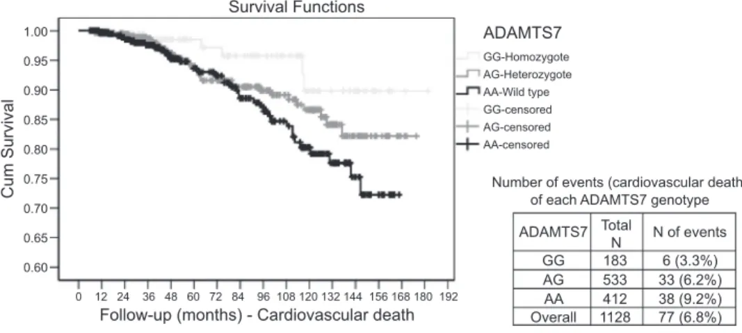
Kaplan-Meier survival-time plot for CV death by ADAMTS7 genotype is shown in Fig. 1. A significant differ-ence in survival by ADAMTS7 genotype was observed (log-rank statistic 6.48, P⫽ 0.039). At the end of follow-up period, the survival probability was 89.8% for the GG genotype carriers, 82.2% for heterozygote individuals (AG) and 72.3% for the wild-type (AA). Within each ADAMTS7 genotype, 3.3% (6/183) of events (CV deaths) occurred for patients carrying the polymorphic genotype (GG), 6.2% (33/533) among the heterozygote (AG) carriers, and 9.2% (38/412) occurred for patients who had the wild-type genotype (AA) as shown in Fig. 1.
Table 3.
Group Wild Type (AA) Heterozygote (AG) Homozygote (GG) WT (A) Allele Variant (G) Allele
A: All patients/All deaths
Survivors (1,019) 364 (35.7%) 484 (47.5%) 171 (16.8%) 1,212 (59.5%) 826 (40.5%) Deaths (109) 48 (44.0%) 49 (45.0%) 12 (11.0%) 145 (66.5%) 73 (33.5%) Total (1,128) 412 (36.5%) 533 (47.3%) 183 (16.2%) 1,357 (60.2%) 899 (39.8%) B: CV deaths/CV survivors No CV death (1,051) 374 (35.6%) 500 (47.6%) 177 (16.8%) 1,248 (59.4%) 854 (40.6%) CV deaths (77) 38 (49.4%) 33 (42.9%) 6 (7.8%) 109 (70.8%) 45 (29.2%) Total (1,128) 412 (36.5%) 533 (47.3%) 183 (16.2%) 1,357 (60.2%) 899 (39.8%)
For A, death by genotype contingency tables gives P⫽ 0.134 (2). For B, CV death by genotype gives P⫽ 0.021 2), P⫽ 0.017 (likelihood ratio) or P ⫽ 0.006 (linear-by-linear association).CV, cardiovascular; WT, wild type.
Table 4.
Variables B SE Wald df Hazard Ratio (CI 95%) P Value
Age 0.049 0.016 10.131 1 1.051 (1.019–1.083) 0.001
AHT ⫺0.455 0.245 3.462 1 0.634 (0.393–1.025) 0.063
ADAMTS7 5.481 2 0.065
AG 0.690 0.444 2.412 1 1.993 (0.835–4.760) 0.120
AA 0.986 0.440 5.016 1 2.681 (1.131–6.353) 0.025
Using forward Wald conditional Cox regression method (SPSS v 19.0), entering age (yr), male sex, diabetic status, history of hypertension, history of dyslipidemia, renal failure, smoking and genetic variant ADAMTS7. P value to exclude variables was 0.10. B, beta coefficient; CI, 95% confidence interval.
A second Cox regression model, adjusted for the same covariates, was performed for predicting all-cause mortality, as shown in Table 5. During the follow-up period, the only independent predictors of all-cause mortality were age (HR 1.05 P ⬍ 0.0001), Diabetes (HR ⫽ 1.469, P ⫽ 0.05), and smoking habit (HR ⫽ 1.58, P ⫽ 0.026). Neither carriage of homozygous (AA) nor heterozygous (AG) genotypes were independent predictors of all-cause mortality (HR ⫽ 1.424,
P⫽ 0.276, HR ⫽ 1.604, P ⫽ 0.147 respectively) (Table 5).
DISCUSSION
In our study, patients with angiographically documented CAD who were carriers of the polymorphic G variant of ADAMTS7 gene had increased survival from CV causes. For instance, at the end of the follow-up period, patients with ADAMTS7 GG showed approximately three times fewer events of CV mortality in relation to AA carriers.
We consider that the carriers of the G allele had reduced migratory ability with lower atherosclerosis progression and severity, reduced proteinases secretion to influence the stability of the complex plaque, and therefore better prognostic than patients carrying the wild-type A of the ADAMT7 gene.
Similarly, Pu and colleagues (13), observed an inverse association between the ADAMTS7 rs3825807 GG genotype and atherosclerosis, this being the genotype associated with lower carotid atherosclerosis prevalence and severity (score). The same study demonstrated that VSMCs of the protective GG genotype had decreased migratory activity compared with those with the AA genotype, and conditioned media of VSMCs of the GG contained less of the cleaved form of COMP, an ADAMTS7 substrate that had been shown to be produced by VSMCs and inhibit its migration (13). Also, You et al. (19) recently demonstrated a strong and direct association between the proportion of CAD patients with three-vessel disease and increasing ADAMTS7 rs3825807 gene dosage of the risk
variant (A). This condition was investigated in the cohort of our study, but no significant association was found between AA genotype and most severe atherosclerosis (three-vessel disease), although there was a strong trend (data not shown).
However, to date, an association between the CAD progno-sis and this polymorphism has not yet been found. As far as we know, this study is the first attempt to establish this association in a cohort of south European descendent.
Previously, investigation of the physiological roles of
ADAMTS7 has largely focused on its association with the
pathogenesis of arthritis and disk diseases (6a). ADAMTS structure contains the thrombospondin type-1 repeats and acts as a negative regulator of endplate chondrocyte differentiation because it downregulates the expression of collagen type II, collagen type X, and SOX9, which are early and late marker genes for chondrogenesis (11). Furthermore, suppression of
ADAMTS7 expression in human chondrocytes markedly
pre-vents COMP degradation (9). ADAMTS7 mRNA is found in normal human bone, cartilage, synovia, tendons, spleen, heart, and brain, as well as in the meniscus, skeletal muscle, and fat at lower levels (10). However, the physiological implications of this widespread tissue expression of ADAMTS7 remain to be determined. Furthermore, cumulative evidence has demon-strated that ADAMTS7 facilitates VSMC migration and neoin-tima formation through degradation of vascular COMP (18). Consistently, genetic deletion of ADAMTS7 reduces neointimal thickening after wire injuries (4).
In our study, it’s interesting to note that besides ADAMTS7 AA genotype, the hypertension status and age were indepen-dent predictors of CV mortality; being elderly and lower tensional status both correlated with CV death. The indepen-dence of CV death predictors from other traditional risk factors like dyslipidemia, diabetes, and renal failure emphasizes the ADAMT7 gene’s mechanistic role being associated with vas-cular cleavage and migration of VSMCs not currently covered
1.00 0.95 0.90 0.85 0.80 0.75 0.70 0.65 0.60 0 12 24 36 48 60 72 84 96 108 120 132 144 156 168 180 192 GG-Homozygote AG-Heterozygote AA-Wild type GG-censored AG-censored AA-censored ADAMTS7 ADAMTS7 Survival Functions
Follow-up (months) - Cardiovascular death
Cum Survival Number of events (cardiovascular death)of each ADAMTS7 genotype
GG AG AA Overall 183 533 412 1128 6 (3.3%) 33 (6.2%) 38 (9.2%) 77 (6.8%) Total N N of events
Fig. 1. The log-rank statistic was 6.480, P⫽ 0.039. There are 6 events (3.3%) among 183 patients carrying the homozygote, 33 events (6.2%) among the patients who had the het-erozygote type, and 38 events (9.2%) among the patients who had the wild type.
Table 5.
Variables B SE Wald df Hazard ratio (CI 95%) P Value
Age 0.050 0.014 13.405 1 1.051 (1.023–1.080) 0.000
Diabetic status 0.384 0.197 3.826 1 1.469 (0.999–2.159) 0.050
Smoking 0.456 0.204 4.973 1 1.578 (1.057–2.355) 0.026
Using forward Wald conditional Cox regression method (SPSS v 19.0), entering age (yr), male sex, diabetic status, history of hypertension, history of hyperlipidemia, renal failure, smoking, and genetic variant ADAMTS7. P value to exclude variables was 0.10. B, beta coefficient; CI, 95% confidence interval.
by updated therapies including statins, ACE inhibitors, angio-tensin-aldosterone-antagonists, B-blockers, and antidiabetic medication. Monoclonal antibodies targeting several ADAMTS gene family members are on the way in several clinical scenarios, and the ADAMTS7 gene seems a highly favorable candidate for use against for CAD because of the gene’s already established association with the development of CAD. Future genetic studies in CAD disease should focus on disease progression and prognosis rather than disease development. So far, established risk factors of atherosclerosis are not able to fully account for CV death due to different pathophysiological mechanisms involved into plaque formation and plaque events. CV death might be associated with the critical mechanisms of plaque stabilization/thrombosis and arrhythmia in CAD pa-tients and gene associated with vascular migration/stabilization are promising targets (14).
Nevertheless, in our study the AA genotype was found to be unassociated with all-cause mortality and associated only with CV mortality, which strengthens the hypothesis that ADAMTS7’s being a regulator of the gene maturation, COMP cleavage, and VSMC migration would translate into fewer CV events, including CV death in patients with a reduced function of the ADAMTS gene (G) variant.
Further research in this area could lead to personalized medicine based on identification of ADAMTS7 genetic vari-ants in patients with atherosclerosis who could benefit from strategies targeting this proteolytic pathway (2).
Study Strengths and Limitations
Very few studies have yet addressed common gene variants and outcomes and long-term prognosis, and even the 9p21 gene failed to demonstrate consistent association with long-term myocardial infarction and CV mortality (17). No report on the specific association of ADAMTS7 gene variants with CV mortality in CAD patients has been published.
However, this study is only moderate in size, and the number of events in the follow-up is limited, so that the confidence intervals for CV survival are quite broad.
A significant limitation of the study was the lack of ejection fraction data at admission for determining the relative protec-tive effect of the variant as a function of the ejection fraction. Patients deceived during the coronary angiogram were also excluded from our study. Those patients probably had severe atherosclerotic coronary disease, and their DNA sample would be useful to enlarge our knowledge of this variant role in CV death. Expression studies of these ADAMTS7 gene variants on the arterial wall of the atherosclerotic plaque or thrombus would also further information from animal studies already published.
In conclusion: Our study emerges as a novel finding in a south European population and provides new evidence impli-cating ADAMTS7 with CAD prognosis. Its spectrum of interest as a therapeutic target may grow once its association with outcomes and CV mortality is clarified. Our results, although promising, should be amplified in larger and long-term studies that focus on CV prognosis rather than disease development. If validated, they suggest that ADAMTS7 genotyping may pro-vide useful information for prognosis and survival in CAD patients.
ACKNOWLEDGMENTS
We are very grateful to Elsa Sousa who performed the phone calls to the subjects and all the administrative procedures.
GRANTS
This study was supported by the European Regional Development Fund’s Operational Programme for the Enhancement of Economic Potential and Territorial Cohesion for the Autonomous Region of Madeira (INTERVIR⫹).
DISCLOSURES
No conflicts of interest, financial or otherwise, are declared by the author(s).
AUTHOR CONTRIBUTIONS
A.P., R.P.d.R., and M.I.M. conception and design of research; A.P., R.R., A.C.S., S.G., I.O., A.I.F., G.G., C.F., and M.I.M. performed experiments; A.P., S.B., E.H., M.R., S.F., and M.I.M. analyzed data; A.P. and M.I.M. interpreted results of experiments; A.P., S.B., E.H., M.R., S.F., and M.I.M. drafted manuscript; A.P., R.P.d.R., S.B., A.B., D.P., and M.I.M. edited and revised manuscript; A.P., R.P.d.R., and M.I.M. approved final version of manuscript; E.H., M.R., and S.F. prepared figures.
REFERENCES
1. Arner EC. Aggrecanase-mediated cartilage degradation. Curr Opin
Phar-macol 2: 322–329, 2002.
2. Arroyo AG, Andrés V. ADAMTS7 in cardiovascular disease: from bedside to bench and back again? Circulation 131: 1156 –1159, 2015. 3. Asmar R, Benetos A, Topouchian J, Laurent P, Pannier B, Brisac AM,
Target R, Levy BI. Assessment of arterial distensibility by automatic
pulse wave velocity measurement: validation and clinical application studies. Hypertension 26: 485–490, 1995.
4. Bauer RC, Tohyama J, Cui J, Cheng L, Yang J, Zhang X, Ou K,
Paschos GK, Zheng XL, Parmacek MS, Rader DJ, Reilly MP.
Knock-out of Adamts7, a novel coronary artery disease locus in humans, reduces atherosclerosis in mice. Circ 131: 1202–1213, 2015.
5. Chobanian AV, Bakris GL, Black HR, Cushman WC, Green LA, Izzo
JL Jr, Jones DW, Materson BJ, Oparil S, Wright JT Jr, Roccella EJ; National Heart, Lung, and Blood Institute Joint National Committee on Prevention, Detection, Evaluation, and Treatment of High Blood Pressure; National High Blood Pressure Education Program Coordi-nating Committee. The seventh report of the Joint National Committee
on Prevention, Detection, Evaluation and treatment of High Blood Pres-sure: the JNC7 report. JAMA 289: 2560 –2572, 2003.
6. Coronary Artery Disease (CAD) Genetics Consortium. A genome-wide association study in Europeans and South Asians identifies five new loci for coronary artery disease. Nat Genet 43: 339 –344, 2011. 6a.Expert Committee on the Diagnosis and Classification of Diabetes
Mellitus. Report of the expert committee on the diagnosis and
classifica-tion of diabetes mellitus. Diabetes Care 26, Suppl 1: S5–S20, 2003. 7. Expert Panel on Detection Evaluation, and Treatment of High Blood
Cholesterol in Adults. Executive Summary of the Third Report of the
National Cholesterol Education Program (NCEPT) Expert Panel on De-tection Evaluation and Treatment of High Blood Cholesterol in Adults (Adult Treatment Panel III). JAMA 285: 2486 –2497, 2001.
8. Gopalakrishnan K, Kumarasamy S, Abdul-Majeed S, Kalinoski AL,
Morgan EE, Gohara AF, Nauli SM, Filipiak WE, Saunders TL, Joe B.
Targeted disruption of Adamts16 gene in a rat genetic model of hyper-tension. Proc Natl Assoc Sci USA 109: 20555–20559, 2012.
9. Guo NH, Krutzsch HC, Inman JK, Roberts DD. Thrombospondin 1 and type I repeat peptides of thrombospondin 1 specifically induce apoptosis of endothelial cells. Cancer Res 57: 1735–1742, 2012.
10. Hanby HA, Zheng XL. Biochemistry and physiological functions of ADAMTS7 metalloprotease. Adv Biochem 1: 43–50, 2013.
11. Kuno K, Matsushima K. ADAMTS-1 protein anchors at the extracellular matrix through the thrombospondin type I motifs and its spacing region. J
Biol Chem 273: 13912–13917, 1998.
12. National Institute of Health, National Heart, Lung, and Blood
Insti-tute North American Association for the Study of Obesity. The
Prac-tical Guide: Identification, Evaluation, and Treatment of Overweight and Obesity in Adults. NHLBI Obesity Education Initiative, NIH Publication
13. Pu X, Xiao Q, Kiechl S, Chan K, Ng FL, Gor S, Poston RN, Fang C,
Patel A, Senver EC, Shaw-Hawkins S, Willeit J, Liu C, Zhu J, Tucker AT, Xu Q, Caulfield MJ, Ye S. ADAMTS7 cleavage and vascular
smooth muscle cell migration is affected by a coronary-artery-disease-associated variant. Am J Hum Genet 92: 366 –374, 2013.
14. Reilly MP, Li M, He J, Ferguson JF, Stylianou IM, Mehta NN,
Burnett MS, Devaney JM, Knouff CW, Thompson JR, Horne BD, Stewart AF, Assimes TL, Wild PS, Allayee H, Nitschke PL, Patel RS; Myocardial Infarction Genetics Consortium; Wellcome Trust Case Control Consortium; Martinelli N, Girelli D, Quyyumi AA, Anderson JL, Erdmann J, Hall AS, Schunkert H, Quertermous T, Blankenberg S, Hazen SL, Roberts R, Kathiresan S, Samani NJ, Epstein SE, Rader DJ; Myocardial Infarction Genetics Consortium; Wellcome Trust Case Control Consortium. Identification of ADAMTS7 as a novel locus
for coronary atherosclerosis and association of ABO with myocardial infarction in the presence of coronary atherosclerosis: two genome-wide association studies. Lancet 377: 383–392, 2011.
16. Schunkert H, König IR, Kathiresan S, Reilly MP, Assimes TL, Holm
H, Preuss M, Stewart AF, Barbalic M, Gieger C, Absher D, Aherrah-rou Z, Allayee H, Altshuler D, Anand SS, Andersen K, Anderson JL, Ardissino D, Ball SG, Balmforth AJ, Barnes TA, Becker DM, Becker LC, Berger K, Bis JC, Boekholdt SM, Boerwinkle E, Braund PS, Brown MJ, Burnett MS, Buysschaert I; Cardiogenics Carlquist JF, Chen L, Cichon S, Codd V, Davies RW, Dedoussis G, Dehghan A, Demissie S, Devaney JM, Diemert P, Do R, Doering A, Eifert S, Mokhtari NE, Ellis SG, Elosua R, Engert JC, Epstein SE, de Faire U, Fischer M, Folsom AR, Freyer J, Gigante B, Girelli D, Gretarsdottir S, Gudnason V, Gulcher JR, Halperin E, Hammond N, Hazen SL, Hofman A, Horne BD, Illig T, Iribarren C, Jones GT, Jukema JW, Kaiser MA, Kaplan LM, Kastelein JJ, Khaw KT, Knowles JW, Kolovou G, Kong A, Laaksonen R, Lambrechts D, Leander K, Lettre G, Li M, Lieb W, Loley C, Lotery AJ, Mannucci PM, Maouche S, Martinelli N, McKeown PP, Meisinger C, Meitinger T, Melander O,
Merlini PA, Mooser V, Morgan T, Mühleisen TW, Muhlestein JB, Münzel T, Musunuru K, Nahrstaedt J, Nelson CP, Nöthen MM, Olivieri O, Patel RS, Patterson CC, Peters A, Peyvandi F, Qu L, Quyyumi AA, Rader DJ, Rallidis LS, Rice C, Rosendaal FR, Rubin D, Salomaa V, Sampietro ML, Sandhu MS, Schadt E, Schäfer A, Schil-lert A, Schreiber S, Schrezenmeir J, Schwartz SM, Siscovick DS, Sivananthan M, Sivapalaratnam S, Smith A, Smith TB, Snoep JD, Soranzo N, Spertus JA, Stark K, Stirrups K, Stoll M, Tang WH, Tennstedt S, Thorgeirsson G, Thorleifsson G, Tomaszewski M, Uit-terlinden AG, van Rij AM, Voight BF, Wareham NJ, Wells GA, Wichmann HE, Wild PS, Willenborg C, Witteman JC, Wright BJ, Ye S, Zeller T, Ziegler A, Cambien F, Goodall AH, Cupples LA, Querter-mous T, März W, Hengstenberg C, Blankenberg S, Ouwehand WH, Hall AS, Deloukas P, Thompson JR, Stefansson K, Roberts R, Thor-steinsdottir U, O’Donnell CJ, McPherson R, Erdmann J; CARDI ConsortiumoGRAM, Samani NJ. Large-scale association analysis
iden-tifies 13 new susceptibility loci for coronary artery disease. Nat Genet 43: 333–338, 2011.
17. Virani SS, Brautbar A, Lee VV, MacArthur E, Morrison AC, Grove
ML, Nambi V, Frazier L, Wilson JM, Willerson JT, Boerwinkle E, Ballantyne CM. Chromosome 9p21 single nucleotide polymorphisms are
not associated with recurrent myocardial infarction in patients with estab-lished coronary artery disease. Circ J 76: 950 –956, 2012.
18. Wang L, Zheng J, Bai X, Liu B, Liu CJ, Xu Q, Zhu Y, Wang N, Kong
W, Wang X. ADAMTS-7 mediates vascular smooth muscle cell
migra-tion and neointima formamigra-tion in balloon-injured rat arteries. Circ Res 104: 688 –698, 2009.
19. You L, Tan L, Liu L, Shen R, Chaugai S, Wang DW, Cui W. ADAMTS7 locus confers high cross-race risk for development of coro-nary atheromatous plaque. Mol Genet Genomics 242: 351–356, 2015. 20. Zhang Y, Lin J, Wei F. The function and roles of ADAMTS-7 in