Department of Chemistry
Health Science Research Center
T
T
h
h
e
e
r
r
o
o
l
l
e
e
o
o
f
f
P
P
K
K
C
C
δ
δ
a
a
s
s
a
a
r
r
e
e
g
g
u
u
l
l
a
a
t
t
o
o
r
r
o
o
f
f
t
t
h
h
e
e
N
N
o
o
x
x
1
1
c
c
o
o
n
n
t
t
r
r
i
i
b
b
u
u
t
t
i
i
o
o
n
n
f
f
o
o
r
r
t
t
h
h
e
e
m
m
e
e
c
c
h
h
a
a
n
n
i
i
s
s
m
m
o
o
f
f
p
p
a
a
r
r
a
a
q
q
u
u
a
a
t
t
i
i
n
n
d
d
u
u
c
c
e
e
d
d
d
d
o
o
p
p
a
a
m
m
i
i
n
n
e
e
r
r
g
g
i
i
c
c
n
n
e
e
u
u
r
r
o
o
t
t
o
o
x
x
i
i
c
c
i
i
t
t
y
y
Joana Ribeiro Barata
Dissertation presented to the University of Beira Interior for purpose of Master Degree in Biochemistry.
Supervisor: Prof. Doutora Graça Baltazar Co-supervisor: Ana Clara Cristóvão
Universidade da Beira Interior
Departamento de Química
Centro de Investigação em
Ciências da Saúde
P
P
a
a
p
p
e
e
l
l
d
d
a
a
P
P
K
K
C
C
δ
δ
n
n
a
a
r
r
e
e
g
g
u
u
l
l
a
a
ç
ç
ã
ã
o
o
d
d
a
a
N
N
o
o
x
x
1
1
e
e
s
s
u
u
a
a
c
c
o
o
n
n
t
t
r
r
i
i
b
b
u
u
i
i
ç
ç
ã
ã
o
o
n
n
o
o
s
s
m
m
e
e
c
c
a
a
n
n
i
i
s
s
m
m
o
o
s
s
d
d
e
e
t
t
o
o
x
x
i
i
c
c
i
i
d
d
a
a
d
d
e
e
d
d
o
o
p
p
a
a
m
m
i
i
n
n
é
é
r
r
g
g
i
i
c
c
a
a
i
i
n
n
d
d
u
u
z
z
i
i
d
d
o
o
s
s
p
p
e
e
l
l
o
o
p
p
a
a
r
r
a
a
q
q
u
u
a
a
t
t
o
o
Joana Ribeiro Barata
Dissertação apresentada à Universidade da Beira Interior para obtenção do grau de Mestre em Bioquímica.
Orientadora: Prof. Doutora Graça Baltazar Co-orientadora: Ana Clara Cristóvão
“Science does not know its debt to imagination”
Foremost I want to thank to my supervisor, Prof. Doutora Graça Baltazar, for her knowledge, help, and patience during this year.
To Ana Clara, I want to thank all of you friendship, your patience and kindness. Thank you for believe in this project as much or more than me. I’m grateful for knowing you, and work with you was a great experience. Thank you for all the endless conversation, about all and nothing… Thank you for everything!
Xica, thanks for sharing ideas and work with me. Thanks for all your help, your tolerance and your friendship! Thank you for all support in my work, and specially thank you for our conversations in the happiness, but mostly in the disappointing moments.
Bruno, thanks for all of your friendship and support along these years. Thanks for your comprehension, encouragement and presence in my life…
Sara, thanks for your friendship. Even on the other side of the laboratory, you were present, and helped me overcome the bad results. Thanks for our long conversations at “Aquarium”…
To all my friends and laboratory colleagues, specially Ana M., Irina, Rita, Sandra e Filipa, that helped and supports me along this year! I know that I’m not easy to understand!
À minha avó e às minhas tias: um grande obrigado por todo o apoio e carinho!
À minha mãe, a quem eu dedico esta tese, por estar sempre presente nos bons, mas principalmente, nos maus momentos. Obrigada por nunca me teres deixado desistir, mesmo quando isso parecia ser a única alternativa…Obrigada pelo interesse, dedicação e carinho. Obrigada!
Finalmente, e não menos importante, à minha irmã. Obrigada pelas longas conversas ao telefone. Obrigada pela tua amizade de mana. Obrigada por acreditares sempre em mim e por me ajudares em tudo! Obrigada por tudo!
All data presented in this dissertation are of my entire responsibility,
____________________________________ (Joana Ribeiro Barata)
List of abbreviations 1
Resumo 2
Abstract 4
1. Introduction 6
1.1.Parkinson’s Disease 7
1.1.1.Genetic mutations and PD 10
1.1.2.Environmental toxins and PD 12
1.2.Oxidative Stress and Parkinson’s disease 15
1.3.NADPH Oxidase System 16
1.4.PKCδ 18
1.4.1.PKCδ and Nox1 20
1.5.Characterization of N27 cell line 21
2. Objectives 22
3. Methodologies 24
3.1. Methods 25
3.1.1. Cell-culture 25
3.1.2. Cell Transfection with siRNA 25
3.1.3. Paraquat Treatment 26
3.1.4. Total RNA Extraction and RT-PCR analysis 26
3.1.5. Western-blot analysis 27
3.1.6. Cell viability assays 27
3.1.7. Determination of cellular ROS content 28
3.1.8. Data analysis and statistics 28
4.1. Selective knockdown of PKCδ expression by siRNA 31 4.2. PKCδ gene silencing decrease the PQ-induced ROS generation 33 4.3. PKCδ knockdown reduces PQ-induced dopaminergic cell death 35 4.4. PKCδ gene silencing decreases PQ-induced Nox1 expression 37
5. Discussion 39
6. Conclusion 42
7. Attachments 44
BBB: Blood Brain Barrier CNS: Central Nervous System DA: dopaminergic;
DAT: Dopamine transporter
DCFDA: 2′,7′-Dichlorodihydrofluorescein Diacetate; DHE: dihydroethidium;
GAPDH: Glyceraldehyde 3-phosphate dehydrogenase; LDH: Lactate dehydrogenase; Lewy Bodies: LBs LPS: lypopolysaccharide MB: Maneb MPP+: 1-methyl-4-phenylpyridinium; MPTP: 1-methyl-4-phenyl-1,2,3,6-tetrahydropyridine; mRNA: Messenger RNA
MTT: 3-(4,5-dimethylthiazal-2-yl)-2,5-diphenyl-tetrazolium bromide; NADPH: Nicotinamide adenine dinucleotide phosphate;
Nox: NADPH oxidase; PD: Parkinson’s disease PKCδ: Protein kinase Cδ PQ: paraquat;
RNA: Ribunucleic acid
ROS: Reactive oxygen species;
RT-PCR: Reverse Transcriptase Polymerase Chain Reaction; SEM: standard error of the mean
siRNA: small interfering Ribonucleic acid SN: substantia nigra;
A doença de Parkinson (DP) é uma doença neurodegenerativa caracterizada pela perda progressiva de neurónios dopaminérgicos (DA) da substantia nigra (SN)
pars compacta (SNpc). Apesar de a etiologia da doença não ser totalmente conhecida
sabe-se que pode ser desencadeada por factores como o envelhecimento, mutações génicas, e por factores ambientais. Vários estudos provaram que existe um aumento da incidência da DP em zonas rurais, e levantaram a hipótese de que a utilização de pesticidas, como o paraquato (PQ), podia conduzir à morte dos neurónios DA.
O PQ pode ser reduzido pela enzima nicotinamida adenina dinucleótido fosfato (NADPH) oxidase (Nox) e induzir assim a sua toxicidade através da produção de aniões superóxido, e acumulação de espécies reactivas de oxigénio (ROS) nas células. Resultados obtidos por Matsuzaki e colaboradores mostraram que a as ROS produzidas pela exposição ao PQ induzem a agregação da α-sinucleína, e consequentemente a formação de corpos de Lewy, característicos na DP. A Nox utiliza o oxigénio molecular para produzir ROS. Alguns estudos têm sugerido que a Nox1 tem um papel fundamental no stress oxidativo e consequente morte de neurónios DA, em células expostas ao PQ. Foi já demonstrado que a proteína cinase Cδ (PKCδ) está envolvida na sinalização de vias apoptóticas através da fosforilação e activação de várias moléculas sinalizadoras da morte celular. Estudos demonstraram que em neurónios DA expostos ao PQ há um aumento das ROS e consequentemente da morte neuronal.
O presente trabalho tem como objectivo entender o papel da PKCδ na regulação da produção de ROS pela Nox1, em células expostas ao PQ. Para analisar de que forma a PKCδ influencia a expressão da Nox1, assim como a produção de ROS e a morte de neurónios DA, inibiu-se especificamente a PKCδ utilizando uma sequência de RNA de interferência (siRNA).
Nas células transfectadas com o siRNA específico para a PKCδ observou-se uma diminuição de 32% do RNA mensageiro (mRNA), e de 54% na expressão da proteína. Vimos ainda que os níveis de ROS e morte celular de células expostas ao PQ, foi significativamente menor nas células transfectadas com o siRNA da PKCδ, em comparação com as células não transfectadas. Observámos ainda que em neurónios DA, expostos ao PQ e transfectados com siRNA da PKCδ, houve uma diminuição da Nox1, a nível do mRNA e proteína. Estes resultados parecem confirmar a hipótese de que a PKCδ está envolvida na regulação das vias de expressão da Nox1, em neurónios DA expostos ao PQ.
Parkinson’s disease (PD) is a progressive neurodegenerative disorder characterized by the selective loss of dopaminergic (DA) neurons in the substantia nigra (SN) pars compacta (SNpc), to which contribute the combination of aging and genetic and environmental factors. Several studies clearly showed an increase in the incidence of PD in rural environments, and hypothesized the involvement of pesticides, like paraquat (PQ), in the mechanisms of neurodegeneration.
PQ can be reduced by the nicotinamide adenine dinucleotide phosphate (NADPH) oxidase (Nox) enzyme and can induce its toxicity through the production of superoxide anions, which leads to the accumulation of reactive oxygen species (ROS) in the cell. Matsuzaki and its collaborators showed that ROS formation induced by PQ facilitate α-synuclein association, a biochemical hallmark of PD. The Nox system generates ROS in a regulated manner, producing ROS in various tissues. It has been suggested that Nox1 subunit plays a role in plays a role in the oxidative stress and subsequent DA cell death elicited by PQ. Protein kinase C delta (PKCδ) has been showed to be involved in apoptotic signaling pathways, by phosphorylation and activation of several inducers of cell death. PQ induces an increase of ROS production and death of DA neurons.
In the present work we aimed to investigate in PQ exposed cells the possible relationship between ROS generation by Nox1 and the putative regulatory effect of PKCδ in the process. To investigate whether PKCδ influences ROS production, DA cell death and Nox1 expression, short interference RNA (siRNA) was used to specifically knockdown PKCδ. Transfection with PKCδ-siRNA decrease mRNA levels by 32% and protein levels by 54%. We observed that ROS and DA cell death induced by PQ were significantly reduced by PKCδ knockdown, in N27 cells transfected with PKCδ-siRNA. This study also revealed that, in DA neurons, the upregulation of Nox1 mRNA and protein induced by PQ exposure is reversed when PKCδ is knockdown, suggesting a possible involvement of PKCδ in the regulation of Nox1 expression pathways affected by PQ.
1.1.
Parkinson’s Disease
In 1817, James Parkinson described the clinical features of one of the most neurodegenerative disorders affecting the nigrostriatal pathways involved in movement control, named Parkinson Disease (PD) (Parkinson, 1817; Toulouse and Sullivan, 2008). PD primarily affect areas of the brain involved in motor control, leading to the debilitating motor signs include resting tremor, rigidity, bradykinesia (slowness of movement), gait disturbances and postural instability. During the progression of the disease, non-motor areas of the brain become affected, potentially leading to depression, sleep disorders and cognitive impairment (Olanow and Tatton, 1999).
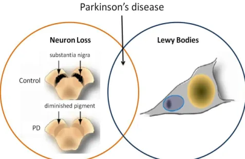
PD results primarily from the progressive and selective loss of dopaminergic (DA) neurons in the substantia nigra pars compacta (SNpc), with a resultant decrease of dopamine levels in the striatum (an area of the brain critical for movement control). When clinical symptoms appear, 50% of DA neurons in the SNpc and 80% of the striatal dopamine levels have already been lost (Dauer and Przedborski, 2003; Fahn, 2003) Another important pathological feature of PD is the presence of neuronal cytoplasmatic inclusions known as Lewy Bodies (LBs) which are intracytoplasmic inclusions of cytoplasmic proteins such as ubiquitin and α-synuclein (Figure 1) (Matsuzaki et al. 2004).
Figure 1. Relationship between cell death and LBs formation in PD. DA cell death (orange circle) is characterized by loss of neurons in the SNpc and LBs (blue circle) Adapted from Cookson et al. 2008.
PKCδ: a mediator in Nox1 regulation Introduction ____________________________________________________________________________________
Several hypotheses were raised to explain the loss of DA neurons occurring in PD (Figure 2). It has been postulated that increased oxidative environment on DA neurons might be a crucial event in the pathogenesis of PD. In DA neurons dopamine is rapidly sequestered within vesicles by the vesicular monoamine transporter, where the acidic pH significantly delays the oxidation of dopamine. However, an oxidative environment can be created if dopamine remains in the cytosol, where it can be oxidize at physiological pH to generate reactive ortho-quinones, as well as superoxide and hydrogen peroxide (LaVoie and Hastings 1999). Another hypothesis is that it seems to be a relation between abnormal protein accumulation or degradation, oxidative stress and mitochondrial dysfunction (Figure 2).
In transgenic mice, overexpressing α-synuclein, an impairment of mitochondrial function increases oxidative stress and enhances the nigral pathology induced by 1-methyl-4-phenylpyridinium (MPTP) (Lin and Beal 2006). This hypothesis is supported by the finding that 1-methyl-4-phenylpyridinium ion (MPP+) could induce a parkinsonian condition in humans, non human primates and rodents. Once mitochondrial dysfunction compromises the levels of ATP, the cell try to produce energy by other secondary pathways, which lead to the generation of ROS (ROS), and thus increase the toxicity within the cell. The exposure to environmental toxins has also been proposed as a
Figure 2. Schematic representation of possible mechanisms responsible for DA neurodegeneration in PD. Increased ROS production resulting from several different causes, may induce cellular damage and death through the modification of nucleic acids, lipids, and proteins. Protein oxidation may lead to proteolytic dysfunction and enhanced protein aggregation, which could in turn be toxic to DA neurons. Adapted from (Hald and Lotharius 2005).
cause for developing PD pathogenesis, and is associated to increased ROS levels and the consequent DA neurons lost. Matsuzaki and its collaborators (2004) showed that ROS could induce aggregation of cytoplasmic proteins, like α-synuclein. This theory is sustained by studies revealing that a significant defect in the proteasome, the principal machinery responsible for protein degradation, leads to protein aggregation and increased cell death. The accumulation of aggregated proteins, may lead to the formation of LB, which are a feature of PD. Ultimately, the selective loss of DA neurons in PD has been attributed to high levels of ROS within the DA neurons that could be generated by: the increased of dopamine oxidation in citosol; mitochondrial dysfunction and the consequent decrease in ATP levels; exposure to environmental toxins and the increase of protein aggregates inside the neuron (Figure 2) (reviewed by Hald and Lotharius 2005).
PKCδ: a mediator in Nox1 regulation Introduction ____________________________________________________________________________________
1.1.1. Genetic mutations and PD
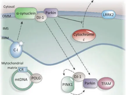
Despite the fact that the majority of PD cases are sporadic, PD pathogenesis can also be caused by familial genetic mutations. At present six genes, from thirteen genetic loci, have been established as a causative factor of familial PD: α-synuclein (PARK1/4), parkin (PARK2), PINK1 (PARK6), DJ-1 (PARK7), LRRK2 (PARK8), and ATP13A2 (PARK9) (Figure 3) (reviewed by Malkus et al. 2009). Mutations in mtDNA-encoded complex I subunits, 12SrRNA, and POLG also cause parkinsonism (Figure 3) (Lin and Beal 2006).
The first discovered mutation associated with PD occurs in the SNCA gene, and encodes for α-synuclein, contained within LBs (Cookson 2005). The pathogenic mutation A53T leads to the increase of mitochondrial dysfunction and thus ROS levels (Martin et al. 2006). α-Synuclein is a protein with a natural tendency to aggregate into oligomers that can then further aggregate into fibrils that are deposited as LBs, and the mutations on SNCA variably produce either oligomers/proto- fibrils or fibrils (Cookson 2005).
Leucine-rich repeat kinase 2 (LRRK2) has been recognized as a cause of an autossomal dominant late-onset form of familial PD. Once the LRRK2 kinase is localized in mitochondria, a mutation of LRRK2 affects the capacity of the cell to handle
Figure 3. Schematic representation of several proteins susceptible to mutations that have been associated with PD. Outer mitochondrial membrane (OMM), Intermembrane space (IMS), Internal mitochondria membrane (IMM) Adapted from Lin and Beal 2006.
oxidative stress and consequently leads to a decrease in cell viability (Lin and Beal 2006; Liou et al. 2008).
Parkin gene mutations were also identified as contributers to an increased lifetime risk for sporadic PD. The parkin protein is an ubiquitin-E3 ligase localized to the mitochondria that has a role in controlling protein degradation (Kuroda et al. 2006). Parkin also protects against citochrome c release when it associates with the outer mitochondrial membrane (Figure 3). It may also associate with mitochondrial-transcription-factor A (TFAM) and enhance mitochondrial biogenesis (Lin and Beal 2006).
PTEN-induced kinase 1 (PINK1) is a mitochondrial associated protein whose mutations lead to a recessive form of hereditary early onset PD. Functionally, it is believed that PINK1 phosphorylate mitochondrial proteins in response to cellular stress and thus protects against mitochondrial dysfunction (Cookson et al. 2008).
Mutations and deletions in the gene encoding DJ-1 have been linked to recessive familial PD. DJ-1 is a mitochondrial-associated protein, which when oxidized is translocated to mitochondria (intermembrane space and matrix), downregulates the PTEN-tumour suppressor, and protects the cell from ROS-induced cell death (Taira et
al. 2004; Lin and Beal 2006). Diverse methodologies have identified Cys106 as the
essential amino acid for DJ-1 mediated protection against oxidative stress; consequently, mutation at Cys106 make the protein incapable to protect the cells from oxidant insults (Canet-Aviles et al. 2004). Physical associations have been reported between DJ-1 and α-synuclein, DJ-1 and parkin, and DJ-1 and PINK1, and there is genetic evidence that DJ-1, PINK1 and parkin function sequentially in the same pathway (Figure 3) (Lin and Beal 2006).
PKCδ: a mediator in Nox1 regulation Introduction ____________________________________________________________________________________
1.1.2. Environmental toxins and PD
Although a fraction of PD pathogenesis occurrence is related to mutations in genes over 90% of PD is likely linked to environmental causes, in part due to pesticide exposure (Hatcher et al. 2008). One of the most remarkable evidences into the processes involved in PD came from the observation of rapid-onset motor impairments that replicated most of the features of sporadic PD in individuals accidentally exposed to 1-Methyl-4-phenyl-1,2,3,6-tetrahydropyridine (MPTP) (Langston et al. 1983). Additional epidemiological studies have suggested that exposure to other pesticides and environmental toxins increase the risk of PD development. Once they have an implied ability to target mitochondria, disrupt dopamine metabolism, and participate in the formation of oxidants, these toxins initiate a cascade of events that can cause the progressive degeneration observed in PD (Olanow and Tatton 1999).
To support the idea of the involvement of environmental causes in PD development, several studies started to examine the pathogenesis of nigrostriatal degeneration using the exposure to environmental toxins, with a specific emphasis on the utilization of pesticides. The most used pesticides are rotenone, paraquat (PQ) and maneb (MB) (Cicchetti et al. 2009).
1.1.2.1. Rotenone
Rotenone, a chemical that belongs to the family of isoflavones naturally found in the roots and stems of several plants, is used as a broad-spectrum pesticide and in organic food farming. Once it is highly lipophilic, easily crosses the blood brain barrier (BBB), and does not depend on the dopamine transporter (DAT). Once in the cell, rotenone accumulates at mitochondrial complex I, and thus increased ROS production (Figure 4). ROS generation has been associated with complex I dysfunction induced by rotenone, which may produce in DA neurons oxidative damage to DNA and proteins (Dauer and Przedborski 2003; Richardson et al. 2005).
1.1.2.2. Paraquat
PQ (1,1’-dimethyl-4,4’-bipyridinium), widely used as a non-selective herbicide, may damage the nigrostriatal DA system and therefore contribute to the neuropathology of PD. Although positively charged, PQ is actively transported across the BBB by the same neutral amino acid transporter used by L-valine and L-dopa, and once inside the neurons induces ROS by producing superoxide anions in part through redox cycling (Manning-Bog et al. 2002). PQ is selectively taken up into terminals of DA neurons in the SN through DAT (Miller et al. 2009). Within DA neurons, PQ can produce ROS, including hydrogen peroxide, superoxide anion, and hydroxyl radicals (Figure 4). In DA neurons, PQ can be reduced by Nox system to give rise to PQ radicals and can induce its toxicity through the production of superoxide anions by Nox1 (Suntres 2002; Cristóvão et al. 2009; Miller et al. 2009). PQ has most often been implicated as a potential neurotoxin and it is also the only pesticide for which a dose-dependent relationship has been reported between lifetime cumulative exposure and increased PD risk (Liou et al. 1997). MPP+ and PQ share similar chemical structures Figure 4. Mechanisms of action of rotenone, paraquat and maneb within a DA neuron. Adapted from (Cicchetti et al. 2009).
PKCδ: a mediator in Nox1 regulation Introduction ____________________________________________________________________________________
Figure 5. Chemical structures of PQ and MPP+.
PQ strongly reduced glial cell viability, being its toxic effect attenuated by a PKCδ selective inhibitor (rottlerin), an antioxidant (α-tocopherol), and an Nox inhibitor (DPI), demonstrating that ROS plays an important role in PQ-induced glial toxicity. ROS production by PQ could be mediated via at least two sources: redox-cycling reactions of PQ with molecular oxygen and NADPH-dependent formation of superoxide anions (Kim et al. 2008). In addition to these mechanisms of action, recent reports show that PQ-induced apoptosis may involve Bak protein, a pro-apoptosis Bcl-2 family member, and thus trigger the cell to apoptosis (Cicchetti et al. 2009).
1.1.2.3. Maneb
Maneb (MB), is used as a fungicide that preferentially inhibits the mitochondrial complex III, but can also induce apoptosis through Bak activation (Cicchetti et al. 2009). Furthermore, non-toxic doses of MB in primary mesencephalic cultures decrease ATP levels, inhibit Nox system, and cause oxidative phosphorylation uncoupling suggesting impairment of normal mitochondrial function (Figure 4) (Drechsel and Patel 2008).
1.2.
Oxidative Stress and Parkinson’s disease
In the central nervous system (CNS) oxidation and reduction reactions are very important for the formation and metabolic processing of catecholamines, production of signaling molecules such as nitric oxide, and for the metabolism of xenobiotics. The brain is particularly susceptible to ROS-induced damage because of the high oxygen demand of this organ, the abundance of redox-active metals (iron and copper), the high levels of polyunsaturated fatty acids, and the fact that neurons are post-mitotic cells (reviewed Wang and Michaelis 2010). Occasionally, when an inappropriate reduction of molecular oxygen occurs, it results in production of superoxide and hydrogen peroxide, i.e., ROS. To their own protection, neurons have several defensive mechanisms that include lipid turnover, protein re-folding or degradation, and DNA base excision and repair. When these mechanisms are compromised, i.e., when an imbalance occurs between ROS production and the detoxification processes of these highly reactive species, the ROS levels increase. The excessive ROS production has been implicated in several neurodegenerative disorders, including PD. Also, it has been found in postmortem brain tissue of PD patients a substantial increase in oxidized proteins, lipids, and DNA, as compared to age-matched disease-free subjects (Olanow and Tatton 1999). The accumulation of ROS in the SNpc may lead to dopamine oxidation, and formation of 6-hydrodopamine (6-OHDA), which is oxidized to the quinone form with the generation of the superoxide radical (Heikkila and Cohen 1973). This signaling cascade of events could be amplified by the redox cycle of the quinone, leads to significant increases in ROS and ultimately the demise of the DA in SNpc. Previous studies showed that ROS formation induced by PQ, facilitate the α-synuclein association, observed in PD (Manning-Bog et al. 2002).
PKCδ: a mediator in Nox1 regulation Introduction ____________________________________________________________________________________
1.3. NADPH Oxidase System
The Nox complex consists of a membrane-integrated catalytic component (flavocytochrome b558), composed of gp91phox and p22phox, and four cytosolic regulatory components (p47phox, p67phox, p40phox and Rac). The Nox enzyme generates ROS in a regulated manner, producing reactive oxygen in various cells and tissues in response to growth factors, cytokines and calcium signals. To permit superoxide anion generation, the cytosolic subunits have to move from the cytosol to the membrane where they assemble with the catalytic subunit (Lambeth 2004; Nitti et al. 2010). The catalytic subunit of this protein is known as Nox2, or gp91phox. For the activation of Nox2 system, in phagocytes, (Figure 6) the cytosolic oxidase component p47phox must be phosphorylated by several kinases. Consequently, the conformational changes induced by phosphorylation p47phox results in translocation of a p47phox/p67phox complex to the membrane, where it interacts with the integral membrane protein, flavocytochrome b558 (cytochrome b) to form an active enzyme complex. The final step for Nox2 activation is the binding of GTP to Rac, and its translocation to the membrane. The formation of this protein complex allows electrons to flow from NADPH to FAD, and then from FAD to the heme of cytochrome b and finally to molecular oxygen, whose reduction leads to the formation of superoxide anion (Bokoch and Diebold 2002).
The Nox family now consists of seven members: Nox1, Nox2, Nox3, Nox4, Nox5, Dual Oxidase1 and 2, each with a distinct tissue distribution, including CNS (Jaquet et al. 2009).
Figure 6. Regulation of gp91phox (Nox2)-mediated ROS generation in phagocytes. Adapted from (Lambeth
Nox1 was the first of the novel NADPH oxidase catalytic subunits to be cloned. Nox1 mRNA is most highly expressed in colon epithelia, but is also expressed at lower levels in vascular smooth muscle cells (VSMCs), endothelial cells, uterus, placenta, prostate, osteoclasts, retinal pericytes, neurons, astrocytes, and microglia (Brown and Griendling 2009). It was shown that in the CNS, Nox1-knockout mice exhibit a reduction in the augmented sensitivity to pain that accompanies inflammation. In microglia, lypopolysaccharide (LPS) has been shown to activate Nox1, which suggests a role in host defense (Gervais et al. 2008). At the protein level, Nox1 is activated by forming a complex with cytosolic activators in a similar manner to Nox2, but is most highly activated by the p47phox and p67phox homologues, NoxO1 and NoxA1. Additionally, the association with p22phox and Rac are necessary for Nox1-mediated ROS production. In contrast to the cytosolic localization of p47phox in resting cells, NoxO1 is constitutively associated with Nox1 which may be responsible for some constitutive activity (Lambeth 2004; Brown and Griendling 2009).
PKCδ: a mediator in Nox1 regulation Introduction ____________________________________________________________________________________
1.4.
PKCδ
Several protein kinases including protein kinase C (PKC) and MAPK, have been implicated in the activation of Nox enzymes and therefore contribute to oxidative stress. PKC isoforms are ubiquitously expressed in brain tissue and are involved in the regulation of ion channels and receptors, neurotransmitter release, and synaptic development (Catarsi S 1997; Battaini F 2001).
The PKC family of serine-threonine kinases is activated by diverse stimuli and participates in cellular processes such as growth, differentiation, and apoptosis. This protein family consists of 13 isoforms classified into three distinct subfamilies based on their activation profiles (Brodie and Blumberg 2003). Conventional isoforms of PKCs (cPKC), PKCα, -βI, -βII, and –γ, are activated by intracellular calcium and diacylglycerol,
whereas the novel PKC isoforms (nPKC), PKCδ, -ε, -η, -θ, and -μ, require only diacylglycerol. The atypical PKCs (aPKC), PKCξ and –λ(ι), require neither calcium or phospholipids for activation (Zhang et al. 2007).
Protein Kinase Cδ (PKCδ) was cloned in 1987 from a rat brain cDNA library by Ono and its collaborators (Ono 1987). As a member of the nPKC subfamily, PKCδ contains a carboxy-terminal catalytic domain with two conserved regions, C3 and C4, essential for the catalytic activity and substrate binding. PKCδ has, also, an amino-terminal regulatory domain with an inhibitory pseudo-substrate sequence and two cysteine-rich Zn-finger-like sequences present only in the C1 region, and absent from the C2 region. A disruption of inhibitory intra-molecular interactions, such as those between the C2 and C1 domains, and the C2 and V5 domains, appear to stabilize a conformational change, leading to activation of the enzyme (Gschwendt 1999; Kheifets and Mochly-Rosen 2007).
Some studies suggest that phosphorylation of PKCδ on specific tyrosine residues may regulate the cell functions. Brodie and Blumberg (2003) shows that PKCδ function depends on the cell type and specific stimulus and involves its translocation to distinct cellular compartments. A study, on a DA cell line (N27 cells), demonstrates that low doses of ROS induce a sequential activation of cell signaling events beginning with ROS production, followed by caspase-3 activation and proteolytic activation of PKCδ and DNA fragmentation, which eventually results in apoptotic cell death (Carvour et al. 2008). PKCδ activates specific pathways in the plasma membrane, mitochondria and nucleus. PKCδ has been considerate a sensor for DNA-damage, since this event triggers the translocation of PKCδ to the nucleus and induce its activation as well as its nuclear retention (Basu et al. 2001; DeVries-Seimon et al. 2007). PKCδ also responses
to oxidative stress and translocates to the mitochondria where it activates the caspase-9 pathway (Figure 7).
Once PKCδ is an intermediate in the apoptotic pathway induced by oxidative stress and DNA-damage, its Nuclear Localization Sequence (NLS) and tyrosine phosphorylation is required for PKCδ import to nucleus (Majumder et al. 2001; DeVries-Seimon et al. 2007; Reyland 2007).
Within the nucleus, both c-Abl (Abelson Murine leukemia viral oncogene homolog 1) and PKCδ can down regulate nuclear DNA-dependent protein kinase (DNA-PK), responsible for DNA repair, resulting in more cell death. The activated form of PKCδ binds directly to the C-terminus of DNA-PK and induces its phosphorylation, which results in the dissociation of DNA-PK from DNA, the inhibition of DNA repair and enhanced DNA fragmentation (Yoshida 2008). PKCδ is also a caspase substrate, and caspase 3-mediated cleavage generates the release of the N-terminal regulatory region of the enzyme and of the constitutively active catalytic fragment (PKCδ-CF) that is a potent inducer of apoptosis (Figure 5) (Abbas et al. 2004; Reyland 2007). The PKCδ-CF may serve to amplify downstream events in the apoptotic pathway since the expression of the catalytic fragment alone is enough to induce caspase activation and signaling cell for apoptosis (Dempsey et al. 2000). Recent studies reveal that PKCδ is
Figure 7. Figure 5.DNA-damage and Oxidative stress induce PKCδ activation and its tyrosine phosphorylation. Adapted from (Brodie and Blumberg 2003).
PKCδ: a mediator in Nox1 regulation Introduction ____________________________________________________________________________________ and also cytochrome c release induced by ROS, depends on the activation of PKCδ kinase function (Majumder et al. 2001). Accordingly, it seems like in response to oxidative stress and DNA-damage, that PKCδ has an essential role in activation and signaling cell death.
1.4.1.
PKCδ and Nox1
In non-neuronal cells, like smooth muscle cells, it was shown that PKCδ is able to regulate Nox activity by the upregulation of Nox1 subunit at its mRNA level (Fan et
al., 2005). PKCδ is also involved in the regulation of the increasing levels of ROS
through Nox complex induced by PQ (Miller et al., 2007). A study, on phagocytic cells, reported that PKCδ is involved in the phosphorylation of p47phox and p67phox (NoxO1 and NoxA1 homolog’s), which means that PKCδ has an important role in the activation of Nox1 (Nitti et al. 2010). Since the activation of Nox1 leads to ROS production, when PKCδ is activated by an external stimulus, in phagocytic cells, it can lead to an increasing of ROS levels, and consequently to cell death. Additionally, PKCδ was linked to DA cell death, since in the presence of rottlerin, a PKCδ inhibitor, a neuroprotective against MPTP exposure was observed in DA neurons (Zhang et al. 2007)
1.5. Characterization of N27 cell line
The immortalized DA neuronal cell line (1RB3AN27; hereafter referred to as N27
cells) (Figure 8), was established for the first time by transfecting fetal rat mesencephalic cells with the plasmid vector pSV3neo (Prasad et al. 1994). The N27 cell
line produces dopamine and expresses low activity of the dopamine-synthesizing enzyme tyrosine hydroxylase (TH) and DAT (Prasad et al. 1998). This cell line have functional characteristics similar to DA neurons, therefore they have been used as an
in vitro model to study the effects of various neurotoxins in DA neurons. In that way,
N27 cells are considered a suitable model to study the role of PKCδ in PQ-induced neurotoxicity mechanisms.
B
A
Figure 8. Photomicrographs of N27 immortalized cell line growing in RPMI growth medium. (A) x100, (B) x400.
As cited previously, the cause of sporadic PD is unknown, with uncertainty about the role of environmental toxins and genetic factors. The environmental hypothesis suggests that PD-related neurodegeneration result chronic neurotoxin exposure or by a limited exposure that initiates a cascade of deleterious events. It was also showed that the neurotoxicity induced by environmental toxins could be linked to mitochondrial dysfunction, and to an increase in oxidative stress.
Several studies show that Nox enzymes are important inducers of oxidative stress in PD models. Nox1, an isoform of the Nox system, is involved in the production of ROS that occurs in the presence of diverse toxins, including PQ. PQ is one the toxins that contribute to the pathogenesis of PD and a recent study showed that PQ induces DA toxicity by increasing oxidative stress through Nox1 superoxide generation. PKCδ, a serine-threonine kinase, is known to be involved in several cell functions and in apoptotic pathways. Some studies, in non-neuronal cells, show that PKCδ could be a regulator of the Nox1 expression.
Based on these evidences, we hypothesized that PKCδ could be a regulator of Nox1 expression in DA neurotoxicity induced by PQ. So, the principal aim of this study is the elucidation of the role of PKCδ in Nox1 expression in DA neurons exposed to PQ, and its contribution to DA lesion.
3.1. Methods
3.1.1. Cell-culture
The immortalized rat mesencephalic DA cell line (1RB3AN27, abbreviated here as N27 cells) was grown in RPMI 1640 medium supplemented with 10% fetal bovine serum (FBS), penicillin (100 U/ml), and streptomycin (50μg/ml), and maintained at 37°C in a humidified atmosphere of 5% CO2.
For siRNA transfection experiments, cells were plated at a density of 2x10⁴ cells/well in 96 well culture plates and of 5 x10⁵ cells when plated on 60 mm dishes.
3.1.2. Cell Transfection with siRNA
The sense and anti-sense oligonucleotides targeting to the rat PKCδ cDNA were synthesized chemically, modified into stealth siRNA and purified by Invitrogen. One non-specific siRNA (siRNA-NS) with a similar GC content as PKCδ stealth siRNA was used as negative control.
N27 cells were transfected with 56nM of PKCδ-siRNA#1, #2 and #3 (Figure 9), at 40-50% of confluence. Transfection of siRNAs was performed using Lipofectamine 2000 (Invitrogen) according to the manufacturer’s protocol. N27 cells were plated at a density of 2x10⁵cells/well in 6 well culture plates, and 2x10⁴cells/well density in 96 well culture plates.
Figure 9. Template sequences of PKCδ-siRNAs. The sense and antisense oligonucleotides were syntetized and modified into stealth siRNA to enhance the stability in vitro (Invitrogen).
PKCδ: a mediator in Nox1 regulation Methodologies ____________________________________________________________________________________
3.1.3. Paraquat Treatment
The N27 cell line was stimulated with different concentrations of PQ (500μM, 800μM and 1000μM) 36h after transfection with PKCδ-siRNA. N27 cells were treated for 6h or 18h, to analyze, respectively, the mRNA and protein expression levels of PKCδ and Nox1.
3.1.4. Total RNA Extraction and RT-PCR analysis
Total RNA was extracted from N27 cells using Trizol reagent. Reverse transcription (RT) was performed for 40 min at 42 ºC with 1 g of total RNA using 1 unit/ L of superscript II reverse transcriptase. Random primers were used as primers. The samples were then heated at 94 ºC for 5 min to terminate the reaction. The cDNA (1μl) obtained from 1 g of total RNA was used as a template for PCR amplification. Oligonucleotide primers were designed based on Genebank entries for:
rat Nox1 (sense, 5’-TGACAGTGATGTATGCAGCAT-3’ antisense, 5’- CAGCTTGTTGTGTGCACGCTG-3’)
rat PKCδ (sense, 5’-AGCCTCTCCCTCTCTTCCAC-3’ antisense, 5’-GGTGGGCTTCTTCTGTACCA3’
rat GAPDH (sense, 5’- ATCACCATCTTCCAGGAGCG-3’ antisense, 5’- GATGGCATGGACTGTGGTCA-3’).
PCR reactions solutions contained 10 l of 2x PCR buffer, 1.25 mM of each dNTP, 1 pmol each of forward and reverse primers, and 2.5 units of Taq polymerase to a final volume of 20 l. PKCδ, amplification was achieved in 35 cycles of 40 sec at95ºC, 40 sec at 52ºC, and 40 sec at 72ºC. Nox1, amplification was achieved using 38 cycles of 40 sec at 95ºC, 30 sec at 62ºC, and 2 min at 72ºC. After the last cycle, all samples were incubated for additional 7 min at 72ºC. PCR fragments were analyzed on a 1% agarose gel containing ethidium bromide. Results were quantified using the Quantity One Software (Bio-Rad). All values were normalized against the amplified GAPDH. Each primer set specifically recognized only the gene of interest as indicated by amplification of a single band of the expected size.
3.1.5. Western-blot analysis
Cells were washed with ice-cold PBS and lysed with cold RIPA buffer (50 mM Tris/HCl, PH 8.0, 150 mM NaCl, 2 mM sodium orthovanadate, 1% Nonidet-P40, 0.5% sodium deoxycholate, 0.1% SDS and containing 1% of a protease inhibitor mixture (AEBSF, pepstatin A, E-64, bestatin, leupeptin, and aprotinin)). The soluble fraction was obtained and equal amounts of protein were loaded in each lane of a 12,5% SDS polyacrylamide gel. After electrophoresis and transfer onto a polyvinylidene difluoride membrane, specific protein bands were detected using appropriate primary and secondary antibodies followed by enhanced chemifluorescence system detection. The primary antibodies used were: rabbit anti-Nox1 (1:500); rabbit anti-PKCδ (1:500); and mouse anti- -actin (1:20000). The secondary antibodies used were: anti-rabbit (1:10000) or anti-mouse (1:10000) conjugated to alkaline phosphatase.
3.1.6. Cell viability assays
To assess cell viability, the levels of LDH and MTT reduction were measured. The MTT (3-(4,5-dimethylthiazal-2-yl)-2,5-diphenyl-tetrazolium bromide) solution was added to 96-well plates (200μl per well), at a concentration of 0.5 mg/ml in Krebs, and incubated at 37°C. Following the 90 min incubation period, the formazan precipitates were solubilized with acidic isopropanol (0,04M HCl in absolute ispropanol). The absorvance of the solubilized formazan crystals was measure at a wavelength of 570nm using a microplate reader (BioRad).
LDH activity in the cell-free extracellular supernatant was quantified as an indicator of cell death. We use the cytotoxic assay kit (CytoTox-96-NonRadioactive-Cytotoxicity-Assay for LDH activity; Promega bioscience) to evaluate the activity of LDH released to the culture medium. Following transfection on N27 cells with PKCδ-siRNA and/or treatment with PQ, 25μl of cell culture medium were incubated with 0.26 mM NADH, 2.87 mM sodium pyruvate (total volume of 25μl), at room temperature. After 15-30min, 25μl of 100 mM potassium phosphate buffer (pH 7.4) were added to the culture medium to stop the reaction. LDH activity was measured at 490nm using a
PKCδ: a mediator in Nox1 regulation Methodologies ____________________________________________________________________________________
3.1.7. Determination of cellular ROS content
The cellular ROS content of N27 cells transfected with PKCδ-siRNA and exposed to PQ was measured by the DCFDA and DHE assay.
The DCFDA assay is based on the oxidation of 2’,7’-dichlorofluorescein diacetate (H2DCFDA) by ROS. N27 treated cells were incubated with 100μM DCFDA in
culture medium, at 37°C for 1 h. The fluorescence produced was read on a fluorometer (Fluoromax-4 Spectrofluorometer, Horiba) using 485nm as the excitation wavelength and 535nm as the emission wavelength.
The DHE assay is based on the principle that blue fluorescent dihydroethidium (DHE) is dehydrogenated by superoxide to form the red fluorescent compound ethidium bromide. N27 cells transfected with PKCδ-siRNA and treated with PQ were incubated with 100μM of DHE, in culture medium, at 37°C for 4 hours. The fluorescence emitted was measured on a fluorometer (excitation, 590nm; emission, 620nm) (Fluoromax-4 Spectrofluorometer, Horiba).
3.1.8. Data analysis and statistics
Statistical analysis was carried out with GraphPad Prism v.5 (GraphPad Software Inc., San Diego, CA). Data are expressed as percentages of values obtained in control conditions, and are presented as mean ± SEM of at least three experiments, performed in triplicate, in independent cell cultures. Statistical analyses were performed using one-way ANOVA followed by Dunnett’s test or Bonferroni's Multiple Comparison Test as indicated in figure legends. Values of p < 0.05 were considered significant.
3.2. Materials
FBS, RPMI 1640, trypsin/EDTA and penicillin–streptomycin, were purchased from GibcoBRL. Phenylmethylsulfonyl fluoride (PMSF) and Nonidet P-40 (NP-40) were purchased from Sigma Chemicals. Rabbit anti-Nox1 was obtained from Santa Cruz biotechnology. Taq polymerase from Fermentas. ECF Western Blotting Reagent Packs kit and anti-rabbit or anti-mouse alkaline phosphatase-linked secondary antibodies were obtained from Amersham Bioscience (Piscataway, NJ, USA). Trizol reagent, 2′,7′-Dichlorodihydrofluorescein Diacetate (DCFDA), dihydroethidium (DHE), Lipofectamin TM, superscript II reverse transcriptase were purchased from Invitrogen. Paraquat, 3-(4,5-dimethylthiazal-2-yl)-2,5-diphenyl-tetrazolium bromide (MTT) and protease inhibitor cocktail (AEBSF, aprotinin, bestatin hydrochloride, E-64-[N-(trans-Epoxysuccinyl)-L-leucine 4-guanidinobutylamide] were from Sigma-Aldrich. CytoTox-96-NonRadioactive-Cytotoxicity-Assay for LDH activity was from Promega bioscience. All other chemicals of reagent grade were from Sigma Chemicals or Merck.
4.1. Selective knockdown of PKCδ expression by siRNA
Previous studies from our group, showed that PQ-induced increase of Nox1 mRNA and protein levels in N27 DA cells treated with PQ was prevented by rottlerin a chemical inhibitor of PKCδ (see figures S1and S2 in supplements).A recent study suggested that rottlerin was ineffective and inappropriate to inhibit PKCδ (Soltoff, 2007). Based on this we decide to specifically knockdown PKCδ using short interference RNA (siRNA).
To analyze the ability of three siRNAs to suppress the endogenous PKCδ expression, we transfected N27 cells with 56nM of siRNA#1, siRNA#2, siRNA#3 and with a nonspecific siRNA (siRNA-NS), used as a negative control. Cell lysates were extracted 36h post transfection and PKCδ mRNA and protein were determined by RT-PCR and western-blot, respectively.
In order to perform semi-quantitative RT-PCR to analyze PKCδ mRNA levels, we have first optimized the PCR conditions by evaluating which annealing temperature (Ta) and the number of cycles should be used in order to amplify a fragment of PKCδ. As depicted in Figure 10A and 10B, respectively, the optimal Ta was 52°(C), and the number of cycles was 35.
Figure 10. Determination of the optimal PCR enzymatic reaction to the amplification of PKCδ. (A) Electrophoresis on a 1% agarose gel showing the fragment of PKCδ amplified at a Ta of 52°C and the negative control (NC). (B) Electrophoresis on a 1% agarose gel showing the amplified fragment of PKCδ amplified using 32 to 42 cycles (upper panel). Exponential phase showing PKCδ mRNA levels at different cycles obtained by RT-PCR and quantified by Quantity One software from Bio-rad.
PKCδ: a mediator in Nox1 regulation Results ______________________________________________________________________________________
Figure 11. Knockdown of PKCδ in N27 cells transfected with specific siRNAs. (A) RT-PCR analysis of PKCδ mRNA expression levels of cells transfected with siRNA#1, siRNA#2, siRNA#3 and siRNA-NS. (B) Immunoblotting analysis of PKCδ protein expression of cells transfected with siRNA#1, siRNA#2, siRNA#3 and siRNA-NS, and in non-transfected cells (Non-tx). GAPDH and β-actin were used as internal controls for RT-PCR and immunoblotting, respectively. PKCδ mRNA (A) and protein (B) levels were quantified using Quantity One software from Bio-rad. The results are expressed as percentage of non-transfected cells (Non-tx). Data are shown as the mean ± SEM. Statistical analysis was performed using one-way ANOVA followed by Dunnett’s Multiple Comparison Test. *P<0.05; **P<0.01 and ***P<0.001, when compared with Non-Tx cells.
Using the determined conditions, the RT-PCR analyses revealed that even though all siRNA sequences reduce PKCδ mRNA levels only siRNA#3 significantly reduce PKCδ mRNA levels (Figure 11A). The siRNA#1 reduce PKCδ mRNA level by 18±11%, and siRNA #2 by 22±4%. The most effective siRNA sequence was siRNA#3, that significantly reduce the PKCδ mRNA levels by 32±11%, as compared to non-transfected cells.
The
evaluation of the effectiveness of our siRNA sequences in reducing PKCδ protein levels showed that all siRNA sequences significantly reduced the levels of PKCδ protein. A decrease of 29±10%, of 37±10%, and of 55±10% in protein levels was observed after transfection with siRNA#1, siRNA#2 and siRNA#3, respectively, when compared with non-transfected cells (Figure 11B). Transfection with siRNA-NS induced no changes in both RNA and protein levels of PKCδ.
These results showed that even thought all the sequences were able to knockdown PKC , the siRNA#3 was the more effective sequence and based on this it was selected to use in the further studies.
4.2.
PKCδ gene silencing decrease the PQ-induced ROS
generation
In order to investigate the role of PKCδ in PQ-induced ROS generation by DA cells, N27 cells were transfected with PKCδ siRNA and treated with PQ during 36h after transfection. N27 cells were exposed to 500μM, 800μM and 1000μM PQ. ROS levels were measured 24h after with PQ treatment using DHE and DCFDA assays.
The analysis with DCFDA assay (Figure 12A) revealed that increase of 127 ±5% in ROS level observed in cells treated with1000μM PQ was decreased to 96 ±4% in cells where PKCδ was knockdown.
The DHE assay (Figure 12B) revealed that the transfection of N27 cells with PKCδ siRNA caused a significant decrease in ROS levels when compared with the values obtained in control cells exposed to PQ. We observed a significant decrease of superoxide, from 149±5% and 162±7% in cells treated with 800μM and 1000μM of PQ,
Figure 12. PKCδ knockdown reversed PQ-induced ROS generation in N27 cells. (A) ROS levels in N27 cells transfected with PKCδ siRNA and treated with PQ (500, 800 and 1000μM) for 24h, measured using DCFDA or (B) DHE. The results are expressed as percentage of non-transfected cells. Data are shown as the mean ± SEM of three independent experiments performed in triplicate. Statistical analysis was performed using one-way ANOVA followed by Dunnett’s Multiple Comparison Test and Bonferroni’s Multiple Comparison Test. *P<0,05; **P<0,01 and ***P<0,001 when compared with Non-tx cells. ΔP<0,05; ΔΔP<0,01; ΔΔΔP<0,001, when compared with cells exposed to PQ alone.
PKCδ: a mediator in Nox1 regulation Results ______________________________________________________________________________________
These results suggest a possible involvement of PKCδ as a mediator in the pathway responsible for the generation of ROS induced by PQ.
Figure 13. Inhibition of paraquat-induced N27 cell death by PKCδ-siRNA. (A) The MTT assay was performed for analysis of cell viability. N27 cells were transfected with PKCδ-siRNA for 36h, and then were treated with 500, 800 and 1000μM of PQ for 24 h. (B) The cell death was quantified by measuring the release of LDH from the cells to the culture medium. N27 cells transfected or not with PKCδ-siRNA were treated with paraquat (500, 800 and 1000μM). The results are expressed as percentage of non-transfected cells. Data represent the mean ± SEM of three independent experiments performed in triplicate. Statistical analysis was performed using one-way ANOVA followed by Dunnett’s Multiple Comparison Test and Bonferroni’s Multiple Comparison Test. *P<0,05; **P<0,01 and ***P<0,001 when compared with Non-tx cells. ΔP<0,05; ΔΔP<0,01; ΔΔΔP<0,001, when compared with cells exposed to PQ alone.
4.3.
PKCδ knockdown reduces PQ-induced dopaminergic
cell death
To evaluate the effect of PQ on DA cell death after PKCδ knockdown, we investigated the levels of MTT reduction and LDH release of N27 cells transfected with PKCδ-siRNA for 36h, and then treated with PQ (500μM, 800μM and 1000μM) for 24h.
As shown in Figure 13A, in comparison with non-tx cells a significant decrease in MTT reduction levels was induced by the different concentration of PQ (67 ±1%, 53 ±4% and 39 ±5% for 500μM, 800μM or 1000μM of PQ, respectively), being this effect reversed by PKCδ knockdown (99 ±4%, 96 ±8% and 91 ±14% for 500μM, 800μM and 1000μM of PQ, respectively).
PKCδ: a mediator in Nox1 regulation Results ______________________________________________________________________________________
Similar results were obtained with the LDH assay, measuring LDH levels released to the medium (Figure 13B). The LDH levels of N27 treated by PQ were significantly increased compared with non-tx cells to 132±4%, 140±14% and 153±15% for 500μM, 800μM and 1000μM, respectively. PKC knockdown significantly reduced LDH release induced by 500μM, 800μM and 1000 M of PQ, to 107 ±7%, 98 ±9% and 114 ±15%, respectively, when compared to non-tx cells.
4.4.
PKCδ gene silencing decreases PQ-induced Nox1
expression
A previous report showed that PKCδ appears to activate signaling pathways, which leads to up-regulation of Nox1 gene expression, on VMSCs(Fan et al. 2005). Recently, it was demonstrated that PQ might induce its toxic effects on DA neurons by activating Nox1, and by the involvement of PKCδ (Miller et al. 2007; Cristóvão et al. 2009). Based on these facts, we decided to investigate whether PKCδ knockdown influence Nox1 expression, on DA neurons.
After treatment of N27 cells with PQ, for 6 h, a significantly increased in Nox1 mRNA levels was observed (Figure 14A). However, PKCδ knockdown significantly reduce
Figure 14. Reduction of PQ-induced Nox1 expression by PKCδ knockdown. (A) RT-PCR analysis of Nox1 mRNA expression of cells transfected with PKCδ-siRNA and siRNA-NS, and treated with PQ.(B) Immunoblotting analyses of Nox1 protein expression of cells transfected with PKCδ-siRNA and siRNA-NS. GAPDH and β-actin were used as internal controls for RT-PCR and immunoblotting, respectively. Nox1 mRNA (A) and protein (B) levels were quantified using Quantity One software from Bio-rad. The results are expressed as percentage of non-transfected cells (Non-tx). Data are shown as the mean ± SEM. Statistical analysis was performed using one-way ANOVA followed by Dunnett’s Multiple
PKCδ: a mediator in Nox1 regulation Results ______________________________________________________________________________________
the increase of Nox1 mRNA levels in 34±8% and 40±1%, for 800μM and 1000μM of PQ, respectively, as compared with cells exposed to PQ alone.
Next we evaluated Nox1 protein levels in N27 cells transfected with PKCδ-siRNA, for 36 h and treated with PQ, for 18 h. As shown in Figure 14B, PKCδ knockdown significantly reduced Nox1 protein levels (13 ±2%, 21 ±9% and 44 ±7% for , 800μM and 1000μM of PQ, respectively, as compared with cells exposed to PQ only).
In DA neurons, the PKCδ knockdown reversed the increase of mRNA and protein levels of Nox1, induced by PQ, suggesting that PKCδ can be an important link in the PQ-mediated up-regulation of Nox1.
PKCδ: a mediator in Nox1 regulation Discussion ____________________________________________________________________________________
DA neurons are known to be highly susceptible to ROS-induced cell damage, due to their high rate of oxygen consumption and oxidative phosphorylation. PQ, a widely used herbicide, has been shown to cause oxidative stress and selective death of DA neurons, reproducing the primary neurodegenerative feature of PD (Peng et al. 2005). It is known that Nox generates ROS in a regulated manner, producing ROS in response to inflammatory agents (Lambeth 2004). A previous study from our group has shown that Nox1 is a key player in PQ induced toxicity to DA neurons (Cristóvão et al. 2009). Recently it was demonstrated, in non-neuronal cells, that PKCδ is involved in regulating the phosphorylation of Nox components (Bey et al. 2010). Based on these facts, we decide to investigate in DA neurons if PKCδ is also involved in the regulation of Nox1 expression and the consequent ROS production and cell death induced by PQ.
In order to clearly understand the role of PKCδ in PQ DA neurons toxicity, we used a siRNA strategy instead to the chemical inhibitor rottlerin. In a recent study, rottlerin did not inhibit PKCδ activity but was instead able to inhibit other kinase and non-kinase proteins in vitro. Rottlerin is also a mitochondrial uncoupler, and the consequent decrease in ATP levels affects mitochondrial production of ROS. Thus, the decline of ATP levels can reduce PKCδ tyrosine phosphorylation and activation, and generates secondary changes similar to the inhibition of kinase (Soltoff 2007). For this reasons, rottlerin was considered inappropriate and as an unspecific inhibitor of PKCδ activity. The knockdown of PKCδ protein expression using siRNA is a strategy used to investigate the specific function of genes in biological response (Agrawal et al. 2003).
With the established siRNA strategy we were able to decrease PKCδ levels by 54%. In this way, this method demonstrates to be the more convenient to perform our study than the use of rottlerin. The three siRNA sequences transfected in N27 cells, decrease mRNA and protein PKCδ levels. PKCδ knockdown was more effectively detected at protein level than mRNA levels. In spite that all siRNAs were effective in knockingdown the PKCδ gene, the most effective sequence was siRNA#3, which allowings a decrease of 32% in mRNA levels, and 54% in protein expression. Based on these results we choose the siRNA#3 sequence to continue the PKCδ gene silencing strategy.
Therefore, we knockdown PKCδ and treated N27 cells with different doses of PQ. We observed a significant decrease ROS production in cells transfected with PKCδ-siRNA. N27 cell death induced by PQ was also decreased by PKCδ knockdown. These results strongly suggest that PKCδ could be directly or indirectly involved in PQ-induced ROS production and DA cell death pathways. PKCδ has been implicated in
several signaling pathways for ROS production and cell death. A previous study, showed that treatment of cells with H2O2 is associated with the translocation of PKCδ
to the mitochondria and with the loss of mitochondrial transmembrane potential, cytochrome c release and apoptosis suggesting that these processes are mediated by PKCδ activation (Majumder et al. 2001). Another report showed, that in glial cells, PKCδ may be involved in PQ-induced ROS generation and, as a consequence, in cell death (Kim et al. 2008). It was previously demonstrated that PQ induces the increase of ROS levels in DA neurons (Cristóvão et al. 2009). In order to determine if this effect was dependent of PKCδ, we transfected N27 cells with PKCδ-siRNA. So, we observed a reduction in ROS levels (116 ±12% and 117 ±12%, to 800μM and 1000μM, respectively), and a decrease of DA cell death (96 ±8% and 91 ±14% for 800μM and 1000μM of PQ, respectively), compared to non-transfected cells. Our data reveal that PKCδ is an important mediator of PQ-induced ROS production and cell death in DA neurons, since the PKCδ knockdown showed a neuroprotective effect on DA neurons against PQ toxicity. PKCδ could be a mediator in PQ-induced ROS production, and in this way, signaling apoptosis pathways.
A previous study, in non-neuronal cells, demonstrates that PKC is able to regulate NADPH oxidase activity by up-regulating Nox1 subunit at its mRNA level (Fan
et al. 2005). Miller and collaborators showed that PQ toxicity on microglia cells involves
increase of ROS levels mediated by the Nox complex, in a process regulated by PKCδ (Miller et al., 2007). Preliminary results of our group have shown that inhibiting PKCδ with rottlerin induces a decrease in Nox1 mRNA levels, ROS production as well as a decrease cell death (see figures S1 and S2 in supplements). Therefore, we hypothesize that PKCδ may regulate Nox1 expression. In fact, in the present work we showed that Nox1 up-regulation induced by PQ is reversed when PKCδ is knockdown. Therefore, these findings present powerful evidence that PKCδ is a molecular mediator that may directly, or indirectly, activate Nox1 expression and ROS production, in DA neurons exposed to PQ.
The pathogenesis of PD involves the loss of DA neurons in the nigrostriatal pathway. Several studies show that the exposure to PQ might be an environmental factor contributing to this neurodegenerative disorder. Although the biochemical mechanism through which PQ causes neurodegeneration in PD is not yet fully understood, it was demonstrated that PQ induces the production of ROS in DA neurons. Nox enzymes are known to play a role in inflammation and generation of ROS. In non-neuronal cells, PKCδ acts as a regulator of Nox1 expression, and consequently promote increase in ROS levels and cell death.
In this study, we showed that PKCδ knockdown reduced PQ-induced ROS production and PQ-induced DA cell death. Thus, our results suggest that PKCδ may be involved, directly or indirectly, in ROS generation as well as in PQ-induced DA neurons apoptosis. Accordingly, we decide to investigate if Nox1 expression was regulated by PKCδ. Interestingly, PKCδ-knockdown reverse the increase of Nox1 expression induced by PQ. These results show that the exposure to PQ leads to PKCδ activation, and this protein may in turn contribute to the regulation of Nox1 expression.
In conclusion, it is likely that PQ sequentially induces PKCδ activation, Nox1 expression, and, finally, ROS production. However, further in vivo studies are necessary to elucidate the precise mechanisms underlying the Nox1 activation by PKCδ in PQ-induced toxicity, and the relevance of the current findings for the etiology and pathogenesis of PD.
Figure S2. Decreases in PQ-mediated ROS levels and N27 DA cell death by a PKC inhibitor, rottlerin (A and B). Rottlerin significantly reduced PQ-mediated ROS generation by N27 cells. ROS levels were measured using the NBT assay (A), and cell death levels were assessed by measuring MTT reduction (B), in N27 cells pre-treated with 5 M of rottlerin for 3 hrs and then treated with various concentrations of PQ (100, 500, 800 or 1000 M) for 24 hr. The results are expressed as percentage of their controls. Data are shown as the mean ± SEM of three independent experiments performed in triplicate. Statistical analysis was performed using one-way ANOVA followed by Bonferroni’s Multiple Comparison Test. *P<0.05, **P<0.01 and ***P<0.001 vs. control cultures. P<0.05, and P<0.001 vs. cultures not pre-treated with rottlerin. Solid bars, cells pre-treated with rottlerin; open bars, cells without rottlerin treatment.
Figure S1. Increased Nox1 mRNA levels in N27 DA cells treated with PQ was prevented by the inhibition of PKCδ with rottlerin. Nox1 mRNA level was detected by RT-PCR in N27 cells treated with 500 μM of PQ for 6hr in the presence or absence of rottlerin. Cells exposed to rottlerin were pre-treated with the inhibitor 2 hrs before PQ treatment.