R E S E A R C H
Open Access
Purification procedure for the isolation of a
P-I metalloprotease and an acidic phospholipase
A
2
from
Bothrops atrox
snake venom
Danilo L. Menaldo, Anna L. Jacob-Ferreira, Carolina P. Bernardes, Adélia C. O. Cintra and Suely V. Sampaio
*Abstract
Background:Snake venoms are complex mixtures of inorganic and organic components, mainly proteins and peptides. Standardization of methods for isolating bioactive molecules from snake venoms is extremely difficult due to the complex and highly variable composition of venoms, which can be influenced by factors such as age and geographic location of the specimen. Therefore, this study aimed to standardize a simple purification methodology for obtaining a P-I class metalloprotease (MP) and an acidic phospholipase A2(PLA2) fromBothrops atroxvenom, and biochemically characterize these molecules to enable future functional studies.
Methods:To obtain the toxins of interest, a method has been standardized using consecutive isolation steps. The purity level of the molecules was confirmed by RP-HPLC and SDS-PAGE. The enzymes were characterized by determining their molecular masses, isoelectric points, specific functional activity and partial amino acid sequencing.
Results:The metalloprotease presented molecular mass of 22.9 kDa and pI 7.4, with hemorrhagic and fibrin(ogen)olytic activities, and its partial amino acid sequence revealed high similarity with other P-I class metalloproteases. These results suggest that the isolated metalloprotease is Batroxase, a P-I metalloprotease previously described by our research group. The phospholipase A2showed molecular mass of 13.7 kDa and pI 6.5, with high phospholipase activity and similarity to other acidic PLA2s from snake venoms. These data suggest that the acidic PLA2is a novel enzyme fromB. atroxvenom, being denominated BatroxPLA2.
Conclusions:The present study successfully standardized a simple methodology to isolate the metalloprotease Batroxase and the acidic PLA2BatroxPLA2from the venom ofB. atrox, consisting mainly of classical
chromatographic processes. These two enzymes will be used in future studies to evaluate their effects on the complement system and the inflammatory process, in addition to the thrombolytic potential of the
metalloprotease.
Keywords:Snake venoms,Bothrops atrox, Toxins, Metalloprotease, Phospholipase A2, Isolation, Characterization, Chromatography
Background
Envenomation caused by snakes is a serious public health problem worldwide, especially in tropical and subtropical countries [1–4]. In 2012, according to the Brazilian Minis-try of Health, around 28,000 cases of snake envenoma-tions were reported, with the following predominance of
genera responsible for accidents:Bothrops(72 %),Crotalus
(7.6 %),Lachesis(4.5 %) andMicrurus(0.8 %) [5].
Snake venoms consist of inorganic compounds – in-cluding sodium, zinc, calcium and other ions –and or-ganic components such as biogenic amines, amino acids, carbohydrates, citrates, nucleosides, as well as proteins and peptides, which correspond to more than 90 % of the dry weight of the venom. The protein components include enzymes such as phospholipases A2(PLA2s),
L-amino acid oxidases (LAAOs), serine proteases (SVSPs) and metalloproteases (SVMPs) [6, 7]. These toxins and * Correspondence:[email protected]
Departamento de Análises Clínicas, Toxicológicas e Bromatológicas, Faculdade de Ciências Farmacêuticas de Ribeirão Preto, Universidade de São Paulo, (USP), Avenida do Café, s/n, Ribeirão Preto, SP, CEP 14040-903, Brasil
other components of snake venoms can act independ-ently or synergistically to cause local or systemic tissue damage and various other toxic effects [8, 9].
In order to isolate specific proteins from snake venoms, which are highly complex and may present more than 100 protein components [10], usually two or more chromato-graphic steps are needed, which may include steps of molecular exclusion, ion exchange, affinity, reverse phase, among others. The choice of chromatography type de-pends on the specific characteristics of each protein to be isolated.
The composition of snake venoms results from the interaction of several factors such as genetics, age, sex, feeding and geographic location of the specimen [11, 12]. Thus, standardization of methods for the isolation of bioactive molecules from these venoms is extremely diffi-cult to achieve since they may vary widely in their compo-sitions, even within the same snake species. Proteomic studies on venoms ofBothrops atrox, for example, showed significant variations in their protein compositions when venoms were from specimens in different stages of matur-ation or different geographic locmatur-ations [13–15].
The snake species B. atrox is responsible for the ma-jority of snakebites in the Brazilian Amazon region. In humans, envenomations by this snake cause local effects such as edema, necrosis and local hemorrhage, as well as systemic effects, including changes in blood coagula-tion and various bleeding sites along the bite [13]. Prote-omic analyses of venoms from specimens located in Brazil have shown that metalloproteases account for more than 70 % of their protein content (~23 % of the P-I class and 49 % of the P-III class), followed by PLA2
with approximately 14 % (~12 % of Asp49 PLA2s and
~2 % of Lys49 PLA2s) [14].
In this context, the present study aimed to standardize a method of isolation to obtain a metalloprotease of the P-I class and an acidic phospholipase A2from the crude
venom ofB. atrox, as well as to characterize and identify these molecules to enable future functional studies.
Materials and methods
Venom and other materials
The venom of B. atrox, collected from specimens found in the region of Peri Mirim, state of Maranhão, was ac-quired from the Center for Extraction of Animal Toxins (CETA, Morungaba, SP). Equipment and other materials used in this study are described in each specific section of the article, and reagents not otherwise specified were of analytical grade.
Animals
Male BALB/c mice (18–22 g) were provided by the animal facilities at the University of São Paulo (USP), Ribeirão Preto, SP, Brazil, and maintained on a 12
hour-cycle at room temperature (22-25 °C) with free access to standard chow and water. Animal care procedures were performed according to the Brazilian College of Animal Experimentation (COBEA) guidelines and the experi-mental protocols were approved by the Committee for Ethics on Animal Use (CEUA) from FCFRP-USP (proto-col number: 13.1.336.53.4).
Isolation of toxins fromBothrops atroxvenom
Chromatographic fractionation ofB. atrox venom to ob-tain the toxins of interest began with a molecular exclu-sion step on Sephacryl S-200, followed by anion exchange chromatography on DEAE Sepharose. The fraction containing the metalloprotease (MP) was then ultrafiltered in a concentrator tube with membrane of MWCO 3,000, Vivaspin® 20 (Sartorius, Germany), while the fraction containing the phospholipase A2(PLA2) was subjected to
a C18 reverse phase column using ÄKTA™purifier system. The classical chromatography resins as well as the reverse phase column and the ÄKTA™system were obtained from GE Healthcare (USA).
The absorbance of the chromatographic fractions were measured at a wavelength of 280 nm, using a spectropho-tometer Thermo Scientific™ GENESYS 10 UV (Thermo Fisher Scientific, Inc., USA) or the UNICORN™ 5.11 software for the ÄKTA™ purifier system (GE Healthcare, USA). Then, data were plotted on graphs using Origin 8 software for the obtainment and analysis of the chromato-graphic profiles.
Molecular exclusion chromatography on Sephacryl S-200
Crude and crystallized venom fromB. atrox(350 mg) was suspended in 2 mL of 0.2 M ammonium bicarbonate buf-fer (AMBIC), pH 7.8, followed by centrifugation at 10,000 ×gfor ten minutes at room temperature. The clear supernatant obtained was applied to a chromatography column containing Sephacryl S-200 resin (100 × 2.6 cm), previously equilibrated and eluted with 0.2 M AMBIC buffer, pH 7.8. Fractions of 3 mL were collected per test tube, at a flow rate of 20 mL/hour at room temperature. All eluted fractions were assessed for their hemorrhagic activity and on SDS-PAGE, as described below. Chroma-tographic fraction S3 was selected based on its protein profile in gel and by presenting hemorrhagic activity, being lyophilized and submitted to the next chromato-graphic step.
Ion exchange chromatography on DEAE Sepharose
concentration gradient of AMBIC from 0.05 M to 0.5 M, pH 7.8 (150 mL), and finally 100 mL of 1 M AMBIC, pH 7.8. Fractions of 3 mL were collected per test tube at a flow rate of 30 mL/hour at room temperature. All eluted fractions were assessed for their hemorrhagic and phospholipase activities and on SDS-PAGE, as described below. The chromatographic fraction that showed hemorrhagic activity (D4) was selected and lyophilized, and then subjected to ultrafiltration on Vivaspin® 20. The fraction with phospholipase activity (D3) was lyophilized and subjected to a third chromatographic step on a C18 reverse phase column.
Ultrafiltration on Vivaspin® 20
A pool of D4 fractions obtained in the chromatographic step on DEAE Sepharose was diluted in 15 mL of Milli-Q water, and desalinated by ultrafiltration on Vivaspin® 20 system, with polyethersulfone membrane with 3,000 MWCO cutoff, by centrifugation at 8,000 ×g (5804R centrifuge, Eppendorf, Germany) for 20 minutes. The pool was ultrafiltered until the material passing through the iltration membrane showed an optical reading lower than 0.1 Abs at 280 nm, thereby freeing the sample of salts, peptides and other low molecular mass components. Then, the sample had its protein concentration measured by the method of Bradford as described below, and was separated in 1.5 mL conical tubes in volumes equivalent to 1 mg/tube and lyophilized.
Reverse phase chromatography (RP-HPLC) on C18 column
Fraction D3 obtained in the chromatographic step on DEAE Sepharose was subjected to a C18 reverse phase column (4.6 mm ID × 25 cm, CLC-ODS, Shimadzu, Japan) using ÄKTA™purifier system (GE Healthcare, USA). The column had been previously equilibrated with a solution of 0.1 % trifluoroacetic acid (TFA) (solvent A), and about 10 mg of fraction D3 was diluted in the same solvent and applied to the system using a 500 μL loop. Elution was performed at a flow rate of 0.5 mL/minute with a linear concentration gradient solution containing 70 % aceto-nitrile and 0.1 % TFA (Solvent B): 0-100 % solvent B in ten column volumes. All eluted fractions were assessed for their phospholipase activity and on SDS-PAGE, as described below. The fraction that showed phospholipase activity was pooled, lyophilized and rechromatographed in the same column, this time using a segmented concentra-tion gradient of 0-60 % solvent B in three column volumes, 60-80 % in five column volumes, and 80-100 % in one column volume.
The purity level of the fraction D4 after Vivaspin® 20 was also evaluated in this reverse phase column using a linear concentration gradient of 0-100 % solvent B in five column volumes.
Sodium dodecyl sulfate-polyacrylamide gel electrophoresis (SDS-PAGE)
Chromatographic fractions and isolated toxins were evalu-ated by SDS-PAGE, performed on 12 % gels using a Mini VE 10 × 10 cm Vertical Gel Electrophoresis System (GE Healthcare, USA), according to Laemmli [16]. Samples were prepared using reducing buffer containing SDS and β-mercaptoethanol, followed by heating at 100 °C for three minutes. After running (15 A, 120 V), the gels were stained with Coomassie brilliant blue R250. The molecular mass standard used was either Spectra Multicolor Broad Range Protein Ladder (10–260 kDa, Thermo Fisher Scien-tific, Inc., USA) or Unstained Protein Molecular Weight Marker (14.4-116 kDa, Thermo Fisher Scientific, Inc., USA).
Protein quantification
Dosages of proteins were performed using Bradford re-agent (Sigma-Aldrich, USA), according to the manufac-turer instructions, whereas the absorbance of samples was determined at 595 nm in a microplate reader (PowerWave XS2, BioTek, USA). The standard curve was determined from different concentrations (0.1 to 1.5 mg/mL) of bovine serum albumin (BSA).
Characterization of B. atrox toxins Molecular mass determination
The molecular masses of B. atrox toxins were initially estimated according to their SDS-PAGE profile, by inter-polating a linear logarithmic curve of the relative mo-lecular mass of standard proteins versus the distance of migration of sample proteins in the gel.
MALDI-TOF mass spectrometry analyses were also performed to determine the molecular mass of intact proteins, using an AXIMA Performance MALDI-TOF/ TOF mass spectrometer (Shimadzu, Japan) previously calibrated with known molecular mass standards. Mass spectra were acquired in linear mode, evaluating the range from 5,000 to 50,000 m/z. The samples were di-luted in 50μL of Milli-Q water, mixed in a 1:1 ratio with a matrix consisting of sinapinic acid (10 mg/mL) in 50 % acetonitrile and 0.1 % TFA, and applied on the MALDI plate using the dried-droplet method.
Isoelectric focusing
the same conditions. The isoelectric focusing was per-formed for approximately four hours (settings: 1500 V, 30 mA, 5 W). Focusing was completed when the voltage reached 1500 V and the current was 2 mA or less. The pH gradient was determined after the current was switched off by cutting sections of the gel (1 × 2 cm) along the gel sides, immersing them individually in 0.5 mL of Milli-Q water for two hours, and measuring their pH. The remaining gel containing the proteins was stained with Coomassie brilliant blue G250. The pI of samples was calculated from the curve of pH versus the distance of mi-gration in the gel.
In situ gel digestion and mass spectrometry analysis
MP bands separated by 12 % SDS-PAGE were subjected to in situ gel digestion with 0.5 μg of modified trypsin (Promega Co., USA) [18]. The tryptic peptides obtained were desalted in a microtip filled with POROS R2 (Per-septive Biosystems, USA) and eluted with 5 % formic acid in 60 % methanol for analysis. Samples were dried and re-dissolved in 5μL of α-cyano-4-hydroxycinnamic acid (10 mg/mL), then 2 μL was applied to the MALDI target using the dried-droplet method, followed by analysis by MALDI-TOF MS (AXIMA Performance, Shimadzu Biotech, UK) in the automatic data acquisition mode.
N-terminal amino acid sequencing
PLA2 sample from RP-HPLC was lyophilized and
sub-mitted to Edman degradation [19]. N-terminal amino acid sequencing was performed using a PPSQ-33A auto-matic sequencer (Shimadzu, Japan). Phenylthiohydantoin (PTH) derivatives of amino acids were identified using an online RP-HPLC by comparison with the retention times of PTH-amino acids of a standard mixture.
Amino acid sequence alignment
The amino acid sequences obtained by MALDI-TOF MS and Edman degradation were compared using multiple sequence alignment with other sequences obtained from the NCBI database (http://blast.ncbi.nlm.nih.gov/), using the software ClustalX version 2.0.11. (http://www.clusta-l.org/).
Fibrinolytic activity
The fibrinolytic activity of MP was assessed on fibrin clots formed in Petri dishes, prepared with 0.95 % agar-ose solution containing 0.3 % fibrinogen and 1 mg/mL thrombin in 50 mM Barbital buffer, pH 7.8, according to Leitãoet al.[20]. Samples (25μL) of phosphate-buffered saline (PBS, negative control), B. atrox crude venom (20 μg, positive control) or MP (4, 6, 8 and 10μg) were added to cavities (5 mm diameter) made on the fibrin gel, and incubated at 37 °C for 24 hours. The fibrinolytic
activity was evaluated visually and quantified according to the halo diameter (mm).
Fibrinogenolytic activity
The ability of MP to digest fibrinogen was evaluated according to the method published by Edgar and Prentice [21], with modifications. Briefly, 25μL of fibrinogen solu-tion (3 mg/mL in 2 mM Tris–HCl, pH 7.4) was incubated with MP (1 μg) at 37 °C for one hour. The reaction was stopped with 15μL of 50 mM Tris–HCl, pH 6.8, contain-ing 10 % glycerol (v/v), 4 % SDS (w/v), 0.05 % bromophe-nol blue (v/v) and 4 %β-mercaptoethanol (v/v), followed by heating at 100 °C for five minutes. After denaturation, one third of the samples (final volume of 75 μL) was assayed by 10 % SDS-PAGE.
Hemorrhagic activity
The hemorrhagic activity of chromatographic fractions and the isolated metalloprotease was evaluated by the method described by Nikai et al. [22]. Briefly, 50 μL of samples or PBS (negative control) was injected intrader-mally into the back of BALB/c mice. Inhibition of this activity was evaluated by pre-incubation of enzyme with 5 mM EDTA (ethylenediamine tetraacetic acid) for 30 minutes at 37 °C. After three hours, the animals were euthanized in a CO2 chamber, and had their skins
re-moved in order to observe the presence or absence of hemorrhagic halos. The minimum hemorrhagic dose (MHD) was defined as the minimum dose of sample re-quired to induce the formation of a halo with 10 mm diameter.
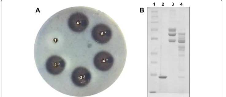
Phospholipase activity
The phospholipase activity of chromatographic fractions and of the isolated phospholipase A2 was evaluated on
plates, as described by Gutiérrezet al.[23], changing the agarose for agar and without using erythrocytes. Briefly, a gel containing 0.01 M CaCl2, egg yolk diluted in PBS
(pH 7.2) in the ratio 1:3 (v/v), 1 % bacteriological agar and 0.005 % sodium azide was formed in Petri dishes. Then, holes of approximately 5 mm in diameter were made in the gel, and samples were applied at a final vol-ume of 40μL, followed by incubation at 37 °C overnight. The formation of translucent halos around the holes in the gel was considered to be indicative of phospholipase activity, which was quantified by the measurement of each hole in millimeters. The minimum phospholipase dose (MPD) was defined as the minimum dose of the sample required to induce the formation of a halo with 20 mm diameter.
Modification of residue His48 of the PLA2 with
4-bromophenacyl bromide (BPB) was carried out based on previously described methodologies [24]. Briefly, the PLA2
bicarbonate, pH 8.0, and 10μL of BPB (1 mg/mL in etha-nol) was added. The mixture was incubated for 24 hours at 25 °C. After that period, the phospholipase activity of BPB-PLA2(2μg) was evaluated as described above.
Results and Discussion
Purification ofB. atroxtoxins
The toxins of interest, a P-I class metalloprotease (MP) and an acidic phospholipase A2 (PLA2), were isolated
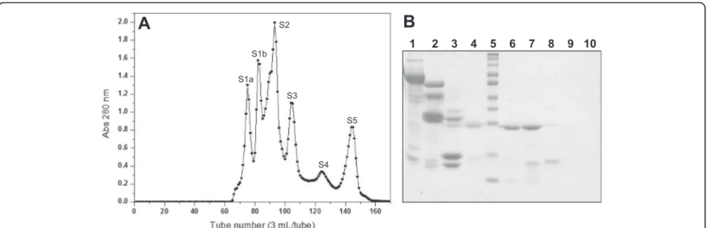
from B. atrox venom by consecutive chromatographic steps, starting the process by performing chromatog-raphy on Sephacryl S-200. The chromatographic profile obtained showed six well-defined fractions, identified as S1a, S1b, S2, S3, S4 and S5 (Fig. 1a). Analysis of these fractions by SDS-PAGE showed that fractions S1a, S1b and S2 mainly displayed protein components with mo-lecular masses above 30 kDa. The protein profile of frac-tion S3 presented bands with molecular masses around 25 kDa and 15 kDa, while fractions S4 and S5 seemed to consist only of components with molecular masses below 15 kDa (Fig. 1b). Fraction S3 was chosen based on its protein profile in SDS-PAGE and the hemorrhagic activity observed, being then subjected to an anion ex-change chromatography.
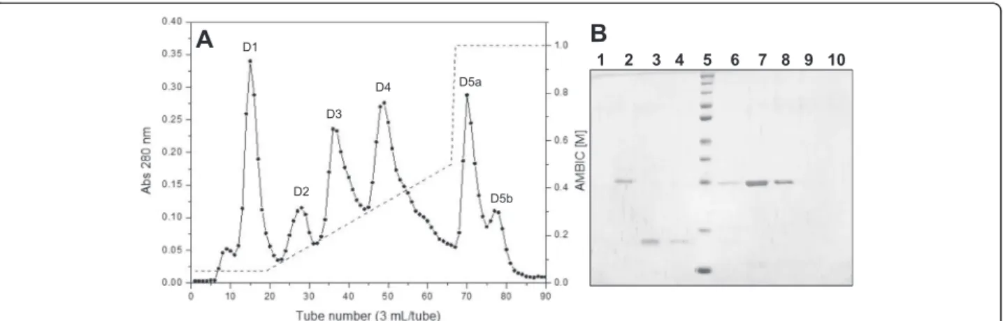
DEAE Sepharose chromatography of fraction S3 re-sulted in six fractions, denominated D1, D2, D3, D4, D5a and D5b (Fig. 2a). Several of these fractions (D1, D5a and D5b) showed no visible bands on SDS-PAGE, indicating the presence of only low-molecular-mass components. Fraction D2 showed a protein band with molecular mass slightly above 25 kDa, whereas fractions D3 and D4 showed single bands around 15 kDa and 25 kDa, respectively (Fig. 2b). Based on these protein profiles and the hemorrhagic and phospholipase activ-ities, it was determined that the PLA2was in fraction D3
and the MP in fraction D4. Thereafter, two separate iso-lation procedures were used to obtain each enzyme.
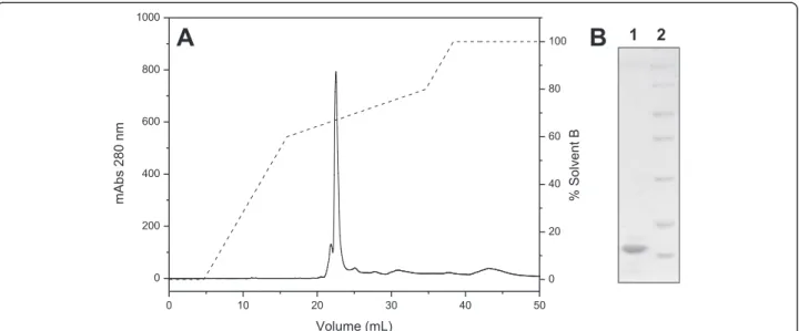
Fraction D3, which contained the PLA2, was applied
to a C18 reverse phase column, resulting in six major fractions (Fig. 3a). This chromatographic step enabled the separation of the acidic and catalytically active PLA2
from other fractions containing low molecular mass peptides (fractions 1, 4 and 5) which do not appear on 12 % SDS-PAGE, and from phospholipases A2 without
catalytic activity (fractions 2 and 3) (Fig. 3b) The fraction that showed phospholipase activity (fraction 6), indica-tive of catalytic activity related to the residue Asp49, was then rechromatographed on the same column to assess its purity level. The chromatographic profile shows a fraction eluted around 65 % solvent B (Fig. 4a), which appeared as a single band of approximately 14 kDa on SDS-PAGE (Fig. 4b).
Fraction D4 containing the MP was ultrafiltered on a Vivaspin® 20 system, which was used as a third step of isolation and may be considered a molecular exclusion step, since it enabled clearance of the fraction of low-molecular-mass compounds (such as peptides) and salts from the anion exchange chromatographic step. After this ultrafiltration step, the MP purity level was evi-denced by RP-HPLC using a C18 column, eluting with ~90 % solvent B (Fig. 5a), and appearing as a single band of molecular mass around 25 kDa on SDS-PAGE (Fig. 5b). Usually, reverse phase chromatographic steps are used only to verify the purity levels of metallopro-teases, since these enzymes lose their proteolytic activity when exposed to organic solvents such as TFA and acetonitrile, possibly due to denaturation of the mole-cules promoted by the low pH of solvents.
Purification of P-I class metalloproteases is commonly performed using two to three chromatographic steps,
Fig. 1aChromatographic profile of the crude venom ofB. atroxon a Sephacryl S-200 molecular exclusion column. Elution was performed with 0.2 M ammonium bicarbonate buffer (AMBIC), pH 8.0, collecting 3 mL/tube at a flow rate of 20 mL/hour.b12 % SDS-PAGE. Lanes: 1–tube 74
(S1a), 2–tube 82 (S1b), 3–tube 92 (S2), 4–tube 99 (S2 valley), 5–molecular mass standard (260, 140, 100, 70, 50, 40, 35, 25, 15, 10 kDa),
with a predominance of molecular exclusion and ion ex-change steps. A purification process employing a single chromatographic step was described for neuwiedase fromB. neuwiedi venom, nevertheless, most procedures usually comprise two steps, as described for BaP1 from
B. asper, leucurolysin-a from B. leucurus, atroxlysin-I from B. atrox and BjussuMP-II from B. jararacussu
venom [25–29]. Some studies also show the isolation of P-I metalloproteases using three chromatographic steps, as described for BmooMP-α from B. moojeni and for BpirMP from B. pirajai venom, which were isolated by combining molecular exclusion, ion exchange and affin-ity steps [30, 31].
Although our research group had already proposed a method for the isolation of a P-I metalloprotease denominated Batroxase from B. atrox venom using a molecular exclusion step on Sephadex G-75 and an
anion exchange chromatography on ES-502 N 7C col-umn [32], the new method described in the present study was standardized so that an acidic phospholipase A2could also be obtained from this venom. By using this
novel methodology, a P-I metalloprotease and an acidic PLA2 were successfully isolated from B. atrox venom
using the same two initial chromatographic steps and a third distinct one for each enzyme.
Acidic PLA2s from Bothrops venoms are commonly
purified by a combination of chromatographic methods, including molecular exclusion, ion exchange, RP-HPLC and hydrophobic steps. Cogo et al. [33] isolated two acidic PLA2s fromB. insularis venom using a single
RP-HPLC step. Other enzymes were isolated using two chromatographic steps, as described for BthA-I-PLA2
from B. jararacussu, BpirPLA2-I from B. pirajai,
BL-PLA2fromB. leucurus, BmooPLA2fromB. moojeniand Fig. 2aChromatographic profile of fraction S3 on a DEAE Sepharose anion exchange column. Elution was initiated with 0.05 AMBIC, pH 7.8, followed by a gradient of AMBIC from 0.05 M to 0.5 M, pH 7.8, and finally 1 M AMBIC, pH 7.8. Fractions of 3 mL/tube were collected at a flow rate of 30 mL/hour.b12 % SDS-PAGE. Lanes: 1–tube 15 (D1), 2–tube 28 (D2), 3–tube 36 (D3), 4–tube 40 (D3), 5–molecular mass standard
(260, 140, 100, 70, 50, 40, 35, 25, 15, 10 kDa), 6–tube 44 (D3-D4 valley), 7–tube 49 (D4), 8–tube 55 (D4), 9–tube 71 (D5a), 10–tube 77 (D5b)
Fig. 3aChromatographic profile of fraction D3 on a C18 reverse phase column. Elution was performed using a RP-HPLC system at a flow rate of 0.5 mL/minute with a linear concentration gradient of 0-100 % solvent B (70 % acetonitrile and 0.1 % TFA) in ten column volumes.b12 % SDS-PAGE. Lanes: 1–fraction 1; 2–fraction 2; 3–fraction 3; 4–fraction 4; 5–fraction 5; 6–fraction 6; 7–molecular mass standard (116, 66.4,
BaSPIIRP4 fromB. alternatusvenom [24, 34–37]. There are also reports of the combination of three or four chromatographic steps for obtaining some acidic PLA2s
fromBothropsvenoms [38–40].
After the isolation of B. atrox toxins, some biochem-ical and functional experiments were performed in order to identify the enzymes of interest, including the deter-mination of molecular masses, isoelectric points, partial amino acid sequences and evaluation of characteristic functional activities for metalloproteases and phospholi-pases A2.
Characterization of the MP
MP showed molecular mass of 22.9 kDa by MALDI-TOF MS and 26.2 kDa when estimated by SDS-PAGE. P-I SVMPs present variable molecular masses ranging from 20 to approximately 30 kDa, e.g. neuwiedase (20 kDa), BthMP (23 kDa), leucurolysin-a (23 kDa), atroxlysin-I (23 kDa), BpirMP (23 kDa), BaP1 (24 kDa), BnP1 (24 kDa), BH2 (26 kDa), Batroxase (27 kDa by SDS-PAGE and 22.9 kDa by MALDI-TOF MS) and BaltMP-I (29 kDa) [25–28, 31, 32, 41–44]. Nevertheless, it should be taken into account that some of those Fig. 4aChromatographic profile of fraction 6 on a C18 reverse phase column. Elution was performed using a RP-HPLC system at a flow rate of 0.5 mL/minute using a segmented concentration gradient of 0-60 % solvent B in three column volumes, 60-80 % in five column volumes and 80-100 % in one column volume.b12 % SDS-PAGE. Lanes: 1–PLA2, 2–molecular mass standard (116, 66.4, 45, 35, 25, 18.4, 14.4 kDa)
Fig. 5aEvaluation of the purity level of fraction D4 after Vivaspin® 20 on a C18 reverse phase column. After having been subjected to ultrafiltration on a Vivaspin® 20 system, fraction D4 was evaluated by RP-HPLC using a C18 column with a linear concentration gradient of 0-100 % solvent B in five column volumes.b12 % SDS-PAGE. Lanes: 1–MP, 2–molecular mass standard (260, 140, 100, 70, 50, 40, 35, 25,
differences in the molecular masses could be attributable to the different sensitivity of the methodologies used for their resolution, e.g. SDS-PAGE or MALDI-TOF MS.
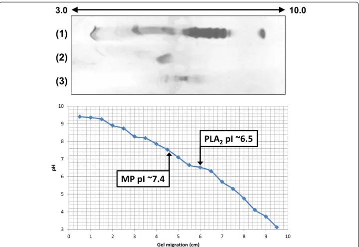
The isoelectric focusing showed that MP is a neutral protein with pI of approximately 7.4 (Fig. 6). P-I metallo-proteases may present pI values ranging between 5 and 8, and thus may show acidic character such as neuwie-dase, BjussuMP-II and BH2, neutral character as Batrox-ase and BthMP or basic character as BJ-PI2 [25, 29, 32, 41, 43, 45].
Partial amino acid sequencing revealed high similarity between MP and other P-I metalloproteases previously isolated from B. atrox venom, such as atroxlysin-I and Batroxase, with 100 % identity with the latter enzyme (Fig. 7) [28, 32]. SVMPs are classified according to their structural domains: P-I class, presenting only the metal-loprotease domain; P-II class, presenting the metallopro-tease domain and the disintegrin domain; P-III class, containing the disintegrin domain, cysteine rich domain and the metalloprotease domain [46]. Thus, P-I SVMPs belong to the simplest class of metalloproteases, with
lower molecular masses and an average of 200–210 amino acid residues [47]. The zinc binding catalytic site of these molecules is formed by the consensus sequence HEXXHXXGXXH, with the conserved Met-turn se-quence CI/VM adjacent to the site [48, 49]. Although these portions were not determined in the partial sequen-cing of the MP from B. atroxvenom, the multiple align-ment (Fig. 7) and the biochemical and functional characteristics confirm that it is a P-I class metalloprotease.
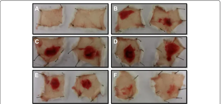
In relation to the functional characterization, MP showed high fibrin(ogen)olytic activity, with low doses inducing significant fibrinolysis halos (Fig. 8a) and pref-erential degradation of the Aα chains of fibrinogen, although it also induced degradation of the Bβ chains (Fig. 8b).
Most P-I SVMPs are fibrinogenolytic enzymes that preferentially degrade the Aα chains of fibrinogen, while also degrading the Bβchains at slower ratios [6]. Exam-ples of fibrinogenolytic metalloproteases from Bothrops
venoms include BthMP, BmooMP-α, atroxlysin-I, Batrox-ase, BaltMP-I, BaP1, neuwiedase and BpirMP [25, 26, 28,
30–32, 41, 44]. Some SVMPs also showed fibrinolytic ac-tivity, including BnP1, bothrojaractivase, BthMP, BpirMP, atroxlysin-I and Batroxase [28, 31, 32, 41, 42, 50].
Recent studies have been exploring the potential of fibri-n(ogen)olytic SVMPs as thrombolytic agents, since these enzymes may act directly on fibrin clots, and also promote depletion of fibrinogen molecules and thus prevent the formation of new clots. For this reason, fibrin(ogen)olytic SVMPs with low or no hemorrhagic activity, such as fibro-lase and its recombinant form alfimeprase, have been eval-uated as potential drugs for the treatment of patients with vascular diseases [51].
The MP fromB. atroxvenom showed high hemorrhagic activity at doses up to 20μg, with MHD close to 5μg and significant inhibition observed after incubation with 5 mM EDTA (Fig. 9). This inhibition promoted by chelat-ing agents such as EDTA and 1,10-phenanthroline on the proteolytic and hemorrhagic activities of SVMPs is associ-ated with the chelation of the zinc ion, which is essential for the catalytic activity of these molecules [26, 43].
The hemorrhage induced by SVMPs is related to the hydrolysis of basal membrane of capillaries [52]. In rela-tion to hemorrhagic potential, P-III SVMPs are the most potent among the three classes, being capable of inducing Fig. 7Comparison of the partial amino acid sequence obtained for the MP fromB. atroxvenom with two other P-I metalloproteases previously isolated from the same venom: Batroxase and atroxlysin-I. Multiple sequence alignment was made using the program ClustalX v. 2.0.11. (*) indicates positions with fully conserved amino acid residues
Fig. 8Fibrin(ogen)olytic activity of the MP isolated fromB. atroxvenom.aEvaluation of the fibrinolytic activity on fibrin gel. Samples were applied on plates containing fibrin gels, and incubation was performed at 37 °C for 24 h. Samples (halo diameter): 1. PBS (0 mm), 2.B. atrox
venom 20μg (15 mm), 3. MP 4μg (15 mm), 4. MP 6μg (15.5 mm), 5. MP 8μg (16 mm), 6. MP 10μg (16.5 mm).bEvaluation of the
fibrinogenolytic activity by SDS-PAGE. Samples were incubated at 37 °C for 1 h and then evaluated on 10 % polyacrylamide gel under denaturing conditions. Lanes: 1 Molecular mass standard (260, 140, 100, 70, 50, 40, 35, 25, 15 kDa), 2. MP 5μg, 3. Fibrinogen 25μg, 4. Fibrinogen
not only local but also systemic hemorrhages, while P-I SVMPs induce mostly local hemorrhaging [53]. Addition-ally, differences in the hemorrhagic potential of P-I SVMPs can also be observed [54, 55], with some enzymes presenting this activity [26, 28, 32, 41, 56] while others do not [25, 29, 30]. The comparison between structures of hemorrhagic and non-hemorrhagic P-I SVMPs showed small variations in loop regions around the catalytic site, suggesting that the variations of hemorrhagic potential may be related to such areas [57].
Some P-I metalloproteases have already been de-scribed from B. atrox venom, e.g. HI-5, atroxlysin-I, BaTx-I and Batroxase [28, 32, 56, 58]. As not all of these enzymes had their amino acid sequences elucidated, it is possible that some of them are the same molecule de-scribed by different authors. It is also possible that the different geographical locations of B. atrox specimens used as venom sources in these different studies (i.e. Brazil, Peru and Colombia) exerted some type of influ-ence on the structural and functional differinflu-ences re-ported for these enzymes, as shown by proteome studies on venoms of this snake species [14].
Considering the characteristics found for the MP from
B. atroxdescribed in the present study, it was noted that this enzyme showed significant similarities to Batroxase, including partial amino acid sequence, molecular mass (determined by both SDS-PAGE and MALDI-TOF MS methods), neutral pI and fibrin(ogen)olytic and hemorrhagic activities [32]. Thus, our findings suggest that the isolated MP can be identified as Batroxase.
Characterization of the PLA2
B. atrox PLA2presented a molecular mass of 13.7 kDa
by MALDI-TOF MS and 14.4 kDa by SDS-PAGE, and was revealed to be an acidic enzyme with pI ~ 6.5 (Fig. 6). Acidic snake venom PLA2s present molecular masses
around 14 kDa and pI varying from 4.0 to 5.5. Some examples are BthA-I-PLA2 with 13.7 kDa and pI 4.5,
BpirPLA2-I with 13.7 kDa and pI 4.8, Bp-PLA2 with
15.8 kDa and pI 4.3, BmooPLA2with 13.6 kDa and pI 5.2
and BL-PLA2 with 15 kDa and pI 5.4 [24, 34–36, 59]. None of these acidic PLA2showed a pI value superior to
5.5 as shown for the PLA2isolated in this study (pI ~ 6.5).
Although this pI value is close to neutral, alignment with other PLA2s confirmed its high similarity to acidic
pro-teins, including PA2-II from Gloydius blomhoffii venom (gi 129420) and BpPLA2-TX-I fromB. pauloensisvenom
[40] (Fig. 10).
PLA2s are classified into 15 different subgroups
accord-ing to specific features, with snake venom PLA2s
belong-ing to groups I and II [60, 61]. Group IIA PLA2s are
further divided into Asp49 PLA2s, enzymes that usually
exhibit high catalytic activity, and Lys49 PLA2s with low
or no enzymatic activity on artificial substrates [62, 63]. The catalytic site of PLA2s is composed of the residues
Three active site residues (His48, Asp49 and Tyr52), the calcium binding site and six conserved cysteine residues related to the formation of disulfide bonds were deter-mined in the partial amino acid sequencing of B. atrox
PLA2(Fig. 10).
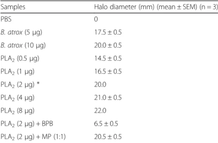
The PLA2showed high phospholipase activity at doses
up to 8 μg, with MPD of 2 μg (Table 1). These results show a high catalytic activity for this enzyme, which is consistent with other acidic PLA2s fromBothropsvenoms,
such as BmooTX-I, with MPD of 1μg, and BpirPLA2-I,
with MPD of 4 μg [34, 38]. Some studies describe the acidic Asp49 PLA2s as catalytically more active than the
basic isoforms [24, 34, 38, 39, 59]. Additionally, treatment with BPB significantly inhibited the phospholipase activity of the PLA2from B. atrox(Table 1), as already described
for other acidic PLA2s [34, 38].
Some phospholipase A2 enzymes were previously
de-scribed from B. atrox venom, e.g. a myotoxin with phospholipase and anticoagulant activity, basic and neu-tral Asp49 PLA2s, named BaPLA2I and BaPLA2III and a
Lys49 PLA2called myotoxin I [67–69]. Recently, a study published by Furtado et al. [70] described the isolation of three PLA2s from B. atroxvenom: a Lys49 (BaTX-I),
an Asp49 (BaTX-II) and an acidic Asp49 myotoxin (BaPLA2). However, as the authors did not perform amino
acid sequencing of the molecules, it is not possible to determine whether these enzymes are novel or the same ones previously described in the literature. Thus, as there are no reports in the literature of acidic PLA2s isolated
from B. atrox venom, we denominated the enzyme
described in the present study BatroxPLA2, an
unprece-dented acidic phospholipase A2.
Synergism
It is well known that snake toxins can act independently or synergistically [8]. To assess this possible synergism between the MP and the PLA2fromB. atrox, the hemorrhagic and
phospholipase activities were evaluated using a mixture of the enzymes at the molar ratio of 1:1, which resulted in no significant changes compared to the isolated activities of each enzyme (Fig. 9 and Table 1), suggesting that those ac-tivities were not enhanced or impaired by the combination of both toxins. Future studies will further evaluate the Fig. 10Multiple alignment of the N-terminal sequence of the PLA2fromB. atroxvenom with other acidic PLA2s from snake venoms. The highlighted amino acid residues are part of disulfide bonds (yellow) and the catalytic site (red). GI numbers (from top to bottom, respectively): 129420, 528050010, 387537880, 387537878, 123907441, 3914270, 23396779, 458601843 and 403399514. (*) indicates positions with fully conserved amino acid residues; (:) indicates conservation of the following groups with high score: K/R/Q/H, S/A, K/N/D, F/L/V/I, E/D/N/Q, T/S/A, I/M/L; (.) indicates conservation of the following groups with lowest score: N/R/G, G/D/N, A/V/T, Q/K/E/R, S/K/G/A, D/K/H, C/S, T/P
Table 1Phospholipase activity ofB. atroxvenom and the isolated PLA2
Samples Halo diameter (mm) (mean ± SEM) (n = 3)
PBS 0
B. atrox(5μg) 17.5 ± 0.5
B. atrox(10μg) 20.0 ± 0.5
PLA2(0.5μg) 14.5 ± 0.5
PLA2(1μg) 16.5 ± 0.5
PLA2(2μg) * 20.0
PLA2(4μg) 21.0 ± 0.5
PLA2(8μg) 22.0
PLA2(2μg) + BPB 6.5 ± 0.5
PLA2(2μg) + MP (1:1) 20.5 ± 0.5
possible synergistic effects of the two toxins on different physiological processes, e.g. inflammation.
Conclusions
The interest in the biochemical and functional charac-terization of toxins isolated from snake venoms is due not only to their relevance in envenomations, but also to their potential use as valuable research tools in different areas of knowledge. Pharmacological and biochemical studies conducted in recent decades have shown the presence of a variety of enzymes, toxins and biologically active compounds in these venoms, as well as the great diversity of their actions. Consequently, numerous at-tempts have been made to use these compounds as tools for research and for applications in the medical field, and as such, the purification and characterization of snake toxins are of utmost importance.
In this context, the present study successfully stan-dardized a purification procedure, mainly composed of classical chromatographic techniques, for the isolation of a P-I metalloprotease identified as Batroxase and a new acidic PLA2 denominated BatroxPLA2 from B. atrox
venom. These two enzymes will be used in future studies to evaluate their effects on the complement system and the inflammatory process, in addition to the thrombo-lytic potential of the metalloprotease.
Ethics committee approval
Animal care procedures were performed according to the Brazilian College of Animal Experimentation (COBEA) guidelines and the experimental protocols were approved by the Committee for Ethics on Animal Use (CEUA) from FCFRP-USP (protocol number: 13.1.336.53.4).
Competing interests
The authors declare that they have no competing interests.
Authors’contributions
DLM and ALJF contributed equally to the accomplishment of this study and wrote the manuscript. CPB and ACOC assisted in the biochemical and functional experiments, while SVS supervised and critically discussed the study. All authors read and approved the final manuscript.
Authors’Information
1Department of Clinical Analyses, Toxicology and Food Sciences, School of
Pharmaceutical Sciences of Ribeirão Preto, University of São Paulo (USP), Ribeirão Preto, SP, Brazil.
Acknowledgments
The authors would like to thank the financial support provided by the São Paulo Research Foundation (FAPESP, grants 2012/11963-1, 2012/21569-9 and 2011/23236-4), the Coordination for the Improvement of Higher Education Personnel (CAPES) and the National Council for Scientific and Technological Development (CNPq process 476932/2012-2). We are also grateful to Dr. Karla C. F. Bordon and Prof. Dr. Eliane C. Arantes from FCFRP-USP for the N-terminal analyses, and to Prof. Dr. José César Rosa and the Protein Chemistry Center, Medical School of Ribeirão Preto-USP, for the MALDI-TOF MS analyses. Thanks are also due to the Center for the Study of Venoms and Venomous Animals (CEVAP) of UNESP for enabling the
publication of this special collection (CNPq process 469660/2014-7).
Received: 26 November 2014 Accepted: 21 July 2015
References
1. Gutiérrez JM, Williams D, Fan HW, Warrell DA. Snakebite envenoming from a global perspective: towards an integrated approach. Toxicon.
2010;56(7):1223–35.
2. Habib A. Public health aspects of snakebite care in West Africa: perspectives from Nigeria. J Venom Anim Toxins incl Trop Dis. 2013;19(1):27.
3. Gutiérrez JM. Current challenges for confronting the public health problem of snakebite envenoming in Central America. J Venom Anim Toxins incl Trop Dis. 2014;20(1):7.
4. Bochner R. The international view of envenoming in Brazil: myths and realities. J Venom Anim Toxins incl Trop Dis. 2013;19:29.
5. Sistema de Informação de Agravos de Notificação - SINAN. Brasília: Ministério da Saúde. http://dtr2004.saude.gov.br/sinanweb/index.php. (2012). Access 25 Jan 2015.
6. Markland FS. Snake venoms and the hemostatic system. Toxicon. 1998;36:1749–800.
7. Mebs D. Snake venom composition and evolution of viperidae. Kaupia Darmstadt. 1999;8:145–8.
8. Doley R, Kini RM. Protein complexes in snake venom. Cell Mol Life Sci. 2009;66(17):2851–71.
9. Vonk FJ, Jackson K, Doley R, Madaras F, Mirtschin PJ, Vidal N. Snake venoms: from fieldwork to the clinic: recent insights into snake biology, together with new technology allowing high-throughput screening of venom, bring new hope for drug discovery. Biogeosciences. 2011;33(4):269–79.
10. Calvete JJ, Juárez P, Sanz L. Snake venomics. Strategy and applications. J Mass Spectrom. 2007;42(11):1405–14.
11. Mackessy SP. The field of reptile toxinology: snakes, lizards, and their venoms, Handbook of venoms and toxins of reptiles. Boca Raton: CRC Press; 2009. p. 3–23.
12. Lourenço Junior A, Zorzella Creste CF, de Barros LC, Delazari dos Santos L, Pimenta DC, Barraviera B, et al. Individual venom profiling ofCrotalus durissus terrificusspecimens from a geographically limited region: crotamine assessment and captivity evaluation on the biological activities. Toxicon. 2013;69:75–81.
13. Guércio RA, Shevchenko A, Shevchenko A, López-Lozano JL, Paba J, Sousa MV, et al. Ontogenetic variations in the venom proteome of the Amazonian snakeBothrops atrox. Proteome Sci. 2006;4:11.
14. Núñez V, Cid P, Sanz L, De La Torre P, Ângulo Y, Lomonte B, et al. Snake venomics and antivenomics ofBothrops atroxvenoms from Colombia and the Amazon regions of Brazil, Perú and Ecuador suggest the occurrence of geographic variation of venom phenotype by a trend towards
paedomorphism. J Proteomics. 2009;73(1):57–78.
15. Calvete JJ, Sanz L, Pérez A, Borges A, Vargas AM, Lomonte B, et al. Snake population venomics and antivenomics ofBothrops atrox: Paedomorphism along its transamazonian dispersal and implications of geographic venom variability on snakebite management. J Proteomics. 2011;74(4):510–27.
16. Laemmli UK. Cleavage of structural proteins during the assembly of the head of bacteriophage T4. Nature. 1970;227(5259):680–5.
17. Arantes EC, Prado WA, Sampaio SV, Giglio JR. A simplified procedure for the fractionation ofTityus serrulatusvenom: isolation and partial characterization of TsTX-IV, a new neurotoxin. Toxicon. 1989;27(8):907–16.
18. Williams KR, Stone KL. Enzymatic cleavage and HPLC peptide mapping of proteins. Mol Biotechnol. 1989;8(2):155–67.
19. Edman P, Begg G. A protein sequenator. Eur J Biochem. 1967;1(1):80–91.
20. Leitão DP, Polizello AC, Rothschild Z. Coagulation and fibrinolysis in capybara (Hydrochaeris hydrocaeris), a close relative of the guinea-pig (Cavia porcellus). Comp Biochem Physiol A Mol Integr Physiol. 2000;125(1):113–20.
21. Edgar W, Prentice CRM. The proteolytic action of ancrod on human fibrinogen and its polypeptide chains. Thromb Res. 1973;2(1):85–95.
22. Nikai T, Mori N, Kishida M, Sugihara H, Tu AT. Isolation and biochemical characterization of hemorrhagic toxin f from the venom ofCrotalus atrox
(western diamondback rattlesnake). Arch Biochem Biophys. 1984;231(2):309–19.
23. Gutiérrez JM, Avila C, Rojas E, Cerdas L. Alternativein vitromethod for testing the potency of the polyvalent antivenom produced in Costa Rica. Toxicon. 1988;26(4):411–3.
25. Rodrigues VM, Soares AM, Guerra-Sá R, Rodrigues V, Fontes MR, Giglio JR. Structural and functional characterization of neuwiedase, a nonhemorrhagic fibrin(ogen)olytic metalloprotease fromBothrops neuwiedisnake venom. Arch Biochem Biophys. 2000;381(2):213–24.
26. Gutiérrez JM, Romero M, Díaz C, Borkow G, Ovadia M. Isolation and characterization of a metalloproteinase with weak hemorrhagic activity from the venom of the snakeBothrops asper(terciopelo). Toxicon.
1995;33(1):19–29.
27. Gremski LH, Chaim OM, Paludo KS, Sade YB, Otuki MF, Richardson M, et al. Cytotoxic, thrombolytic and edematogenic activities of leucurolysin-a, a metalloproteinase fromBothrops leucurussnake venom. Toxicon. 2007;50(1):120–34.
28. Sanchez EF, Schneider FS, Yarleque A, Borges MH, Richardson M, Figueiredo SG, et al. The novel metalloproteinase atroxlysin-I from PeruvianBothrops atrox(Jergón) snake venom acts both on blood vessel ECM and platelets. Arch Biochem Biophys. 2010;496(1):9–20.
29. Marcussi S, Bernardes CP, Santos-Filho NA, Mazzi MV, Oliveira CZ, Izidoro LF, et al. Molecular and functional characterization of a new non-hemorrhagic metalloprotease fromBothrops jararacussusnake venom with antiplatelet activity. Peptides. 2007;28(12):2328–39.
30. Bernardes CP, Santos-Filho NA, Costa TR, Gomes MS, Torres FS, Costa J, et al. Isolation and structural characterization of a new fibrin(ogen)oliytic metalloproteinase fromBothrops moojenisnake venom. Toxicon. 2008;51(4):574–84.
31. Bernardes CP, Menaldo DL, Camacho E, Rosa JC, Escalante T, Rucavado A, et al. Proteomic analysis ofBothrops pirajaisnake venom and characterization of BpirMP, a new P-I metalloproteinase. J Proteomics. 2013;80:250–67.
32. Cintra AC, De Toni LG, Sartim MA, Franco JJ, Caetano RC, Murakami MT, et al. Batroxase, a new metalloproteinase fromB. atroxsnake venom with strong fibrinolytic activity. Toxicon. 2012;60(1):70–82.
33. Cogo JC, Lilla S, Souza GH, Hyslop S, De Nucci G. Purification, sequencing and structural analysis of two acidic phospholipases A2from the venom of
Bothrops insularis(jararaca ilhoa). Biochimie. 2006;88(12):1947–59.
34. Teixeira SS, Silveira LB, da Silva FM, Marchi-Salvador DP, Silva Jr FP, Izidoro LF, et al. Molecular characterization of an acidic phospholipase A2from
Bothrops pirajaisnake venom: synthetic C-terminal peptide identifies its antiplatelet region. Arch Toxicol. 2011;85(10):1219–33.
35. Nunes DC, Rodrigues RS, Lucena MN, Cologna CT, Oliveira AC, Hamaguchi A, et al. Isolation and functional characterization of proinflammatory acidic phospholipase A2fromBothrops leucurussnake venom. Comp Biochem Physiol C Toxicol Pharmacol. 2011;154(3):226–33.
36. Silveira LB, Marchi-Salvador DP, Santos-Filho NA, Silva Jr FP, Marcussi S, Fuly AL, et al. Isolation and expression of a hypotensive and anti-platelet acidic phospholipase A2fromBothrops moojenisnake venom. J Pharm Biomed Anal. 2013;73:35–43.
37. Garcia Denegri ME, Acosta OC, Huancahuire-Vega S, Martins-de-Souza D, Marangoni S, Maruñak SL, et al. Isolation and functional characterization of a new acidic PLA2Ba SpII RP4 of theBothrops alternatussnake venom from Argentina. Toxicon. 2010;56(1):64–74.
38. Santos-Filho NA, Silveira LB, Oliveira CZ, Bernardes CP, Menaldo DL, Fuly AL, et al. A new acidic myotoxic, anti-platelet and prostaglandin I2inductor phospholipase A2isolated fromBothrops moojenisnake venom. Toxicon. 2008;52(8):908–17.
39. Fernández J, Gutiérrez JM, Angulo Y, Sanz L, Juárez P, Calvete JJ, et al. Isolation of an acidic phospholipase A2from the venom of the snake
Bothrops asperof Costa Rica: biochemical and toxicological characterization. Biochimie. 2010;92(3):273–83.
40. Ferreira FB, Gomes MSR, de Souza DLM, Gimenes SNC, Castanheira LE, Borges MH, et al. Molecular cloning and pharmacological properties of an acidic PLA2fromBothrops pauloensissnake venom. Toxins (Basel). 2013;5(12):2403–19.
41. Gomes MS, Mendes MM, de Oliveira F, de Andrade RM, Bernardes CP, Hamaguchi A, et al. BthMP: a new weakly hemorrhagic metalloproteinase fromBothrops moojenisnake venom. Toxicon. 2009;53(1):24–32.
42. Baldo C, Tanjoni I, León IR, Batista IF, Della-Casa MS, Clissa PB, et al. BnP1, a novel P-I metalloproteinase fromBothrops neuwiedivenom: biological effects benchmarking relatively to jararhagin, a P-III SVMP. Toxicon. 2008;51(1):54–65.
43. Borkow G, Gutiérrez JM, Ovadia M. Isolation and characterization of synergistic hemorrhagins from the venom of the snakeBothrops asper. Toxicon. 1993;31(9):137–50.
44. Costa JO, Petri CB, Hamaguchi A, Homsi-Brandeburgo MI, Oliveira CZ, Soares AM, et al. Purification and functional characterization of two fibrinogenolytic enzymes fromBothrops alternatusvenom. J Venom Anim Toxins incl Trop Dis. 2007;13(3):640–54.
45. da Silva IR, Lorenzetti R, Rennó AL, Baldissera Jr L, Zelanis A, Serrano SM, et al. BJ-PI2, a non-hemorrhagic metalloproteinase fromBothrops jararaca
snake venom. Biochim Biophys Acta. 2012;1820(11):1809–21.
46. Fox JW, Serrano SM. Insights into and speculations about snake venom metalloproteinase (SVMP) synthesis, folding and disulfide bond formation and their contribution to venom complexity. FEBS J. 2008;275(12):3016–30.
47. Takeda S, Takeya H, Iwanaga S. Snake venom metalloproteinases: structure, function and relevance to the mammalian adam/adamts family proteins. Biochim Biophys Acta. 2012;1824(1):164–76.
48. Bjarnason JB, Fox JW. Snake venom metalloendopeptidases: reprolysins. Methods Enzymol. 1995;248:345–68.
49. Fox JW, Serrano SM. Structural considerations of the snake venom metalloproteinases, key members of the M12 reprolysin family of metalloproteinases. Toxicon. 2005;45(8):969–85.
50. Berger M, Pinto AF, Guimarães JA. Purification and functional characterization of bothrojaractivase, a prothrombin-activating metalloproteinase isolated fromBothrops jararacasnake venom. Toxicon. 2008;51(4):488–501.
51. Deitcher SR, Toombs CF. Non-clinical and clinical characterization of a novel acting thrombolytic: alfimeprase. Pathophysiol Haemost Thromb. 2005;34(4–5):215–20.
52. Escalante T, Shannon J, Moura-da-Silva AM, Gutiérrez JM, Fox JW. Novel insights into capillary vessel basement membrane damage by snake venom hemorrhagic metalloproteinases: A biochemical and immunohistochemical study. Arch Biochem Biophys. 2006;455(2):144–53.
53. Gutiérrez JM, Rucavado A, Escalante T, Díaz C. Hemorrhage induced by snake venom metalloproteinases: biochemical and biophysical mechanisms involved in microvessel damage. Toxicon. 2005;45(8):997–1011.
54. Ramos OH, Selistre-de-Araujo HS. Comparative analysis of the catalytic domain of hemorrhagic and non-hemorrhagic snake venom metallopeptidases using bioinformatic tools. Toxicon. 2004;44(5):529–38.
55. Escalante T, Rucavado A, Fox JW, Gutiérrez JM. Key events in microvascular damage induced by snake venom hemorrhagic metalloproteinases. J Proteomics. 2011;74(9):1781–94.
56. Patiño AC, Pereañez JA, Núñez V, Benjumea DM, Fernandez M, Rucavado A, et al. Isolation and biological characterization of Batx-I, a weak hemorrhagic and fibrinogenolytic P-I metalloproteinase from ColombianBothrops atrox
venom. Toxicon. 2010;56(6):936–43.
57. Lingott T, Schleberger C, Gutiérrez JM, Merfort I. High-resolution crystal structure of the snake venom metalloproteinase BaP1 complexed with a peptidomimetic: insight into inhibitor binding. Biochemistry.
2009;48(26):6166–74.
58. Petretski JH, Kanashiro MM, Rodrigues FG, Alves EW, Machado OLT, Kipnis TL. Purification and identification of a 25 kDa hemorrhagin fromB. atrox
venom. Protein Peptide Lett. 2001;8(3):187–92.
59. Rodrigues RS, Izidoro LF, Teixeira SS, Silveira LB, Hamaguchi A,
Homsi-Brandeburgo MI, et al. Isolation and functional characterization of a new myotoxic acidic phospholipase A2fromBothrops pauloensissnake venom. Toxicon. 2007;50(1):153–65.
60. Six DA, Dennis EA. The expanding superfamily of phospholipase A2 enzymes: classification and characterization. Biochim Biophys Acta. 2000;1488(1–2):1–19.
61. Burke JE, Dennis EA. Phospholipase A2structure/function, mechanism, and signaling. J Lipid Res. 2009;50(Suppl):S237–42.
62. Gutiérrez JM, Lomonte B. Phospholipase A2myotoxins fromBothropssnake venoms. Toxicon. 1995;33(11):1405–24.
63. Guarnieri MC, Melo ESL, Melo KMS, Albuquerque-Modesto JC, Prieto-da-Silva ARB, Rádis-Baptista G. Cloning of a novel acidic phospholipase A2from the venom gland ofCrotalus durissus cascavella(Brazilian northeastern rattlesnake). J Venom Anim Toxins incl Trop Dis. 2009;15(4):745–61.
64. Arni RK, Ward RJ. Phospholipase A2–a structural review. Toxicon.
1996;34(8):827–41.
65. Kang TS, Georgieva D, Genov N, Murakami MT, Sinha M, Kumar RP, et al. Enzymatic toxins from snake venom: structural characterization and mechanism of catalysis. FEBS J. 2011;278(23):4544–76.
67. Lomonte B, Gutiérrez JM, Furtado MF, Otero R, Rosso JP, Vargas O, et al. Isolation of basic myotoxins fromBothrops moojeniandBothrops atrox
snake venoms. Toxicon. 1990;28(10):1137–46.
68. Kanashiro MM, Escocard RCM, Petretski JH, Prates MV, Alves EW, Machado OLT, et al. Biochemical and biological properties of phospholipases A2from
Bothrops atroxsnake venom. Biochem Pharmacol. 2002;64(7):1179–86.
69. Núñez V, Arce V, Gutiérrez JM, Lomonte B. Structural and functional characterization of myotoxin I, a Lys49 phospholipase A2homologue from the venom of the snakeBothrops atrox. Toxicon. 2004;44(1):91–101.
70. Furtado JL, Oliveira GA, Pontes AS, Setúbal Sda S, Xavier CV, Lacouth-Silva F, et al. Activation of J77A.1 macrophages by three phospholipases A2isolated fromBothrops atroxsnake venom. Biomed Res Int. 2014;2014:683123.
Submit your next manuscript to BioMed Central and take full advantage of:
• Convenient online submission
• Thorough peer review
• No space constraints or color figure charges
• Immediate publication on acceptance
• Inclusion in PubMed, CAS, Scopus and Google Scholar
• Research which is freely available for redistribution