Effects of Human Umbilical Cord Mesenchymal Stem Cells
on Renal Ischaemia-reperfusion Injury in Rats
_______________________________________________
Zhenyu Qiu, Dun Zhou, Dongxiao Sun
Department of Nephrology, the First Affiliated Hospital, Liaoning Medical College, Jinzhou 121001, China
ABSTRACT
ARTICLE
INFO
______________________________________________________________ ______________________
Objective: This study aims to observe the function of umbilical cord-mesenchymal stem cells (UC-MSCs) labelled with enhanced green fluorescent protein (eGFP) in the repair of renal ischaemia-reperfusion (I/R) injury, to determine the effects on inflammatory cascade in an established rat model and to explore possible pathogenesis.
Materials and Methods: Sixty rats were randomly divided into three groups: the sham--operated, I/R and UC-MSC treatment groups. All rats underwent right nephrectomy. Ischaemia was induced in the left kidney by occlusion of the renal artery and vein for 1hour, followed by reperfusion for 24 hours or 48 hours. Kidney samples were collected to observe morphological changes. Immunohistochemistry was performed to assess the expression of intercellular adhesion molecule 1 (ICAM-1) in the renal tissue sample, as well as the number of infiltrating polymorphonuclear neutrophils (PMNLs) and UC--MSCs with positive eGFP.
Results: Renal histopathological damages and the expression of ICAM-1 and PMNL increased significantly in the I/R group compared with those in the sham-operated group, whereas the damages were less conspicuous in the UC-MSC treatment group. Conclusions: Renal ICAM-1, which mediated PMNL infiltration and contributed to re-nal damage, was significantly up-regulated in the I/R group. UC-MSCs were identified to inhibit these pathological processes and protect the kidney from I/R injury.
Key words:
Umbilical Cord; enhanced green fluorescent protein [Supplementary Concept]; Intercellular Adhesion Molecule-1
Int Braz J Urol. 2014; 40: 553-61
_____________________
Submitted for publication: September 14, 2013
_____________________
Accepted after revision: February 07, 2014
INTRODUCTION
Renal ischaemia-reperfusion (I/R) injury is commonly observed in clinic and is considered as the main cause of acute renal failure (ARF). Over the last few decades, blood purification technolo-gy and critical care medicine have significantly developed, but the mortality of acute tubular ne-crosis (ATN) induced by septicaemia, shock, and serious trauma did not decline, maintaining a ran-ge of 30% - 50% (1). More studies have focused on the promotion of the regeneration and repair
in different fields of transplantation therapy, but umbilical cord mesenchymal stem cells (UC-MSCs) are rarely studied and are thus confronted with numerous unsolved problems. Compared with the adult bone marrow, the umbilical cord has nume-rous advantages, such as extensive source, easy collection and transformation of waste into bene-ficial products. The isolation of UC-MSCs from the whole umbilical cord provides an extensive and new source of MSCs, which can significantly pro-mote clinical application.
To study the possibility of applying MSCs to the treatment of tubular injury disease, several researchers used an animal model of I/R to exa-mine BM-MSCs. Results showed that BM-MSCs promote renal tubular damage repair and protect renal function. Moreover, BM-MSCs can differen-tiate into renal tubular epithelial cells and vascu-lar endothelial cells, which are directly involved in the repair of renal tubular injury (9). In addition, BM-MSCs can improve and promote endogenous renal proliferation repair, which depends on the complex-regulated paracrine mechanism (10,11). However, the repair mechanisms of BM-MSCs vary in different laboratories, and the possibility that UC-MSC mechanisms are identical to those of BM-MSCs is rarely investigated.
The inflammatory cascade induced by I/R is an important factor causing renal injury. Vascu-lar endothelial cell dysfunction with high expres-sion of intercellular adheexpres-sion molecule-1 (ICAM-1) and activated white blood cells (WBCs) and tissue macrophages can initiate the inflammatory cascade (12). After transplantation, MSCs hypo-thetically enter the blood circulation and express various adhesion molecules, such as ICAM-1, vas-cular cell adhesion molecule-1 (VCAM-1), L-selec-tin and P-selecL-selec-tin. The neutrophils are activated, reach the specific damage sites and adhere to the vascular endothelium. These neutrophils enter the renal interstitium through the vessels with incre-ased permeability, partially replace the original infiltration site of the neutrophils and reduce neu-trophil infiltration. This mechanism reduces the damage induced by reactive oxygen species, in-flammatory mediators and proteases in the tissue at the acute injury stage. No direct experimental evidence confirms the possibility that UC-MSCs
express similar factors, directly differentiate into renal tubular epithelial cells or vascular endo-thelial cells, or promote endogenous repair either through inflammatory response reduction or with the combination of the two mentioned pathways.
In this study, caudal vein injection of UC--MSCs was performed to treat rats with renal I/R. The repair function of UC-MSCs and their effects on inflammatory cascade after I/R were observed to elucidate the UC-MSC plasticity and enhance the theory of acute tubular necrosis repair. ICAM-1 expression in the renal tissue and WBC infil-tration, and their relationship with UC-MSC ho-ming number, were detected. Moreover, the effect of UC-MSC homing on the inflammatory cascade and on the renal protection mechanism was preli-minarily investigated.
MATERIALS AND METHODS
Animals and grouping
Twenty healthy male Sprague–Dawley (SD) rats with body weight ranging from 250g to 300g were provided by the Laboratory Animal Centre of Jinzhou Medical College. After adaptive breeding for 7 days, the rats with negative uri-ne screening results were randomized into three groups: the sham-operated (sham), I/R and hu-man umbilical cord mesenchymal stem cell (UC--MSCs+I/R) groups. Each group had 20 rats. This study was carried out in strict accordance with the recommendations in the Guide for the Care and Use of Laboratory Animals of the National Insti-tutes of Health. The animal use protocol has been reviewed and approved by the Institutional Ani-mal Care and Use Committee (IACUC) of Liaoning Medical College.
Establishment of rat model of renal I/R and sampling process
of artery clamp removal. The right kidney was re-moved, and abdomen closure was performed layer by layer. In the UC-MSC group, 1 x 10⁶ (0.5mL)
UC-MSCs transfected with enhanced green fluo-rescent protein (eGFP-UCMSCs) (Heze Biotech Co., Ltd., Beijing, China) were injected into the caudal vein of the rats 30 min. after reperfusion. In the sham group, the right kidney of the rats was re-moved, and the left renal pedicle was isolated but not occluded. The same volume of normal saline was injected into the caudal vein of the rats in the I/R and sham groups. Ten rats from each group were executed 24 and 48 hours after injection. The left kidney (approximately three quarters) from each rat was fixed in FAA solution (every 100mL of FAA solution contained 10mL of formalin, 5mL of glacial acetic acid and 85mL of 95% ethanol) for further research, and the rest of the tissue was prepared for conventional frozen section.
Histological study
The renal tissue samples were cut into 2µm slices after fixation in FAA solution, dehydration and paraffin embedding. Hematoxylin and eosin (HE) staining was performed on the slides, and the lesions were examined using a light microsco-pe. Paller’s criterion for grading tubulointerstitial lesion was applied. To record the score, 10 renal tubules were randomly selected from every high--power field. The tubule with apparent expansion and flat or swelling cells scored one point, whereas those with a brush border were injured or fell off and scored one or two points. Moreover, urinary casts scored two points, the necrotic cells (did not form cast or fragments) in the tubule cavity scored one point. The scoring process was performed in 10 random fields (i.e., 100 renal tubules).
Leukocyte counting
Inflammatory cells (per mm2) that infil-trated into the tubule interstitium were analysed by using a computerized medical image analysis system (CMIAS), which indicated the amount of leukocytes in the renal tissue.
ICAM-1detection
Rabbit-anti-rat ICAM-1 and SP working solution kit (Wuhan Boster Biological
Enginee-ring Co., Ltd., Wuhan, China) were applied to de-tect the ICAM-1 expression in the renal tissue by immunohistochemistry. The 4µm tissue sections underwent routine deparaffinization and rehydra-tion. Freshly prepared 3% hydrogen peroxide was used to deactivate endogenous peroxidase. After antigen retrieval by heat, protein block solution was added to block the sample for 20 min. The slides were incubated with rabbit-anti-rat ICAM-1 antibody overnight at 4ºC and with biotinylated secondary antibody at room temperature (RT) for 20 min. Strept Avidin-Biotin Complex was em-ployed at RT for 20 min., followed by DAB deve-loping. The developing time was controlled under a microscope. Finally, hematoxylin was applied for counterstaining, followed by dehydration, transparency and mounting. The negative control received PBS instead of the primary antibody. Re-sults were determined by claybank particles that indicated a positive site. The dark brown area was the strong positive, claybank was positive and light yellow was weak positive. The uncoloured cells were considered negative cells.
Pathological image analysis
An inverted diffraction phase and fluores-cence microscope (Leica Company, Berlin, Ger-many) was used to select 10 high power fields randomly at the lesion site on immunohistoche-mical slides from each group. CMIAS multi-func-tional true colour pathological image analysis system (Motic Image Technology Co., Ltd, Beijing, China) was employed, and computerized image analysis software facilitated computer reading to recognize the immunohistochemically positive signals of the selected slide fields. The expression of ICAM-1 in the renal tissue was assessed semi--quantitatively by calculating the integration of positive-coloured area. This result represented the integrated optical density value for each field, and the average of these 10 values represented the value for each sample. Finally, the values were compared among the groups.
ex-posed to UV (488 ± 15nm). The green cells were the positive eGFP-UCMSCs after transplantation. The number of GFP-positive cells was counted in five high-power fields (400x) for each slide. The percentage of positive cells was calculated as: positive cell % = number of positive cells/ (number of positive and negative cells) x 100%.
Statistical analysis
All values were presented as mean ± standard deviation after quantitative or semi--quantitative analysis and then subjected to one--factor analysis of variance using SPSS 13.0 sof-tware. The linear correlation analysis of indices was employed. Differences were considered sig-nificant at P < 0.05.
RESULTS
Histopathological changes of renal tissue in rats Renal tubules were arranged neatly in the sham group, and no congestion edema was observed in the mesenchyme (Figure-1A). The renal cortex and medulla in the I/R group were
observed to have tinted white colour with mild swelling. Result of HE staining under a micros-cope showed the proximal convoluted tubules in an inordinate and loose arrangement. Epithelial cells apparently swelled and were characterized by an extensive vacuolar degeneration and flaky necrosis. The nuclei of minor epithelial cells were discovered in pyknosis and deep dyeing, and epithelium cells fell off the lumen. Large urinary casts were initially formed in distal con-voluted tubules. Renal interstitium was locally bled and complicated with inflammatory cell in-filtration. Apparent injury was detected in the I/R 24 h group (Figure-1B), that was worse in the I/R 48 hours group (Figures 1C and D). The UC-MSC group showed light tissue injury, mild edema of renal interstitium and normal arrange-ment of renal tubules. Mild swelling of tubular epithelial cells in areas where brush border fell off and urinary casts were occasionally obser-ved. Inflammatory cell infiltration was also less serious in the UC-MSC group compared with the I/R groups (Figures 1E, F and G). The changes in all groups were more severe after 48 hours than after 24 hours.
Figure 1 - HE staining of renal tissue.
A B
D E
C
Scoring of renal tubule and leukocyte counting in renal tissue
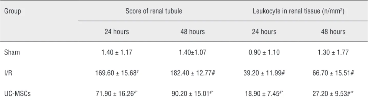
Single factor analysis of variance betwe-en groups was used for the pair-wise compari-son of the renal tubular score among different groups. Results showed that compared with those in the sham group, the renal tubular score and leukocyte number in the renal tissue in I/R group increased significantly (P < 0.01). The renal tu-bular score and leukocyte number in renal tissue in the UC-MSC group were significantly lower than that in the I/R group (P < 0.01). Significant differences were observed after 24 and 48 hours between the other two groups (P < 0.05). Data are summarized in Table-1.
ICAM-1 expression in renal tissue
The sham group showed trace ICAM-1 expression in the renal tissue in Figure-2A. ICAM-1 expression was prominent in the renal vasa recta, peritubular capillaries, glomerulus, proximal convoluted tubule and renal inters-titium, which was significantly up-regulated within 48 hours. The expression of ICAM-1 (Figure-2B) in the I/R 24 h group was higher in the I/R 48 hours group (P < 0.01) (Figure-2C). ICAM-1 expression was significantly lower in the UC-MSC group than that in the I/R group (P < 0.01) (Figure-2D). Significant differences in ICAM-1 expression were observed between 24 and 48 hours in all groups (P < 0.01) (Figure--2E), except for the sham group (Table-2).
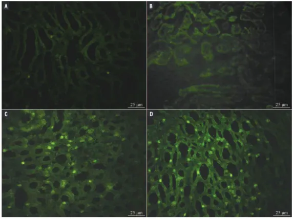
Ratio of positive eGFP-UCMSCs
A fluorescent microscope revealed no GFP-positive UC on the frozen sections of the kid-ney in the sham and I/R groups (Figures 3A and B). GFP-positive MSCs were found among the re-nal tubule cells in the UC-MSC group (Figures 3C and D), and the presence of GFP-positive MSCs was higher in the 48 h group than in the 24 h group (P < 0.01). Data are shown in Table-3.
Correlation analysis
Using linear correlation analysis, the three groups of data were combined to analyze the correlation among the four detected indices. The correlation study of the renal tubule score, leu-kocyte number in renal tissue and ICAM-1 expres-sion showed that renal tubule score of 24 and 48 hours reperfusion was positively correlated with the leukocyte number in renal tissue and ICAM-1 expression (r = 0.89, 0.88, respectively, P < 0.01). The renal tubule score, leukocyte number in re-nal tissue and ICAM-1 expression in the rere-nal tis-sue positively correlated with the ratio of positive eGFP-UCMSCs (r = 0.73, P < 0.01).
DISCUSSION
In this study, caudal vein injection with UC--MSCs was performed for the treatment of renal I/R rats. The rate of eGFP-UCMSC positive cells in the renal tissues and ICAM-1 expression were observed, and the effect of UCMSCs on I/R renal repair and
Table 1 - Renal tubule scoring and renal tissue leukocyte counting in different groups.
Group Score of renal tubule Leukocyte in renal tissue (n/mm2)
24 hours 48 hours 24 hours 48 hours
Sham 1.40 ±1.17 1.40±1.07 0.90 ± 1.10 1.30 ± 1.77
I/R 169.60 ±15.68# 182.40 ± 12.77# 39.20 ± 11.99# 66.70 ± 15.51#
UC-MSCs 71.90 ± 16.26#* 90.20 ± 15.01#* 18.90 ± 7.45#* 27.20 ± 9.53#*
Table 2 - ICAM-1expression in renal tissue at different time in all groups.
Group N 24 hours 48 hours
Sham 10 0.2170 ± 0.1063 0.1919 ±0.0876 I/R 10 5.2569 ± 0.7397# 8.1012 ±1.6640#
UC-MSCs 10 2.2249 ± 0.9637#* 4.4056 ±0.9802#*
Note: #P < 0.01 vs. sham group; *P < 0.01 vs. I/R group.
Figure 2 - Expression of ICAM-1 in renal tissue detected by immunohistochemistry (100x).
A B
E D
C
A) The sham-operated group; B) The I/R group at 24 hours; C) The I/R group at 48 hours; D) The UC-MSCs+I/R group at 24 hours; E) The UC-MSCs+I/R group at 48 hours.
inflammatory cascade after I/R were investigated. Results were theoretically consistent.
In addition, eGFP-UCMSCs were trans-planted to the rat model of renal I/R injury and the homing condition was observed. The GFP that is isolated from jellyfish and stably expressed in mammalian cells is a luminescent protein. This pro-tein has gained considerable attention because of its advantages, such as harmlessness to cell, strongest marker intensity among all marker techniques, no requirement for substrate during detection and long maintenance of fluorescence (13,14). Results sho-wed positive eGFP cells in the renal tissue of
Figure 3 - Green fluorescent protein-marked expression of UC-MSCs in the renal tissue (400x).
Table 3 - Ratio of positive eGFP-UCMSCs in renal tissue at different time in all groups (%).
Group N 24 hours 48 hours
Sham 10 0 0
I/R 10 0 0
UC-MSCs 10 9.8 ±1.2# 21.2 ± 7.3#*
Note: #p < 0.005 vs. sham and I/R group; *p < 0.01 UC-MSCs+I/R 48 hours vs. UC-MSCs+I/R 24 hours I/R group.
A) The sham-operated group; B) The I/R group at 48 hours; C) The UC-MSCs+I/R group at 24 hours; D) The UC-MSCs+I/R group at 48 hours.
A B
D C
injury were decreased, and endogenous repair was promoted by alleviating the inflammatory cascade.
Results of this study showed that ICAM-1 expression in the renal tissue in the I/R group increased significantly. The infiltration degree of inflammatory cells was positively correlated with ICAM-1 expression, and the renal tubular score was also positively correlated to inflammatory cell infiltration degree. This finding indicated that
up--regulation of ICAM-1 expression and mediated leukocyte infiltration and recruitment are the key factors in renal I/R that facilitate the occurrence and development of I/R damage.
al. (11) treated the kidney of I/R rats by BM-MSC transplantation and achieved improvement in in-flammation, vessels and necrotic renal tissue. The function of UC-MSCs is similar to that of BM-MSCs (16). A large number of studies show that UC-MSCs also express multiple receptor molecules, including VCAM-1, L-selectin, P-selectin and platelet endo-thelial cell adhesion molecule (PECAM) (17,18), and that the ICAM-1 expression in MSCs requires induction. These receptor molecules interact with the highly expressed ICAM-1 by through the renal tubule under I/R conditions, resulting in the proli-feration and homing of MSCs (19).
MSCs are adult stem cells that distribute in different tissues of body and are prominent in the bone marrow and UC (2). Considerable attention has been focused on bone marrow-derived MSCs, which have been applied to transplantation thera-py as seed cells. However, few studies were repor-ted on the treatment of renal I/R with UC-MSCs (20). According to a number of laboratory reports, the surface antigens of UC-MSCs have no specifi-city and possess strong self-renewal potency, high proliferation and multi-directional differentiation potential, which differentiate these antigens into any of the three germ layers and define them as the seed cells of various tissues (21). Previous stu-dies on the effects of transplanting MSCs in renal I/R have focused on the function of MSCs in renal colonization and differentiation (22).
The inflammatory cascade induced by re-nal I/R is an important factor leading to rere-nal da-mage (23). Vascular endothelial cell dysfunction, high expression of ICAM-1 and activation of leu-kocytes and macrophages may initiate inflamma-tory cascade (24). Numerous experiments indicate that adhesion and activation of neutrophils is the basis of I/R injury, which further causes mechani-cal obstruction, release of abundant inflammatory substances, activates more inflammatory cells and parenchyma cells and forms inflammatory casca-de. Inhibiting leukocyte activation and blood ves-sel infiltration is the solution to reduce I/R injury, and blocking any link helps to ameliorate the I/R injury (25,26).
In this study, UC-MSC homing was pre-liminarily investigated. Results showed that UC--MSC homing and paracrine regulation may
re-duce inflammation and improve and promote the endogenous renal proliferation. Research on UC--MSC has improved and has provided a new stra-tegy for the treatment of renal tubular damages induced by I/R. However, many questions require further studies to be answered. For instance, in the application of UC-MSCs to the treatment of renal diseases, the consequences of UC-MSC re-ceptor transplantation and its mechanism, as well as chromosome instability and risk of malignancy induced by in vitro UC-MSC amplification in long term (27), remain poorly understood. Therefore, more studies are needed to elucidate the biology of UC-MSCs in vivo and in vitro.
In conclusion, the multilineage differentia-tion capacity of MSCs can repair the renal tissue. Moreover, inflammatory cytokine consumption and inflammatory cell reduction during the MSC homing process contribute to the reduction of re-nal I/R injury and promote rere-nal repair. The trans-plantation of MSCs may be an effective method for the treatment of severe renal tubular injury.
ABBREVIATIONS
BM-MSCs = bone marrow MSCs
eGFP = enhanced green fluorescent protein
eGFP-UCMSCs = UC-MSCs transfected with
enhanced green fluorescent protein
ICAM-1 = intercellular adhesion molecule 1 I/R = renal ischaemia-reperfusion
MSCs = mesenchymal stem cells
PMNLs = polymorphonuclear neutrophils
UC-MSCs = umbilical cord-mesenchymal stem cells
CONFLICT OF INTEREST
None declared.
REFERENCES
1. Rabb H, O’Meara YM, Maderna P, Coleman P, Brady HR: Leukocytes, cell adhesion molecules and ischemic acute renal failure. Kidney Int. 1997; 51: 1463-8.
3. Pittenger MF, Mackay AM, Beck SC, Jaiswal RK, Douglas R, Mosca JD, et al.: Multilineage potential of adult human mesenchymal stem cells. Science. 1999; 284: 143-7. 4. Zvaifler NJ, Marinova-Mutafchieva L, Adams G, Edwards CJ,
Moss J, Burger JA, et al.: Mesenchymal precursor cells in the blood of normal individuals. Arthritis Res. 2000; 2: 477-88. 5. Umezawa A, Makino H: Cell source for regenerative medicine.
Nihon Rinsho. 2008; 66: 865-72.
6. Erices A, Conget P, Minguell JJ: Mesenchymal progenitor cells in human umbilical cord blood. Br J Haematol. 2000; 109: 235-42.
7. Secco M, Zucconi E, Vieira NM, Fogaça LL, Cerqueira A, Carvalho MD, et al.: Multipotent stem cells from umbilical cord: cord is richer than blood! Stem Cells. 2008; 26: 146-50. 8. Safford KM, Hicok KC, Safford SD, Halvorsen YD, Wilkison
WO, Gimble JM, et al.: Neurogenic differentiation of murine and human adipose-derived stromal cells. Biochem Biophys Res Commun. 2002; 294: 371-9.
9. Lin F, Cordes K, Li L, Hood L, Couser WG, Shankland SJ, et al.: Hematopoietic stem cells contribute to the regeneration of renal tubules after renal ischemia-reperfusion injury in mice. J Am Soc Nephrol. 2003; 14: 1188-99.
10. Duffield JS, Park KM, Hsiao LL, Kelley VR, Scadden DT, Ichimura T, et al.: Restoration of tubular epithelial cells during repair of the postischemic kidney occurs independently of bone marrow-derived stem cells. J Clin Invest. 2005; 115: 1743-55. 11. Tögel F, Hu Z, Weiss K, Isaac J, Lange C, Westenfelder C:
Administered mesenchymal stem cells protect against ischemic acute renal failure through differentiation-independent mechanisms. Am J Physiol Renal Physiol. 2005; 289: F31-42.
12. Zhang XL, Selbi W, de la Motte C, Hascall V, Phillips A: Renal proximal tubular epithelial cell transforming growth factor-beta1 generation and monocyte binding. Am J Pathol. 2004; 165: 763-73.
13. Tsien RY: The green fluorescent protein. Annu Rev Biochem. 1998; 67: 509-44.
14. Flynn A, Barry F, O’Brien T: UC blood-derived mesenchymal stromal cells: an overview. Cytotherapy. 2007; 9: 717-26. 15. Gordon D, Glover CP, Merrison AM, Uney JB, Scolding NJ:
Enhanced green fluorescent protein-expressing human mesenchymal stem cells retain neural marker expression. J Neuroimmunol. 2008; 193: 59-67.
16. Liu Y, Dulchavsky DS, Gao X, Kwon D, Chopp M, Dulchavsky S, et al.: Wound repair by bone marrow stromal cells through growth factor production. J Surg Res. 2006; 136: 336-41. 17. Jiang ZS, Gao Y, Mu N: Multipotent adult progenitor cells
from human bone marrow differentiate into hepatocyte-like cells induced by co-culture with human hepatocyte line. Zhonghua Yi Xue Za Zhi. 2007; 87: 414-8.
18. Le Blanc K, Tammik L, Sundberg B, Haynesworth SE, Ringdén O: Mesenchymal stem cells inhibit and stimulate mixed lymphocyte cultures and mitogenic responses independently of the major histocompatibility complex. Scand J Immunol. 2003; 57: 11-20.
19. Gupta S, Verfaillie C, Chmielewski D, Kim Y, Rosenberg ME: A role for extrarenal cells in the regeneration following acute renal failure. Kidney Int. 2002; 62: 1285-90.
20. Sarugaser R, Lickorish D, Baksh D, Hosseini MM, Davies JE: Human umbilical cord perivascular (HUCPV) cells: a source of mesenchymal progenitors. Stem Cells. 2005; 23: 220-9.
21. Lee OK, Kuo TK, Chen WM, Lee KD, Hsieh SL, Chen TH: Isolation of multipotent mesenchymal stem cells from umbilical cord blood. Blood. 2004; 103: 1669-75.
22. Kern S, Eichler H, Stoeve J, Klüter H, Bieback K: Comparative analysis of mesenchymal stem cells from bone marrow, umbilical cord blood, or adipose tissue. Stem Cells. 2006; 24: 1294-301.
23. Singbartl K, Ley K: Leukocyte recruitment and acute renal failure. J Mol Med (Berl). 2004; 82: 91-101.
24. Baer PC, Geiger H: Different effects of growth factors on human renal early distal tubular cells in vitro. Kidney Blood Press Res. 2006; 29: 225-30.
25. Rentsch M, Post S, Palma P, Lang G, Menger MD, Messmer K: Anti-ICAM-1 blockade reduces postsinusoidal WBC adherence following cold ischemia and reperfusion, but does not improve early graft function in rat liver transplantation. J Hepatol. 2000; 32: 821-8.
26. Souza-Moraes MR, David-Filho R, Baptista-Silva JC, Ullian M, Franco MF, Gabriel A Jr, et al.: Effect of antibodies to intercellular adhesion molecule type 1 on the protection of distant organs during reperfusion syndrome in rats. Braz J Med Biol Res. 2003; 36: 605-12.
27. Bobick BE, Tuan RS, Chen FH: The intermediate filament vimentin regulates chondrogenesis of adult human bone marrow-derived multipotent progenitor cells. J Cell Biochem. 2010; 109: 265-76.
_______________________ Correspondence address: