Analysis of the efects of aquatic exercise on the quality of life of
people with chronic venous disease
Análise dos efeitos dos exercícios aquáticos na qualidade de vida de
indivíduos com doença venosa crônica
Michael Augusto dos Santos Aquino1
*
, Larissa Christina Vieira da Paixão1, Flávia de Jesus Leal1, Renata Cardoso Couto1
Abstract
Background: Aquatic exercises have become a very important therapeutic option for chronic venous disease (CVD). here is evidence in the literature showing that this type of exercise is a mechanism that improves venous return and is important in vascular reeducation. hese exercises also help to reduce the venous hypertension caused by CVD, improving patients’ quality of life. Objectives: To analyze the efects of aquatic exercises on the quality of life of patients with CVD. Methods: his was a longitudinal, prospective, interventional pilot study conducted with 16 people with CVD classiied from C1 to C5. Participants were assessed at baseline using a data collection form and administration of two quality of life questionnaires, the SF-36 (Generic) and the AVVQ-Brazil (CVD-speciic), and an Analog Visual Pain Scale (AVPS). hey then undertook a program of 10 sessions of aquatic exercises, three times per week. he quality of life questionnaires and the AVPS were administered once more after all sessions had been conducted. Results: he data collected were subjected to statistical analysis to a signiicance level of p < 0.05. Patients exhibited improved quality of life as measured by the SF-36 in the domains Physical functioning, Physical role limitation and Pain (p < 0.05). he patients’ pain levels reduced after treatment according to the AVPS (p = 0.007). Only scores for the Pain and dysfunction domain of the AVVQ-Brazil questionnaire exhibited signiicant improvement (p = 0.013). Conclusions: Aquatic exercises were capable of improving aspects of quality of life and of reducing pain, demonstrating that they beneit patients with CVD.
Keywords: venous insuiciency; physiotherapy modalities; hydrotherapy.
Resumo
Contexto: O uso dos exercícios aquáticos se tornou uma modalidade terapêutica muito importante na doença venosa crônica (DVC). Tais exercícios têm sido apontados pela literatura como um mecanismo favorável ao retorno venoso, sendo importantes na reeducação vascular. Também contribuem para a diminuição da hipertensão venosa ocasionada pela doença, melhorando a qualidade de vida dos indivíduos acometidos. Objetivos: Analisar os efeitos dos exercícios aquáticos na qualidade de vida de pacientes com DVC. Métodos: Trata-se de um estudo-piloto, interventivo prospectivo longitudinal, composto por 16 indivíduos com DVC classiicados de C1 a C5. Os participantes foram avaliados através de um formulário de coleta de dados e instruídos a responder dois questionários sobre qualidade de vida: SF-36 (Geral) e AVVQ-Brasil (especíico para DVC), além da Escala Visual Analógica da dor (EVA). Em seguida, foram submetidos a 10 sessões de exercícios aquáticos, três vezes por semana, tendo respondido novamente aos questionários de qualidade de vida e EVA após o termino de todas as sessões. Resultados: Os dados coletados foram tratados estatisticamente, com nível de signiicância de p < 0,05. Os pacientes apresentaram melhora na qualidade de vida medida pelo SF-36 nos domínios capacidade funcional, limitação e dor (p < 0,05). O nível de dor nos pacientes tratados reduziu segundo a EVA (p = 0,007). Em relação ao questionário AVVQ-Brasil, apenas o domínio Dor e Disfunção apresentou melhora signiicativa (p = 0,013). Conclusão: Os exercícios aquáticos foram capazes de melhorar aspectos da qualidade de vida e de reduzir a dor, demonstrando trazer benefícios para pacientes com DVC.
Palavras-chave: insuiciência venosa; modalidades de isioterapia; hidroterapia.
1 Faculdade Estácio de Alagoas – ESTÁCIO-FAL, Maceió, AL, Brazil.
Financial support: None.
Conlicts of interest: No conlicts of interest declared concerning the publication of this article. Submitted: June 27, 2015. Accepted: January 03, 2016.
INTRODUCTION
Aquatic exercises have been used to treat a wide range of pathologies since ancient times. The method is currently becoming a very important therapeutic option, helping to improve many different types of dysfunction, including cardiovascular conditions such as chronic venous disease (CVD).1
There is ample literature showing that aquatic exercises are a mechanism that improves venous return. This activity is an important tool for vascular reeducation because immersion in water up to the neck allows the hydrostatic pressure of the water to act on the entire body, provoking displacement of blood from peripheral vessels to the large thoracic vessels. This reduces the venous hypertension caused by valve incompetence, obstruction of vessels and calf muscle pump dysfunction, or by all of these factors together.2,3
Exercises in an aquatic environment can be performed with the body totally or partially immersed in the water, and the activities performed include hydrokinesiotherapy and aerobic exercises and
speciic activities such as those prescribed by the Bad
Ragaz, Watsu and Halliwick methods, and a range of equipment can be used to facilitate the treatment.1
This therapeutic modality has become more widespread because of a number of different effects that it is capable of provoking, such as relief from pain and muscle spasms, muscle relaxation, increased blood circulation, maintenance and/or increase of amplitude of movements, muscle reeducation, improved muscle strength and walking function, improvements in patients’ psychological conditions and increased maximum functional independence.1,2
Chronic venous disease involves abnormal function of the venous system caused by valve incompetence which may or may not be combined with obstruction
of venous low, leading to abnormalities of the skin
and subcutaneous tissues, primarily in the lower limbs.
Either or both venous systems, supericial and deep,
can be affected and the condition may be acquired or congenital.4,5
Chronic venous disease has a direct impact on the quality of life (QoL) of those affected, who may complain of pain, tired legs, tingling, dormancy, edema and ulceration in advanced stages of the disease.5-7 The pathophysiologic mechanisms are
venous valve dysfunction and/or calf vein-muscle pump dysfunction, which cause the venous circulation disorder.8 The Clinical signs; Etiology; Anatomic
distribution; Patholophisiology (CEAP) classiication
was developed to provide improved clinical assessment of CVD and has become the standard method.8
While there are few scientiic studies providing
evidence of vascular reeducation as a result of aquatic exercises, it is known that the physical properties of water are contributing factors for reducing pain, mitigating intraarticular impact thanks to the property of buoyancy, improving joint mobility, reducing edema and improving venous return through the action of hydrostatic pressure that compresses vessels and
brings the valve lealets together.1-3,7
Therefore, the QoL of people affected by CVD is altered as a function of signs and symptoms that have negative impacts on their daily lives. The lack of studies correlating aquatic exercises for treatment of CVD with patient QoL and the importance of physiotherapy for prevention of CVD exacerbation mean that studies investigating the subject in depth are needed in order to analyze the effects of aquatic exercises on the QoL of people with CVD, thereby contributing to improved treatment of this disease by adding another resource to the list of those available.
METHODS
This is a longitudinal, prospective, interventional pilot study that was conducted at the Clínica Escola Estácio de Alagoas (ESTÁCIO-FAL) teaching clinic to determine the effects of aquatic exercises on the QoL of people with CVD. The study was approved by the Research Ethics Committee at the Faculdade Estácio de Alagoas (ESTÁCIO-FAL), under protocol
750.433, and was designed to comply with Brazilian
National Health Council (Conselho Nacional de Saúde) resolution number 466/12.
A non-probabilistic sampling technique was used and participants were chosen by convenience. A total of 16 patients were selected from the clinic’s vascular physiotherapy waiting list. An initial assessment found that all 16 patients met the criteria for participation in the study, but only 12 of them were able to take
part. Patients with CVD with clinical classiications
of C1 to C5 (CEAP) were invited to take part in the study and the resulting sample had a non-uniform distribution of these CVD subgroups.
The CEAP classiication is based on a scoring
system developed in 1995 to provide a comprehensive assessment of the clinical, anatomic, etiologic and pathophysiologic elements of the disease and its clinical
classiication is divided into the following categories:
lipodermatosclerosis, C5 – healed ulcer and C6 – active ulcer.9
A total of 16 patients of both sexes were recruited on a voluntary basis, all with CVD diagnosed on the
basis of the CEAP clinical classiication (C1 to C5).
Patients were excluded if they were aged under
18, had speciic concurrent arterial and lymphatic
conditions, severe respiratory or cardiac problems, kidney disease, diabetes, neuropathies, acute deep vein thrombosis or had open ulcers (C6).
Data collection was conducted during August and September of 2014. At the start of the study, all volunteers who had agreed to take part signed free and informed consent forms and the researchers explained all of the procedures involved to them, to ensure that there were no doubts or any type of coercion. All participants received a copy of the form signed by the researchers.
Participants were requested to complete a data collection form designed by the researchers and covering
data such as body mass index (BMI), medications,
physical activity, previous surgery, erysipelas, age, CEAP score, heredity, symptoms and disease duration. Additionally, they were also assessed using the
Medical Outcomes Study 36 - Item Short-Form Health
Survey (SF-36, generic) and the Aberdeen Varicose
Veins Questionnaire (AVVQ-Brazil, CVD-speciic).
The Analog Visual Pain Scale (AVPS) was also administered at baseline and after completion of the
total number of sessions predeined for the study.
The SF-36 is a generic QoL questionnaire and its validity has already been tested with people with CVD with good results. It comprises 36 questions that assess how disease is affecting a person’s life in terms of their ability to perform daily activities and occupational activities and also whether there is emotional compromise because of the disease, bearing in mind that the questionnaire assesses eight
domains of health: Physical Functioning, Physical
Role Limitation, Pain, General health, Vitality, Social
Functioning, Emotional Role Limitation and Mental Health. The inal score ranges from 0 to 100 and the
higher the score the better the respondent’s perceived QoL, offering a very detailed analysis of this aspect and of the patient’s health.10
The AVVQ questionnaire is a QoL scale designed
speciically for people with CVD and provides a
measure of disease severity. The Portuguese version
was validated for Brazil in 2012 by Leal et al.11
It has four domains, Pain and dysfunction, Esthetic appearance, Extent of varicosities and Complications, and comprises 13 questions. The first question consists of a diagram on which the patient illustrates,
by drawing, the sites where they feel each limb is affected by varicose veins, which is then scored from 0 to 100, where 0 indicates no evidence of varicose veins and 100 the most severe problems associated with varicose veins.11
The AVPS was developed to provide a means for measuring and expressing perceived pain, which can be represented in numeric values from 0 to 10, where 0 is for no pain and 10 for the worst pain possible, or by illustrations of facial expressions classifying pain as mild, moderate and intense, or as a combination of both, and is quick and practical to administer, so it can be used in hospital, clinical and research settings.12
After baseline assessments and data collection, patients took part in 10 sessions of aquatic exercises, three times a week. After the end of the 10 sessions
the SF-36 and AVVQ-Brazil questionnaires and the
AVPS were administered again.
Aquatic exercises were administered in group sessions with a maximum duration of 50 minutes each. Warm-up was performed in the swimming pool and consisted of walking with long steps for 10 minutes followed by five minutes walking backwards. A whole-body stretching routine was conducted with an emphasis on the major lower limb muscle groups such as quadriceps, hamstrings, triceps surae, adductors, abductors and tibialis anterior, allowing 20 seconds for each muscle group. Next aerobic exercises designed to improve physical condition were performed, such as cycling with the aid of
“noodle” loats for 2 minutes and walking on the spot
at high speed for 2 minutes. Finally, the participants
performed strengthening exercises, as follows: with
their feet supported on the edge of the swimming pool
with loats below the plantar surface, patients perform lexion-extension of hips and knees (three series of
12 repetitions), squats with legs together (three series of 12 repetitions), strengthening of the calf pump by
plantar lexions with the knees extended (three series of 12 repetitions), lexion and extension of the knees with ankle loats (three series of 12 repetitions), and lexion and extension of hips with knees extended
(three series of 12 repetitions). To end the session, a
ive-minute relaxation period during which patients
were placed in decubitus dorsal, using a cervical
loat jacket, “noodle” loats at the popliteal fossa and
another jacket for the lumbar region, to achieve slow
and smooth loating from side to side.
Once the exercise program had been completed, pretreatment and post-treatment data from the SF-36
WA, United States). The Wilcoxon test was used to analyze results and determine whether or not patients had achieved satisfactory improvements in signs and symptoms and QoL or if they had worsened.
RESULTS
Sixteen patients were invited to take part in the study, but only 12 were able to participate. Four patients did not attend any further physiotherapy sessions because of a variety of reasons related to work commitments, worsening health, withdrawal from treatment and lesion involvement beyond the scope of the study.
As can be seen in Table 1, 11 of the 12 patients who took part in the study were women (91.7%) and one was a man (8.3%). Their ages ranged from 48 to 70 years, with a mean of 60.08 (± 6.75). They had
BMI ranging from 23.34 to 39.54, with a mean of 30.78 (± 4.42). Seven of the 12 participants had BMI
> 30 kg/m2, indicating obesity, and they had had CVD
for periods ranging from 3 to 50 years, with a mean of 28.50 years (± 13.74).
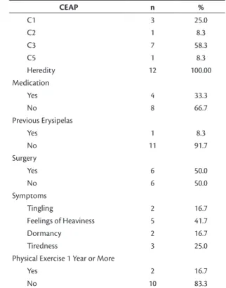
Table 2 shows that, according to the CEAP classification, three patients were in category C1 (25.0%), one was in C2 (8.3%), seven patients
were in C3 (58.3%) and one patient was classiied
in category C5 (8.3%). All participants reported a family history of the disease, while four (33.3%) of them reported taking medication for the disease and eight (66.7%) said they were not taking medication for the disease.
With regard to previous illnesses, one patient (8.3%) had a history of erysipelas and the other 11 (91.7%) did not have a history of this disease. Half of the patients (n = 6, 50%) reported having had surgery for varicose veins in the past and the other six (50%) had not undergone this type of procedure (Table 2).
In response to questions about their symptoms,
two patients (16.7%) reported tingling, ive (41.7%)
feelings of heaviness, two (16.7%) dormancy and three (25%) lower limb tiredness. Additionally, 10 (83.3%) patients were sedentary while two (16.7%) had been engaging in physical activity for more than 1 year (Table 2).
With relation to the distributions of scores for the domains of the SF-36 QoL questionnaire before and after treatment, improvements could be observed in certain domains. Table 3 lists the statistically signiicant differences detected in the domains Physical functioning (p = 0.011), Physical role limitation (p = 0.011) and
Pain (p = 0.017). There were no statistically signiicant
differences for the domains, General Health Status, Vitality, Social Functioning, Emotional Role Limitation
or Mental Health (p > 0.05).
When the domain scores and total scores for the
AVVQ-Brazil QoL questionnaire were compared
for before and after treatment, the only statistically
signiicant difference detected was in the Pain and
Dysfunction domain (p = 0.013) with no differences in total scores or for the domains, Esthetic Appearance, Extent of Varicosities or Complications (p > 0.05) (Table 4).
In order to quantify and analyze improvements that patients exhibited in terms of pain, pre-treatment and
Table 1. Demographic data for the sample.
Variables
Sex
Male 1
Female 11
Age
(Minimum-maximum) (48-70)
Mean ± SD 60.8±6.75
Body Mass Index (BMI)
(Minimum-maximum) (23.34-39.54)
Mean ± SD 30.78±4.42
Disease Duration
(Minimum-maximum) (3-50)
Mean ± SD 28.50±13.74
SD: standard deviation
Table 2. Clinical data for the sample.
CEAP n %
C1 3 25.0
C2 1 8.3
C3 7 58.3
C5 1 8.3
Heredity 12 100.00
Medication
Yes 4 33.3
No 8 66.7
Previous Erysipelas
Yes 1 8.3
No 11 91.7
Surgery
Yes 6 50.0
No 6 50.0
Symptoms
Tingling 2 16.7
Feelings of Heaviness 5 41.7
Dormancy 2 16.7
Tiredness 3 25.0
Physical Exercise 1 Year or More
Yes 2 16.7
No 10 83.3
post-treatment AVPS ratings were compared, revealing
a statistically signiicant difference (p = 0.007), as
shown in Table 5.
DISCUSSION
Chronic venous disease affects thousands of people worldwide, forcing them to change their habits to achieve better control of the signs and symptoms.
Many studies indicate that the disease affects more
women than men,5,9,13,14 which was borne out in this
study, in which females predominated (91.7%) over males (8.3%), and which can be attributed to factors such as pregnancy, use of contraceptives and obesity.14
The prevalence of CVD increases with age. Studies show that 5 to 15% of adults aged 30 to 70 years have this disease, with 1% suffering from venous ulcers.4-9,15 These data were also conirmed in this
study, in which the mean age was 60.08 years.
The risk factors for CVD include elevated BMI.
According to the World Health Organization (WHO),
BMI values greater than 30 kg/m2 indicate obesity.15
In this case, adipose tissue functions as a compressive
factor acting on veins, leading to increased relux,
and to increased vein diameter and venous pressure. Sugerman et al.16 state that severe obesity is also
associated with risk of venous stasis in lower limbs,
pre-tibial ulceration, cellulitis and edema. More than
half of the people who participated in this study had
a BMI indicative of obesity, associated with long
duration disease (28.50 years), demonstrating that this risk factor is present.
With the growing increase in the number of people with CVD and the pressing need to understand its varying clinical forms and analyze the degree of
compromise, such as venous relux, disease severity and deinition of its anatomic distribution, in addition
to designing the best treatment plan for patients, the
CEAP classiication is an excellent tool. In the sample in this study, the CEAP clinical classiication category
with the largest number of patients was C3 (58.3%), followed by C1 (25.0%). In contrast, Gurgel et al.17
reported that in a sample of patients who were treated surgically for venous disease type C2 (60.5%) was most common, followed by C3 (30.2%).
Table 3. Means, standard deviations and p values for SF-36 domains before and after treatment.
Domains Before After P value
Mean SD Mean SD
Physical functioning 40.00 20.67 64.17 18.27 0.011*
Physical role limitation 33.33 41.74 68.75 41.46 0.011*
Pain 49.03 22.58 65.92 21.94 0.017*
General health 47.17 13.17 52.42 11.17 0.284
Vitality 50.42 13.05 52.92 14.37 0.405
Social functioning 61.46 26.36 76.04 28.46 0.048
Emotional role limitation 50.00 48.20 61.11 44.57 0.334
Mental health 61.67 20.92 70.67 22.13 0.140
*Statistical signiicance (p < 0.05); Wilcoxon test. SD: standard deviation
Table 4. Means, standard deviations and p values for AVVQ-BRAZIL domains before and after treatment.
Domains Before After P value
Mean SD Mean SD
Pain and dysfunction 23.33 14.14 8.70 12.04 0.013*
Esthetic appearance 42.35 30.25 24.90 25.24 0.139
Extent of varicosities 28.43 17.13 23.29 16.08 0.336
Complications 5.98 8.20 2.86 2.59 0.104
Total score 20.41 6.22 13.41 7.33 0.080
*Statistical signiicance (p < 0.05); Wilcoxon test. SD: standard deviation
Table 5. Means, standard deviations and p values for AVPS ratings before and after treatment.
Variable Before After P value
Mean SD Mean SD
Analog visual pain scale (AVPS) 4.17 3.10 1.58 1.98 0.007*
In this study, all of the participants (100%) reported a family history of the disease. According to Nogueira et al.,18 the data from their study showed
that 45% of patients had a history of venous disease
in family members. In a study conducted by Morais
and Ferreira,19 63% of the participants reported a
family history and Morais and Ferreira19 also reported
that the majority of their sample (60%) were on medication, which is different from this study, in which just 33.3% of the volunteers reported using this type of treatment.
Souza et al.20 reported that surgical procedures
to correct supericial venous insuficiency lead to
improvements in deep vein system function, improving these patients’ clinical status. The data collected for the present study revealed that half of the patients (50%) reported having undergone this procedure.
With relation to symptoms, a considerable proportion of the participants (41.7%) described feelings of
heaviness in their lower limbs. Berenguer et al.21
reported that 39 of the 53 (73.58%) participants in their study complained of the same symptoms, which is in line with what was observed in the present study.
A study conducted by Salles-Costa et al.22
analyzed 4,030 employees of a University in Rio de Janeiro, detecting that inactivity predominated
among females (59.2%), conirming data from the
present study, in which 10 (83.3%) members of the sample were sedentary and just two (16.7%) had been physically active for more than 1 year. According to Danielsson et al.23 and Scott et al.,24 a lack of physical
activity reduces calf muscle pump function and can be partially responsible for a lack or reduction of venous drainage from the legs.
Many different resources are currently been used to
improve control of CVD, including drug treatments, surgery, compression stockings, physiotherapy and physical activity, with the objective of preventing exacerbation and improving the QoL of those affected.4,5 This study analyzed a proposal for including
aquatic exercise as one type of physical activity in the day-to-day routines of patients and the results show that its effect improved some domains of QoL
according to the SF-36 QoL questionnaire, speciically,
Physical Functioning, Physical Role Limitation and Pain (p < 0.05).
Silva et al.25 conducted a study designed to analyze
improvement in QoL and pain reduction in a group
of ibromyalgia patients after treatment in an aquatic environment, observing signiicant differences in scores
on the SF-36 QoL questionnaire, with improvements in parameters in several health domains, such as Physical Role Limitation, Pain, Vitality, Social
Functioning, Emotional Role Limitation and Mental
Health (p < 0.05). The choice of activities performed in the water was based on the fact that aquatic exercises have been used for several chronic pathologies, but have been investigated little with relation to CVD.
Silva et al.25 attempted to determine whether people
with ibromyalgia would experience a reduction
in pain after treatment in an aquatic environment.
They observed that there was no signiicant improvement
in pain rated with an AVPS, which contrasts with the present study, in which patients with CVD did report a reduction in this symptom (p = 0.007).
Research has shown that increased tolerance of exercise and improved levels of physical resistance lead to an improvement in general condition. When physical condition improves, there is a simultaneous improvement in symptoms, such as presence of pain after effort and muscle weakness.25
According to Alberti et al.,26 physical exercise
increases lower limb muscle tone and as a consequence can modify its effect on the venous system, with a consequent fall in pressure when walking and increased venous return. When exercise is combined with the aquatic environment there is a compound effect from the physical properties of water, such as hydrostatic
pressure, buoyancy, viscosity, low and temperature.
The AVVQ QoL questionnaire is considered an excellent tool for assessment of QoL in venous patients, since it is an easily-understood and easy to apply questionnaire that can be used to analyze people seeking surgical treatment, conservative treatment or other treatments and offering excellent reliability and viability.27
In a study conducted by Klem et al.27 to analyze
post-surgery QoL in patients with whom two different techniques had been employed to treat incompetence of the great saphenous vein, patients in both groups exhibited improvements in their postoperative AVVQ scores. In that study the participants exhibited improvements in Pain and Dysfunction, while the
other domains did not respond signiicantly.
Limitations of this study were the small number of people in the sample and the short data collection period and therefore more in-depth studies are needed
to conirm the true eficacy of aquatic exercises for
CVD.
CONCLUSIONS
Analysis of the effects of aquatic exercises on the QoL of people with CVD showed that these exercises were capable of improving certain aspects of QoL and of reducing pain, demonstrating that they are
REFERENCES
1. Biasoli MC, Machado CM. Hidroterapia: aplicabilidades clínicas. Rev Bras Med. 2006;63(5):225-37.
2. Carregaro RL, Toledo AM. Efeitos fisiológicos e evidências científicas da eficácia da fisioterapia aquática. Rev Movimenta. 2008;1(1):23-7.
3. Candeloro JM, Caromano FA. Efeitos de um programa de hidroterapia na pressão arterial e frequência cardíaca de mulheres idosas sedentárias. Fisioter. Pesqui. 2008;15(1):26-32.
4. França LH, Tavares V. Insuficiência venosa crônica. Uma atualização. J Vasc Bras. 2003;2(4):318-28.
5. Figueiredo MA, Filho AD, Cabral AL. Avaliação do efeito da meia elástica na hemodinâmica venosa dos membros inferiores de pacientes com insuficiência venosa crônica. J Vasc Bras. 2004;3(3):231-7.
6. Santos RFFN, Porfírio GJM, Pitta GBB. A diferença na qualidade de vida de pacientes com doença venosa crônica leve e grave. J Vasc Bras. 2009;8(2):143-7. http://dx.doi.org/10.1590/ S1677-54492009000200008.
7. Fernandes S, Rodrigues E, Vianna DL. Efeito da hidroterapia no edema de membros inferiores. Rev. Mackenzie Educ. Fis. Esporte. 2011;10(1):89-97.
8. Costa LM, Higino WJF, Leal FJ, Couto RC. Perfil clínico e sociodemográfico dos portadores de doença venosa crônica atendidos em centros de saúde de Maceió (AL). J Vasc Bras. 2012;11(2):108-13. http:// dx.doi.org/10.1590/S1677-54492012000200007.
9. Moura RM, Gonçalves GS, Navarro TP, Britto RR, Dias RC. Correlação entre classificação clínica CEAP e qualidade de vida na doença venosa. Rev Bras Fisioter. 2010;14(2):99-105. http:// dx.doi.org/10.1590/S1413-35552010005000007. PMid:20464164.
10. Ciconelli RM, Ferraz MB, Santos W, Meinão I, Quaresma MR. Tradução para a língua portuguesa do questionário genérico de avaliação de qualidade de vida SF-36 (Brasil SF-36). Rev Bras Reumatol. 1999;39(3):143-50.
11. Leal FJ, Couto RC, Pitta GBB, et al. Tradução e adaptação cultural do Questionário Aberdeen para veias varicosas. J Vasc Bras. 2012;11(1):34-42.
12. Andrella GQ, Araújo PM, Lima SM. Estudo comparativo entre duas escalas de dor e a aplicação em doentes. Estudos. 2007;34:21-34. 13. Cataldo JL, Godoy P, Barros J. The use of compression stockings
for venous disorders in Brazil. Phlebology. 2012;27(1):33-7. http:// dx.doi.org/10.1258/phleb.2011.010088. PMid:21765190.
14. Lins EM, Barros JW, Appolônio F, Lima EC, Barbosa M Jr, Eduardo A. Perfil epidemiológico de pacientes submetidos a tratamento cirúrgico de varizes de membros inferiores. J Vasc Bras. 2012;11(4):301-4. http://dx.doi.org/10.1590/S1677-54492012000400008. 15. Seidel CA, Mangolim ASM, Rossetti LP, Gomes JR, Miranda FJ Jr.
Prevalência de insuficiência venosa superficial dos membros inferiores em pacientes obesos e não obesos. J Vasc Bras. 2011;10(2):124-30. http://dx.doi.org/10.1590/S1677-54492011000200006. 16. Sugerman HJ, Sugerman EL, Wolfe L, Kellum JM Jr, Schweitzer
MA, DeMaria EJ. Risks and benefits of gastric bypass in morbidly obese patients with severe venous stasis disease. Ann Surg. 2001;234(1):41-6. http://dx.doi.org/10.1097/00000658-200107000-00007. PMid:11460821.
17. Gurgel GA, Castro AA, Araújo M, Amorim JE, Pitta GB, Miranda F Jr. Evaluation of the venous reflux of the great saphenous vein by duplex scan after surgical treatment of the saphenofemoral junction insufficiency. Rev Col Bras Cir. 2013;40(5):380-5. PMid:24573586. 18. Nogueira GA, Oliveira BG, Santana RF, Cavalcanti AC. Diagnóstico de enfermagem em pacientes com úlcera venosa crônica: estudo observacional. Rev Eletr Enf. 2015;17(2):333-9. http://dx.doi. org/10.5216/ree.v17i2.28782.
19. Morais KC, Ferreira AC. O impacto da insuficiência venosa crônica no desempenho funcional em mulheres. InterScientia. 2014;2(3):29-47.
20. Souza MO, Miranda F Jr, Figueiredo LF, Pitta GB, Aragão JA. Implementação financeira e o impacto do mutirão de cirurgias de varizes, após a criação do Fundo de Ações Estratégias e Compensação (FAEC). J Vasc Bras. 2011;10(4):302-7.
21. Berenguer FA, Lins e Silva DA, Carvalho CC. Influência da posição ortostática na ocorrência de sintomas e sinais clínicos de venopatias de membros inferiores em trabalhadores de uma gráfica na cidade do Recife-PE. Rev Bras Saúde Ocup. 2011;36(123):153-61. http:// dx.doi.org/10.1590/S0303-76572011000100016.
22. Salles-Costa R, Werneck GL, Lopes CS, Faerstein E. Associação entre fatores sócio-demográficos e prática de atividade física de lazer no Estudo Pró-Saúde. Cad Saúde Publica. 2003;19(4):1095-105. http:// dx.doi.org/10.1590/S0102-311X2003000400031. PMid:12973574. 23. Danielsson G, Eklof B, Grandinetti A, Kistner RL. The influence
of obesity on chronic venous disease. Vasc Endovascular Surg. 2002;36(4):271-6. http://dx.doi.org/10.1177/153857440203600404. PMid:15599477.
24. Scott TE, LaMorte WW, Gorin DR, Menzoian JO. Risk factor for chronic venous insufficiency: a dual case-control study. J Vasc Surg. 1995;22(5):622-8. http://dx.doi.org/10.1016/S0741-5214(95)70050-1. PMid:7494366.
25. Silva TFG, Suda EY, Marçulo CA, Paes FHS, Pinheiro GT. Comparação dos efeitos da estimulação elétrica nervosa transcutânea e da hidroterapia na dor, flexibilidade e qualidade de vida de pacientes com fibromialgia. Fisioter Pesq. 2008;15(2):118-24.
26. Alberti LR, Petroianu A, Corrêa D, Silva TF. Efeito da actividade física na insuficiência venosa crônica dos membros inferiores. Acta Med Port. 2008;21(3):215-20. PMid:18674413.
27. Klem TM, Schnater JM, Schutte PR, Hop W, van der Ham AC, Wittens CH. A randomized trial of cryo stripping versus conventional stripping of the great saphenous vein. J Vasc Surg. 2009;49(2):403-9. http://dx.doi.org/10.1016/j.jvs.2008.09.016. PMid:19028042.
*
Correspondence
Michael Augusto dos Santos Aquino Av. Antônio José da Costa, 132 – Bairro Novo CEP 57480-000 – Delmiro Gouveia (AL), Brazil Tel.: +55 (82) 9 9972-5491 E-mail: [email protected]
Author information
MASA – Physiotherapy student, Faculdade Estácio de Alagoas (ESTÁCIO-FAL). LCVP – Physiotherapy student, Faculdade Estácio de Alagoas (ESTÁCIO-FAL). FJL and RCC – Physiotherapist. MSc in Sciences from Universidade Federal de São Paulo (UNIFESP); Assistant professor, Faculdade Estácio de Alagoas (ESTÁCIO-FAL).
Author contributions
Conception and design: MASA, LCVP, FJL, RCC Analysis and interpretation: MASA, LCVP, FJL, RCC Data collection: MASA, LCVP Writing the article: MASA Critical revision of the article: MASA, LCVP Final approval of the article*: MASA, LCVP, FJL, RCC Statistical analysis: MASA, LCVP, FJL, RCC Overall responsibility: MASA, LCVP, FJL, RCC