Um Comentário sobre a
Laringos-copia
Senhor Editor,
Embora novos equipamentos e materiais tenham aparecido nos últimos 50 anos, a laringoscopia com o laringoscópio ain-da é o mais rápido e confiável método de inserção de um tubo na traquéia. Tem a vantagem de permitir visão direta das cor-das vocais e evitar intubação traumática. Muitos livros e arti-gos têm sido escritos para ensinar a intubação com o larin-goscópio, mas falta-lhes informação visual adequada1-4. A posição olfatóriarecomendada para facilitar a visão da larin-ge não é anatomicamente definida5. O melhor treinamento pode ser conseguido em cirurgias eletivas, nas quais o paci-ente pode ser previampaci-ente examinado sobre suas condições clínicas e abordagem da via aérea6. Depois de um treina-mento adequado pode-se manipular situações difíceis até mesmo num caso deintubação difícil.
são precárias. A posição do anestesiologista é desconfortá-vel, a laringoscopia pode ser inadequada devido a dificulda-des de posicionamento da cabeça do paciente e o material de aspiração nem sempre é eficiente. Contudo, em condições normais, a maior parte das intubações difíceis são conseqüência do mau uso do laringoscópio.
Desde a construção da lâmina curva proposta por Macintosh7 ela se tornou o equipamento mais popular do mundo para la-ringoscopia.
Este artigo apresenta uma “nova” revisão da intubação tra-queal com o laringoscópio de Macintosh sem o trauma comu-mente relacionado com fratura de dentes8-10.
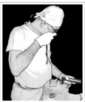
Introdução:Desde 1965 tenho usado esta técnica, que me foi ensinada por Professor Bento Gonçalves, no Hospital Pe-dro Ernesto (Universidade do Estado do Rio de Janeiro). Ele dizia tê-la aprendido diretamente de Sir Robert Macintosh durante sua visita ao Brasil in 1954: A primeira coisa a apren-der é o significado de posição olfatória. A posição olfatória é aquela assumida por uma pessoa de pé cheirando uma flor. Para ver esta posição, deve-se ficar de pé com as costas na parede, mas didaticamente prefiro usar a borda de uma por-ta. Pela flexão completa do pescoço sobre o tronco e defle-xão da cabeça até que o eixo da face recupere sua posição vertical pode-se imaginar uma pessoacheirando uma flor. Isto está muito bem descrito num artigo de Maggil de 193011.
Preparo do paciente: Durante a visita pré-anestésica deve-se solicitar ao paciente que mova seu pescoço e cabe-ça para evitar surpresa no momento do procedimento. Para indução da anestesia, qualquer indutor e relaxante muscular são suficientes.
Altura da mesa (na sala de cirurgia):A altura da mesa DEVE ser baixa, idealmente na altura de seu púbis, incluindo o colchão da mesa. A cabeça do paciente ficará na altura de sua bexiga. Se você é baixo, deve usar um tablado ou degrau (como o das instrumentadoras) e colocar a mesa na altura desejada.
Escolhendo o laringoscópio:Um bom laringoscópio não depende do material de que é feito. Depende do raio da cur-vatura da lâmina. Existem várias lâminas curvas que não são lâminas de Macintosh. Conheço quase todas e posso dizer que a melhor delas é fabricada pela Narcosul Aparelhos de Anestesia Ltda. Verifique a sua. O padrão foi introduzido há muitos anos7.
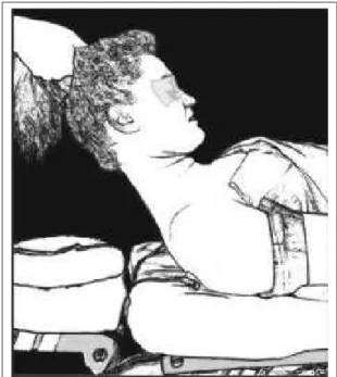
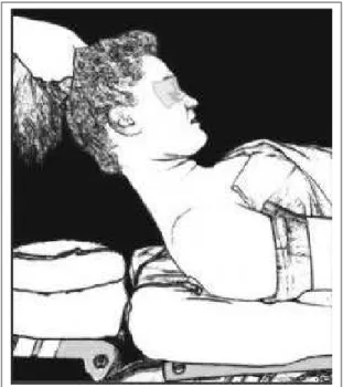
Posicionamento do paciente:O paciente deve ser posicio-nado de tal forma que sua cabeça alcance a beirada da cabe-ceira da mesa que deve estar na horizontal. Depois de uma boa pré-oxigenação e indução apropriada, feche os olhos do paciente com fita adesiva para prevenir lesão da córnea por manipulação da face. Levante a cabeça do paciente até fle-xão completa do pescoço sobre o tronco (Figura 1). Em se-guida, defletir sua cabeça mantendo-a alta (Figura 2) e en-cha o espaço abaixo do crânio com sacos de areia de tama-nhos adequados (Figura 3). Não 5 cm ou 10 cm de altura. Po-nha o que for preciso. Sustente a cabeça do paciente em po-sição, mantendo o eixo da face paralelo ao chão da sala. Com seu próprio corpo, empurre a cabeça do paciente no sentido
dos pés (do próprio paciente) para mantê-la nesta posição (Figura 3).
Revista Brasileira de Anestesiologia 695
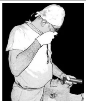
Figura 2 - Deflexão da cabeça sobre o pescoço, man-tendo sua altura da mesa. Não importa quanto. Para cada paciente uma altura
Como usar o laringoscópio:Mantendo o eixo da face para-lelo ao chão abre-se a boca do paciente. Neste momento a cabeça do paciente está sendo empurrada por seu corpo, na altura de seu umbigo e lhe permite aplicar a lâmina de Macin-tosh de tamanho adequado, deslizando-a contra a superfície da língua, longe dos dentes superiores, até que ela atinja a valécula. Com sua mão esquerda, segure o cabo do laringos-cópio e o queixo do paciente juntos (Figura 4). Um força sua-ve é aplicada no sentido anterior (isto é, para cima), puxando a mandíbula e a lamina juntas, abrindo a glote. Esta manobra foi proposta nos anos 5012. Em seguida, outra força move o
cabo do laringoscópio no sentido dos pés do paciente. Este movimento (ou força) abre amplamente a boca do paciente e mantém a lamina longe dos dentes superiores, evitando os traumas tão comentados8-10. A vista é tão ampla que dá para tirar fotos (Figura 5) e mostrar a glote sem aplicar a lâmina nos incisivos do paciente (Figura 6). Esta paciente está usan-do uma dentadura superior que não foi removida, mostranusan-do a segurança da técnica.
A inserção do tubo segue a linha de visão, isto é, vertical, do teto para o chão, ao invés de da cabeça para os pés ou a 45º como sugerido em artigos13,14.
Numa Carta ao Editor, Kurien15descreveu uma boaposição olfatória, mas falhou ao não recomendar manobras neces-sárias para mantê-la fixa. Neste caso, a possibilidade de fra-tura dentária pode ser esperada.
Figura 3 - Enchendo o espaço entre a cabeça e a mesa. Nem cinco nem dez centímetros, mas o que for necessário
Figura 5 - A boca se abre amplamente e permite excelentes fotos com câmeras comuns
Figura 4 - Mantendo fixa a posição da cabeça por pressão exercida com seu corpo contra o crânio (mantendo horizontal o eixo da face) e segurando o queixo e o la-ringoscópio juntos para produzir um movimento ante-rior (para cima) e um movimento para baixo (para os pés). Pode-se ver daqui que o laringoscópio não toca nos dentes superiores da paciente
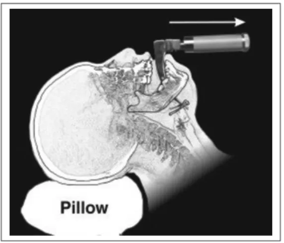
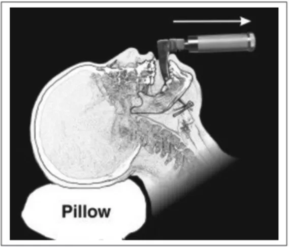
No início de 1991 fiz algumas Xerorradiografias durante pro-cedimentos de intubação para verificar a relação da lâmina do laringoscópio com os dentes superiores e o ponto de vista do anestesiologista (Figura 7).
De uma visão vertical obtém-se a melhor vista da glote, não interessa o que se possa ver. Se for encontrado um índice de Cormack Lehane III ou IV, tente usar um guia, ou uma sonda nasogástrica comum inserida no laringe através de qualquer coisa que possa ser visto da rima glótica e tente passá-la por trás da epiglote e faze-la descer até a traquéia, como propos-to por Macinpropos-tosh em 194616. Aí então ela poderá ser usada como um guia para o tubo traqueal. Neste caso você poderá dizer quefoi diferente, mas não poderá dizer quefoi difícil.
Comentários:Além de atender a estas regras, deve-se ma-nipular a altura da mesa. Um grande erro é subir a mesa até a altura do abdome superior, como sugerido17. Nesta altura, todas as manobras obrigam o anestesiologista a usar o larin-goscópio como uma alavanca, orientando o cabo para o teto, aplicando força nos dentes. Isto é má técnica e favorece a acidentes. Outro erro é colocar a mesa muito baixa14. O cor-po do anestesiologista deve permanecer de pé, elegante, mantendo a cabeça do paciente por pressão aplicada no sen-tido anterior e obtendo um panorama da glote vista de cima (Figura 4). Os mais baixos devem usar um tablado para corri-gir sua posição em relação à mesa. Do tablado a mesa pode ser levantada até a posição desejada para atingir o nível de sua bexiga. Isto favorece à manutenção da cabeça do paci-ente em boa posição, abrir sua boca, inserir a lâmina de Ma-cintosh, levá-la no sentido dos pés do paciente sem levantar o cabo para o teto, o que evita apoio nos dentes incisivos. A linha de visão deve ser de cima para baixo e nunca da cabe-ça para os pés ou a 45º, como freqüentemente mostrado13,14. Qualquer visão diferente da vertical leva à manobras de ala-vanca com a lâmina e a força é aplicada nos incisivos. Um
dente quebrado é sempre o resultado de uma laringoscopia deficiente.
Em 38 anos de Anestesiologia eu nunca retirei uma dentadu-ra da boca de um paciente. Chego mesmo a considedentadu-rar que ela é a razão para manutenção de técnica apropriada de não tocar nos dentes com a lâmina do laringoscópio. Se for remo-vida você pode se sentir livre para puxar o cabo para o teto e lesar as gengivas, o que torna difícil a colocação da dentadu-ra na boca no paciente após a cirurgia.
Se você seguir estas recomendações eu acredito que fará um grande progresso em sua técnica e que resolverá muitas de suasintubações difíceis.
Dr. M. A. Gouveia,TSA
Chefe do Serviço de Anestesiologia Hospital Central do IASERJ
Rua São Clemente, 185/1304-2 Botafogo 22260-001 Rio de Janeiro, RJ
E-mail: [email protected]
A Comment on the Laryngoscopy
Mr. Editor,
Although new equipment, new devices and new techniques have appeared in the last 50 years, laryngoscopy with a laryn-goscope still is the most reliable and fastest way to insert a tube into the trachea. It holds the advantage of permitting a di-rect view of the vocal cords and prevents a traumatic intubation. Many books and articles have been written to teach tracheal intubation with a laryngoscope, but they lack adequate visual information1-4. Thesniffing position recom-mended to facilitate the view of the larynx is not anatomically defined5. The best training can be achieved in connection with scheduled surgery, in which the patient can be previously examined for clinical condition and airway approach6. After proper training one can manage difficult situation even in a case of adifficult intubation.
In an emergency situation, either in an in-hospital case or at the site of an accident, conditions for a lifesaving intubation are usually poor: The position of the anesthesiologist is un-comfortable, laryngoscopy may be inadequate because of poor positioning facilities for the patient’s head and suction equipment may prove inefficient. However, in normal condi-tions, most difficult intubations depend on inadequate use of the laryngoscope.
Since the construction of the curved blade proposed by Macintosh 7, it has become the most popular device for laryngoscopy in the world.
This article is to present a “new” review of tracheal intubation with the Macintosh laryngoscope blade without the trauma al-ways reported on fractures of teeth8-10.
Revista Brasileira de Anestesiologia 697
Introduction: Since 1965 I have been using a technique, which I was taught by Professor Bento Gonçalves, at Hospital Pedro Ernesto (University of the State of Rio de Janeiro, Brazil). He use to say he learned it directly from Sir Robert Macintosh during his visit to Brazil in 1954: The first thing is to learn the meaning of thesniff position. The sniff position is the one assumed by astanding person sniffing a flower. To see this position, one must stand straight with his back on the wall but for the purpose of teaching I prefer the person standing against the edge of a door. By full flexion of the neck against the trunk and deflexion of the head until the axis of the face re-covers the vertical position you can imagine this patient is sniffing a flower. This is well described in the article of 1930 by Maggil11.
Patient preparation:During the pre-anaesthetic interview you must ask the patient to move his/her neck and head to avoid disappointment at the moment of the procedure. For in-duction of anaesthesia any sleeping drug and muscle relax-ant are sufficient.
Table height (in the OR): The height of the table MUST be low, preferably the height of your pubis, including the mat-tress. The head of the patient will be at the level of your blad-der. If you are a short person you would better stand on a step (like the one used by scrub nurses) and level the table to the desired the height.
Choosing a laryngoscope: A good laryngoscope blade does not depend on the material it is made of. It depends on the radius of the arch of its blade. There are many curved blades that are not Macintosh blades. I know almost all brands of laryngoscopes and I can say that the best one is m a n u f a c t u r e d i n B r a z i l b y N a r c o s u l A n a e s t h e s i a Equipments. Check yours. It must be shaped curved, neither straight nor half curved. The standard was introduced many years ago7.
Patient positioning:The patient must be positioned in a way that his/her head reaches the edge of the table on the headrest. Horizontal leveling of the table is recommended. After a good pre-oxygenation and appropriate induction, close the patient’s eyes with adhesive tape to prevent corneal abrasion from mishandling of the face. Lift the head of the pa-tient until full flexion of the neck over the trunk (Figure 1). De-flect his/her head keeping it high (Figure 2) and fill the space under the skull with sand bags of appropriate size (Figure 3). Not 5 cm or 10 cm high. Just the tailored size. Keep the pa-tient’s head in position, maintaining the axis of his/her face parallel to the floor. To maintain this position push the head to-ward the feet with your own body (Figure 3).
How to use the laryngoscope:By keeping the axis of the face parallel to the floor the patient opens his/her mouth. At this moment the patient’s head is pushed by your body at the height of your umbilicus, which permits you to apply the Macintosh laryngoscope blade of appropriate size slipping the blade against the surface of the tongue, far away from the upper teeth, until its tip reaches the valecula while displacing the tongue to the left. With your left hand, hold the handle of the laryngoscope and the chin together (Figure 4). A gentle force is then applied by pulling the jaw and the blade anteriorly
(that meansup), opening the glottis. This maneuver has been proposed in the early 50’s12. Then, another force moves the laryngoscope handle to the toes of the patient. This
move-Figure 2 - Deflexion of the head over the neck, main-taining its height from the table. No matter how high. For each patient one height
ment (or force) opens the mouth wide and keeps the laryngo-scope blade away from the teeth, preventing the trauma usu-ally spoken of8-10. The view is so great that you can even take pictures (Figure 5) and shows the glottis without applying the laryngoscope blade on the incisor teeth of the patient (Figure 6). This patient is using a superior tooth plate that was not re-moved to show the safety of the technique.
Insertion of the tube follows the line of vision, that means, it must be vertical,from ceiling to floor, instead offrom head to footor at 45º as suggested elsewhere13,14.
In a letter to the editor, Kurien15described a goodsniffing po-sitionbut failed in not recommending the necessary maneu-vers to keep the head fixed. In this case the possibility of a bro-ken tooth can be anticipated.
In the beginning of the year 91 I took some Xeroradiography during intubation procedures to check the relation of the la-ryngoscope blade with the teeth and the point of view of the anesthesiologist (Figure 7).
From a top-down view you can achieve the best picture of the glottis, no matter how much you see of it. If you see a Cormack Lehane III or IV just try using a guide, such as an ordinary plastic nasogastric tube inserted into the larynx in whatever you can see of the rima glottidis or try to direct the guide be-hind the epiglottis and thread it into the trachea as proposed by Macintosh in 194916. Then you can use it as a guide to the
Revista Brasileira de Anestesiologia 699
Figure 3 - Filling the space between the table and the head’s height. Nor 5 cm neither 10 cm. Fill it with what it is necessary
Figure 5 - The mouth opens wide and permits excel-lent pictures with ordinary cameras
Figure 4 - Maintaining the position of the head fixed by pressure of your body against the skull (maintaining the horizontal axis of the face) and holding the chin and laryngoscope together to produce an anterior
(up) movement and ato the toes(downward) move-ment. you can see here that the laryngoscope does not touch the upper teeth
tracheal tube. In this case you can say the intubation was dif-ferent, but you cannot say it was difficult.
Comments- Besides complying with these rules, one must also work the table height. A common mistake is to lift the ta-ble until the height of the upper abdomen as suggested17. At this height all maneuvers lead the anesthesiologist to use the laryngoscope blade as a lever and point the handle to the ceil-ing, thus applying pressure on the teeth. This is a bad tech-nique and prone to accident. The other fault is to back from the table and bend over the patient when the table height is too low 14. The body of the anesthesiologist must stand straight, elegant, holding the head of the patient by pressure applied forward and looking to the glottis from top (Figure 4). In the case of a short anesthesiologist a wooden step (such as the one used by scrub nurses) must be used to correct his/her position.
Standing on a step she/he can then lift the table as much as necessary to adjust its height to her/his bladder. This ensues the possibility of holding the patient’s head in a good position, open his/her mouth wide, insert the Macintosh laryngoscope blade, push it to the toes of the patient without lifting its handle to the ceiling and avoids touching the incisor teeth. The line of sight must be from the ceiling to the floor, and never from head to feet or a 45-degree line, as shown13,14. Any sight different from a top-down line leads to the lever maneuver with the laryngoscope blade and a force is then applied upon the incisor tooth. A broken tooth is the result of a faulty laryngoscopy.
In 38 years of anaesthesia Ineverremoved a denture plate of a patient. I even consider it a reason to keep the appropriate technique of not touching the teeth with the laryngoscope
blade. If you remove it you may feel free to pull the handle to the ceiling and hurt the gums of the patient, what makes it ter-rible to put his denture plate in the mouth after the surgery. If you follow these rules I believe you can have a good im-provement in your intubation technique and that you will re-solve most of yourdifficult intubations.
M. A. Gouveia, TSA, MD
Head of Department of Anaesthesia Hospital Central do IASERJ
Rua São Clemente, 185/1304-2 Botafogo 22260-001 Rio de Janeiro, RJ
E-mail: [email protected]
REFERÊNCIAS-REFERENCES
01. Magill IW - Endotracheal Anaesthesia. Am J Surg, 1936;34: 450-455.
02. Benumof JL - Conventional (Laryngoscopic) Orotracheal and Nasotracheal Intubation (single lumen type), em: Benumof JL -Clinical Procedures in Anaesthesia and Intensive Care. J.B. Lippincott Co, 1992;115-148 Chapter 6 pages.
03. Gillespie NA - Endotracheal anaesthesia. The University of Wis-consin Press, 1948;83-129.
04. Benumof JL - Management of the difficult adult airway. Anaesthesiology, 1991;75:1087-1110.
05. Benumof JL - Difficult laryngoscopy: obtaining the best view. Can J Anaesth, 1994;41:361-365.
06. Mallampati DR, Gugino LD, Desai SP et al - A clinical sign to pre-dict difficult tracheal intubation: a prospective study. Can Anaesth Soc J, 1985;32:429-434.
07. Macintosh RR - A new Laryngoscope. Lancet 1943;13:205. 08. Solazzi RW, Ward RJ - The spectrum of medical liability cases.
Int Anaesthesiol Clin, 1984;22:43-59.
09. Owen H, Waddell-Smith I - Dental trauma associated with an-aesthesia. Anaesth Intensive Care, 2000;28:133-145. 10. Bucx MJL, Snijders CJ, van Geel RTM et al - Forces acting on
the maxillary teeth during laryngoscopy using the Macintosh la-ryngoscope. Anaesthesia, 1994;49:1064-1070.
11. Magill IW - Technique in endotracheal anaesthesia. Br Med J, 1930;ii:817-819.
12. Manzocchi EL - Anestesia endotracheale. Redi, Societá Editrice P.A. Milano. (Early 50’?) Ch III:22-47.
13. Cormack RS, Lehane J - Difficult tracheal intubation in obstet-rics. Anaesthesia, 1984;39:1105-1111.
14. Walker JD - Posture used by anaesthetists during laryngoscopy. Br J Anaesth, 2002;89:772-774.
15. Kurien KM - Maintaining optimal head positioning for laryngoscopy. Anaesth Intensive Care, 1998;26:331.
16. Macintosh RR - An aid to oral intubation. Br Med J, 1949;1:28. 17. Otto CW - Tracheal Intubation, em: Nunn JF, Utting JE, Brown BR - General Anaesthesia. London: Butterworths, 1989; 512-539.