ISSN 0102-695X
DOI: 10.1590/S0102-695X2012005000126 Received 26 Jun 2012
Accepted 11 Sep 2012 Available online 6 Nov 2012
Ipomoea pes-caprae using factorial design
followed by antinociceptive and
anti-inl ammatory evaluation
Daniela Vieira,
1Cristina Padoani,
1Janaína dos S. Soares,
1Jerusa Adriano,
2Valdir Cechinel Filho,
2Márcia M. de Souza,
2Tania M. B. Bresolin,
2Angélica G. Couto
*,21Núcleo de Investigações Químico-Farmacêuticas, Universidade do Vale do Itajaí, Brazil,
2Programa de Pós-graduação em Ciências Farmacêuticas, Universidade do Vale do Itajaí, Brazil.
Abstract: Ipomoea pes-caprae (L.) R. Br., Convolvulaceae, is a medicinal plant that grows abundantly as a pan-tropical stand plant. The 32 (two factors and three levels)
factorial design, was applied to determine the best time and drug/solvent proportion
to maximize the l avonoid content in the hydroethanolic extract by maceration process. The antinociceptive and anti-inl ammatory effects were studied at 5-20 mg/
kg, i.p., using the writhing test and carrageenan-induced pleurisy models in mice. The optimized extract was able to inhibit more than 50% of abdominal writhing at 20 mg/kg, with 55.88%±2.4 of maximum inhibition. Indomethacin, used as positive control, inhibited 64.86% at 10 mg/kg. In the pleurisy model, the extract produced
dose-dependent inhibition of the i rst phase of inl ammation (4 h) in the pleural cavity
induced by injection of carrageenan (1%) in mice. It inhibited 50%±0.82 (p<0.01) of exudation induced by carrageenan, and 60.88%±0.14 (p<0.01) of leukocyte migration to the pleural cavity. In conclusion, the results validate the technological conditions of the maceration process to produce an optimized bioactive herb extract for the
development of analgesic and anti-inl ammatory phytopharmaceuticals using 70 °GL
ethanol, a plant to solvent ratio of 12.5% (w/v), and ten days of maceration.
Keywords:
anti-inflammatory antinociceptive hydroethanolic extract Ipomoea pes-caprae pleurisy
total flavonoids
Introduction
Ipomoea pes-caprae (L.) R. Br., Convolvulaceae, known in English as bayhops, beach morning glory, and goat's-foot-convolvulus, and in Brazil as “salsa-da-praia”, “batateira-da-praia” or “pé-de-cabra”, has been used as a
herb remedy in many countries to treat inl ammation, colic,
diuretic disorders, gonorrhea, pain etc. (Pongprayoon et al., 1989; Assis et al., 1994; Khan et al., 1994).
Previous studies have coni rmed some
pharmacological properties of this plant. Extract of petroleum ether caused concentration-dependant inhibition of the contraction of guinea-pig ileum contractions stimulated by different neurotransmitters (Pongprayoon et al., 1989). Bioassay-guided fractionation of this extract
resulted in the isolation of β-damascenone and E-phytol, which were equipotent to papaverine (Pongprayoon et al., 1992). Another interesting action demonstrated by this
plant was its ability to neutralize or inactivate jellyi sh
venoms (Pongprayoon et al., 1991a). Khan et al. (1994)
reported the insulinogenic and hypoglycemic activities of the alcohol extract of the leaves. Previous studies carried out by our research group have revealed that the methanol
extract from the whole plant exhibits potent and signii cant
antinociceptive properties against acetic acid-induced
abdominal constrictions and neurogenic and inl ammatory
phases of formalin-induced pain, probably associated
to ethyl acetate fraction, as well as to glochidona, α and β-amyrin acetate, betulinic acid and isoquercitrin, being
equipotent to some analgesic drugs (Krogh et al., 1999; Souza et al., 2000).
Considering the popular use of this plant to treat pain and inflammation, as well as the promising previous experimental data and its abundance in Brazil, the first end point of the study was to develop an herb extract using an adequate solvent for industrial scaling up, with lower toxicity, less environmentally hazardous, associated with a simple extraction method. Then, we attempted to develop hydroethanolic extracts by maceration process, using factorial design to find
out the extraction conditions for the highest flavonoid content, and antinociceptive and anti-inflammatory effects analysis of the selected extract.
Material and Methods
Plant material
The botanical material was collected from Esplanada Beach (Jaguaruna-SC, Brazil) in February 2006. A voucher was deposited at the Barbosa Rodrigues Herbarium (Itajaí-SC, Brazil) under number V.C. Filho
009. The leaves, stems and other aerial parts (lower buds, seeds and lowers) were manually separated, cleaned and
dried in an air oven at 35 ºC for eleven days. After drying, each set of each plant material was ground in a hammer mill (outlet sieve =3 mm) and the average size of the particles was determined by sieving.
Previous studies for extraction
The ethanol graduation was first investigated by ethanol extractable matter (EM) from 1.0 g of aerial
parts decocted with 100 g of 40, 50, 60, 70 or 80 °GL
ethanol, under reflux for 10 min. In another set of experiments, an extract (5%, w/w) of each solvent was prepared by maceration for seven days, in a hermetically sealed amber glass bottle, stirred twice a day.
The total flavonoid content (TFC), expressed as quercetin (g%, w/w), was determined spectrophotometrically (420 nm) for 15.0 g of each sample solution, after selective extraction, acid hydrolysis and complexation with AlCl3, following the method based on the Brazilian Pharmacopeia (Farmacopeia Brasileira, 2001).
Factorial design for extraction by maceration
This study was carried out following a 32
factorial model, taking into account the extraction time (5, 10 and 15 days) and the drug/solvent ratio (2.5, 7.5 and 12.5%, w/v) as independent variables, and the total flavonoid content and dry residue as dependent variables.
Chromatographic profile of selected extractive solution
A new batch of extractive solution was produced, according to selected extraction conditions, in order to characterize its chromatographic profile by HPLC, using a Shimadzu LC-10AD HPLC system (Shimadzu, Tokyo, Japan) equipped with a binary pump and a SPD-M10A photo diode array detector, a CTO-10A column oven, and an automatic injection system.
The mobile phase consisted of water pH 3.2 (acidified with formic acid) as solvent A and acetonitrile: methanol (50:50, v/v) as solvent B, gradient system of 75:25 for
30 min, to 60:40 on 20 min, 35 °C and flow rate of 0.8
mL, through a C18 5 μm 100Å (250 x 4.6 mm) column (Phenomenex®, Torrance, USA), with detection at 254
nm and injection of 20 μL.
All the solvents were HPLC grade and were previously degassed. The water was purified using an Easy Pure device (Waltham, Massachusetts, USA). For the LC injections, hydroethanolic extract was diluted in methanol and purified water (50:50) (5 mg/ mL, according to its dry residue weight). Isoquercitrin (Chromadex®, Irvine, CA), diluted in methanol and
purified water (50:50), at six concentrations from 10 to 80 µg/mL, was used as a reference substance to elaborate an analytical curve. All the samples were filtered through a 0.45 µm regenerated cellulose membrane (Millipore Maidstone, Kent, UK) before injection. Each analysis was repeated three times and the calibration curves were fitted by linear regression.
Animals
Male wistar mice, 25-30 g, were used. The animals were kept in a controlled temperature room
(23±2 °C), with 12 h light-dark cycles, and were
allowed free access to food and water. The experiments were performed with the approval of the Committee for the Use of Animals in Experiments of the University of Itajaí Valley (Brazil) (CEUA 407/06), and were carried out in accordance with current guidelines for the care of laboratory animals, and the ethical guidelines for investigations of experimental pain in conscious animals, as specified by Zimmermann (1983). The number of animals (8-10 per group) and intensity of the noxious stimuli used were the minimum necessary to demonstrate the consistent effects of the drug treatments.
Antinociceptive activity
Antinociceptive activity was expressed as the reduction in the number of abdominal constrictions between the control animals (saline treated) and the mice pre-treated with extract. The nociceptive response caused by acetic acid injection was analyzed 30 min after the administration of hydroethanolic extract or saline injection. All experiments were carried out at 20-22
°C.
Anti-inflammatory activity
The carrageenan (Cg) induced pleurisy model was used. Pleurisy experiments were carried out as previously described (Saleh et al., 1996) and carrageenan was used as the phlogistic agent, by a single intrapleural injection of 0.1 mL of Cg (1%). The animals were intraperitoneally treated with different doses of Ipomoea pes-caprae (L.) R. Br., Convolvulaceae, extract (5-20 mg/kg) or vehicle, used to dilute the extracts, or indomethacin (10 mg/ kg) as a reference drug, 30 min before the pleurisy induction. The inflammatory parameters (leukocytes and exudation) were analyzed 4 h after Cg injection.
After 4 h, the animals were killed with an overdose of pentobarbital, the thorax was opened, and the pleural cavity was washed with 1.0 mL of sterile PBS (pH 7.6, NaCl 137 mM:KCl 2.7 mmol and phosphate buffer salts 10 mM) containing heparin (20 IU/mL). Samples of the pleural lavage were collected to determine the exudation and total and differential leukocyte counts (Saleh et al., 1996; Dalmarco, 2002). Total leukocyte counts were performed in a Neubauer chamber after diluting the pleural fluid with Turk solution (1:20). The cytospin preparations of pleural
wash were stained with May-Grunwald Giemsa for the
differential count, which was performed under an oil immersion objective.
In another set of experiments, the animals were treated 30 min before Cg, with a solution of Evans blue dye (2.5 mg/kg, 0.2 mL, i.v.) in order to evaluate the degree of exudation in the pleural space (Calixto &
Campos, 1995). A sample (500 μL) of the fluid leakage
collected from the pleural cavity was stored in a
freezer (-20 °C) to further determine the concentration
of Evans blue dye. To this end, on the day of the experiments, a batch of samples was thawed at room temperature and the amount of dye was estimated by optical density using a spectrophotometer (Pharmacia Biotech-Ultrospec 2000) at 600 nm, by interpolation from a standard curve of Evans blue dye in the range of
0.01 to 50 μg/mL (Cordeiro et al., 1999).
Statistical analysis
The factorial design 32 data were analyzed
through response surface plots from adjusted equations by multiple regressions (Cochran & Cox, 1978; Montgomery, 1991), with the aid of Statgraphics Plus® (software version 5.1).
The pharmacological results are presented as mean±standard error deviation (SEM). The statistical significance between groups was analyzed by means of t test or analysis of variance, followed by the Dunnet’s
with the aid of GraphPad Instat® software. p values<0.05
were considered as indicative of significance.
Results and discussion
The batch of herb raw material used for this study was consisted by 52.24% of stems, 32.24% of leaves and 15.46% of buds, seeds and flowers, with a moisture content of 8.99±0.06, 11.76±0.81 and 8.20±0.46, respectively, and was in accordance with acceptable values for preservation of the raw plant material in storage (Zhi-Chen, 1980). The mean particle size was 0.642 mm for the leaves, 0.657 mm for the stems and 0.698 mm for the buds, seeds and flowers together.
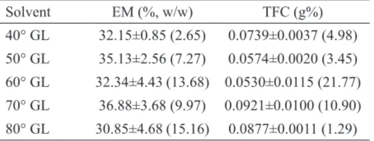
There were no statically differences among all the results of ethanol extractable matter, making this assay poorly discriminative (Table 1). However, the
extraction of flavonoids was higher at 70 or 80 °GL
ethanol. As no statistically differences were observed
between the results, the 70 °GL was chosen due to its
superior microbiological preservation properties.
Table 1. Ethanol extractable matter (EM) and total flavonoid content (TFC) of extractive solutions (1%, w/w and 5%, w/w, for EM and TFC assay, respectively). The results are expressed as mean±standard deviation (variation coefficient).
Solvent EM (%, w/w) TFC (g%)
40° GL 32.15±0.85 (2.65) 0.0739±0.0037 (4.98)
50° GL 35.13±2.56 (7.27) 0.0574±0.0020 (3.45)
60° GL 32.34±4.43 (13.68) 0.0530±0.0115 (21.77)
70° GL 36.88±3.68 (9.97) 0.0921±0.0100 (10.90)
80° GL 30.85±4.68 (15.16) 0.0877±0.0011 (1.29)
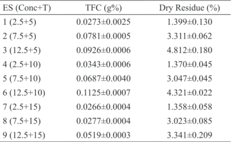
The factorial design was then applied, in order to find out the best drug/solvent proportion and extraction time for the maceration process. Dry residue was investigated for its importance in yield calculations in further studies with technological purposes, such as the drying process. Flavonoids were chosen as chemical markers in this study because of their well-known antinociceptive and anti-inflammatory properties (Muschietti & Martino, 2007). The main flavonoid from this plant, isoquercitrin, proved to be one of the active principles of this plant (Krogh et al., 1999).
or TFC) (Table 2) were adjusted by a second order mathematic model, according to the following equation: Y=β0+β1*Conc+β2*T+β12
Conc*T+β11*Conc2+β22*T2 , where y=studied
response, β0... β22 =coefficients, Conc=concentration
(%, w/v) of I. pes-caprae aerial parts, T=extraction time. The experimental data were adjusted by the quadratic model, resulting in the following equations: y = - 0 . 3 7 2 + 0 . 6 3 7 * C o n c + 0 . 0 7 8 * T- 0 . 0 1 4 * C o n c2
-0.014*Conc*T-0.0015*T2 for dry residue (r2=0.9908)
and y=-0.058+0.010*Conc+0.017*T-4*10-5*Conc2
-4.04*10-4*Conc*T-8.64*10-4*T2 for TFC (r2=0.9143),
where Conc=herb drug concentration (%, w/v) and T=extraction time (days).
Table 2. Total flavonoid content (TFC, g%) and dry residue for extractive solutions (ES) obtained according 32 factorial
design.
ES (Conc+T) TFC (g%) Dry Residue (%) 1 (2.5+5) 0.0273±0.0025 1.399±0.130 2 (7.5+5) 0.0781±0.0005 3.311±0.062 3 (12.5+5) 0.0926±0.0006 4.812±0.180 4 (2.5+10) 0.0343±0.0006 1.370±0.045 5 (7.5+10) 0.0687±0.0040 3.047±0.045 6 (12.5+10) 0.1125±0.0007 4.321±0.022 7 (2.5+15) 0.0266±0.0004 1.358±0.058 8 (7.5+15) 0.0277±0.0004 3.023±0.085 9 (12.5+15) 0.0519±0.0003 3.341±0.209 Conc: herb drug concentration (%; w/v); T: extraction time (day). The results are expressed as mean±standard deviation.
According to the statistical analysis for the dry residue, concentration (p=0.0004) had the highest significance compared to the other variables, like time (p=0.0347) and the interaction of these factors (0.0372) (Figure 1A). For the TFC, only the linear coefficient of concentration demonstrated significance (p=0.0204) as reflected by the surface plot (Figure 1B).
Both response surface plots indicated the best conditions for extraction in order to obtain higher levels of dry residue (Figure 1A) and flavonoid content (Figure 1B): 12.5% (w/v), due to their drug:solvent ratio and extraction time of about seven days.
Through the factorial experiment, it was possible to choice the most favorable extraction conditions, with a
view to achieving greater eficiency in terms of lavonoids and dry residue. Both the total lavonoid content and the dry residue were strongly inluenced by extraction time
and drug:solvent ratio, particularly the latter. According to the response surface plot, the dried, powdered plant matter
of 12.5% (w/v), placed in contact with 70 °GL ethanol over a period of ive to seven days, obtained higher yields
in terms of dry residue and total flavonoid content.
Figure 1. Response Surface plot for evaluating the inluence of
herb drug concentration and extraction time on the dry residue (A); and on the total lavonoid content (TFC) (B).
Chromatographic profile of optimized extractive solution
Another batch was also produced, for the sole
purpose of identifying a possible lavonoid marker. The
new batch was produced using a drug/solvent ratio of 12.5% (g/mL) and extraction time of seven days, taking into account the conditions set by the response surface plot (Figure 1B).
Figure 2 shows the chromatographic profile of ES with six peaks. Only two of them (1 and 2) have a characteristic UV absorbance profile of flavonoids. The second peak was identified as isoquercitrin, since it was eluted at the same retention time (about 17.66 min) and had the same UV absorbance profile (data not show). The isoquercitrin concentration was calculated from the linear regression equation y=48487x-3145.3 (r2=0.9999). In this batch, there was 71.3±7.7 µg/mL of
isoquercitrin, corresponding to 3.1 mg/g, considering its extractive solution dry residue weight of 2.3 g% (w/w).
Pharmacological evaluation
Antinociceptive activity by the Writhing test
The results (Figure 3) demonstrate that I. pes-caprae hydroethanolic extract, given by i.p. 30 min prior to testing, produced dose-related inhibition of acetic acid-induced abdominal constrictions in mice, with MI (Maximum inhibition) and calculated DI50 of 55.88±2.4% and 17.34 (12.23-24.30) mg/kg, respectively. Indomethacin, used in this experiment as positive control, also reduced the number of writhings induced by acetic acid, with inhibition of 64.86% at 10 mg/kg.
Figure 2. Chromatographic profile of Ipomoea pes-caprae
hydroethanolic extract solution (new batch obtained as established by the response surface plot) at 5 mg.mL-1 of
dried residue, monitored at 254 nm (2=isoquercitrin).
Figure 3. Effects of hydroethanolic extract (ES6, Table 2) of
I. pes-caprae on the number of acetic acid induced writhings in mice. Each column represents the average of 8 animals per group and the vertical bars represent the EPMs. The asterisks denote statistical significance (**p<0.01) when compared with the control group (vehicle). ANOVA, complemented by the Dunnnet’s test.
Our results show that the optimized hydroethanolic extractive solution was able to inhibit more than 50% of abdominal writhings at 20 mg/ kg, being more effective than the methanol extract evaluated earlier by Krogh et al. (1999) and Souza et al. (2000), which showed a DI50 of 33.8 mg/kg under the
Figure 4. Effects of hydroethanolic extract (ES6, Table 2) of I. pes-caprae (5-20 mg.kg-1, i.p.) upon inflammatory parameters
same experimental conditions. Moreover, the solvent used in our work presents advantages over other toxic or more polluting organic solvents, being more useful and suitable for the phytopharmaceutical industry.
With respect to the phytochemical constituents isolated from I. pes-caprae related to these activities, Krogh et al. (1999) showed that some triterpenes and isoquercitrin significantly inhibited acetic acid-induced abdominal constrictions in mice. The selected hydroethanolic extractive solution also showed significant inhibition, being more active than isoquercitrin itself and some triterpenes, suggesting the occurrence of synergism, and the fact that flavonoids such as isoquercitrin detected in the extract (Figure 2) are not the only substances responsible for the activity.
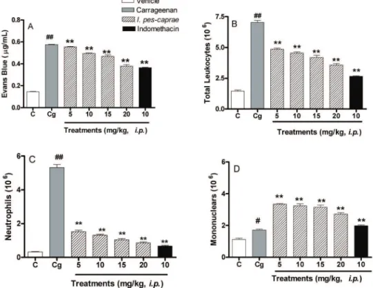
Carrageenan-induced pleurisy
In the present study, it was found that the extract of I. pes-caprae (5, 10, 15 and 20 mg/kg, i.p.) was able
to induce signiicant and dose-dependant inhibition of the irst phase of inlammation (4 h) of the pleural cavity
induced by injection of carrageenan (1%) in mice. It was observed that the extract of I. pes-caprae caused, to a lesser extent, a reduction in carrageenan-induced exudation (% inhibition: 50±0.82) (p<0.01) (Figure 4A). Moreover, the extract produced inhibition of cell migration (number of total leukocytes) to the pleural cavity (% inhibition: 60.88±0.14) (p<0.01) (Figure 4B), mainly represented by neutrophils (% inhibition: 88.90±0.12) (p<0.01) (Figure 4C). However, the extract produced an increase in migration of mononuclear cells (Figure 4D) to the
inlammatory site.
In carrageenan-induced pleurisy in mice, a model of inflammation, carrageenan promotes an increase in the volume of exudate in the pleural cavity, leukocyte migration of the polymorphonuclear type, and participation of some inflammatory mediators such as histamine, serotonin bradykinin, nitric oxide (NO),
TNFα and IL-1β, substance P (SP) and others (Saleh
et al. 1996, 1999; Endo, 2001; Banerjee et al. 2002; Brennan & Mclnnes, 2008).
Regarding the inlammatory process, there are
few reports on I. pes-caprae in the literature. It is known that the plant antagonizes the effects of histamine and also inhibits the release of prostaglandins (Pongrayoon et al., 1991b). Rogers et al. (2000) showed that the methanol extract of this plant potently inhibited ADP induced human platelet [14C]5-HT release in vitro, with levels of inhibition ranging from 62 to 95%.
The increase in migration of mononuclear cells to the inflammatory site is probably related to the immunostimulatory activity, and appears to be independent of the type of plant extract used. Philippi
et al. (2010) demonstrated that I. pes-caprae methanol extract has already been shown to induce human T lymphocyte proliferation.
The pharmacological results performed with I. pes-caprae hydroethanolic extract also suggest analgesic and anti-inflammatory effects similar to those previously demonstrated by methanol extract, ethyl acetate and aqueous fractions of I. pes-caprae (Krogh et al., 1999). Indeed, this work was able to validate the technological conditions of the maceration process to produce a bioactive plant extract for the development of phytopharmaceuticals. Further studies are necessary to elucidate the mechanism of action of this extract.
Acknowledgments
The authors wish to thank FAPESC, CNPq,
and PIPG/PROPPEC, UNIVALI, for their financial
support.
References
Assis C, Toledo CB, Neto SR, Cordeiro I 1994. Nossas Plantas (Mata Atlântica). São Paulo: FTS.
Banerjee T, Van der Vliet A, Ziboh VA 2002. Down regulation
of COX-2 and iNOS by amentolavone and quercetin
in A549 human lung adenocarcinoma cell line. Prostag Leukotr Ess 66: 485-492.
Brennan FM, McInnes IB 2008. Evidence that cytokines play a role in rheumatoid arthritis. J Clin Invest 118: 3537-3545.
Calixto JB, Campos MM 1995. Involvement of B1 or B2 receptors in bradykinin induces rat paw edema. Br J Pharmacol 114: 1005-1013.
Cochran WG, Cox GM. 1978. Experimental designs. John Wiley: New York.
Collier HO, Dinneen LC, Johnson CA, Schneider C 1968. The abdominal response and its suppression by analgesic drugs in the mouse. Br J Pharmacol Chemother 32: 295-310.
Cordeiro PJM, Yariwake JH, Lanças FM 1999. HRGC-MS
analysis of terpenoids from Maytenus aquifolium
Martius and Maytenus ilicifolia Martius (“espinheira santa”). J Brazil Chem Soc 10: 523-526.
Dalmarco EM, Fröde TS, Medeiros YS 2002. Effects of methotrexate upon inflammatory parameters induced by carrageenan in the mouse model of pleurisy. Mediat Inflamm 11: 299-306.
Endo Y 2001. Induction of histidine decarboxylase in inflammation and immune responses. Nihon Yakurigaku Zasshi 118: 5-14.
Farmacopéia Brasileira 2001. São Paulo: Atheneu.
Krogh R, Berti C, Madeira AO, Souza MM, Cechinel-Filho V, Monache, FD, Yunes RA 1999. Isolation and identification of compounds with antinociceptive action from Ipomoea pes-caprae (L). R. Br. Pharmazie 54: 464-466.
Muschietti LV, Martino VS 2007. Atividades biológicas dos flavonóides naturais. In Yunes RA, Cechinel Filho V (orgs) Química de produtos naturais, novos fármacos e a moderna farmacognosia. Itajaí: Univali, p. 185-207.
Montgomery DC 1991. Diseño y Análisis de experimentos. México: Iberoamérica.
Philippi ME, Duarte BM, Da Silva CV, de Souza MT, Niero R, Cechinel Filho V, Bueno EC 2010. Immunostimulatory activity of Calophyllum brasiliense, Ipomoea pes-caprae
and Matayba elaeagnoides demonstrated by human peripheral blood mononuclear cells proliferation. Acta Pol Pharm 67: 69-73.
Pongprayoon U, Bohlin L, Sandberg F, Wasuwat S 1989. Inhibitory effect of extract of Ipomoea pes-caprae on guinea-pig ileal smooth muscle. Acta Pharm Nordica 1: 41-44.
Pongprayoon U, Baeckstrom P, Jacobsson U, Lindstrom M, Bohlin L 1991a. Compounds inhibiting prostaglandin synthesis isolated from Ipomoea pes-caprae. Planta Med 57: 516-518.
Pongprayoon U, Bohlin L, Wasuwat S 1991b. Neutralization of toxic effects of different crude jellyfish venoms by an extract of Ipomoea pes-caprae (L). R. Br. J Ethnopharmacol 35: 65-69.
Pongprayoon U, Baeckstrom P, Jacobsson U, Lindstrom M, Bohlin L 1992. Antispasmodic activity of beta-damascenone and E-phytol isolated from Ipomoea pes-caprae.Planta Med 58: 19-21.
Rogers KL, Grice ID, Griffiths LR 2000. Inhibition of platelet
aggregation and 5-HT release by extracts of Australian plants used traditionally as headache treatments. Eur J Pharm Sci 9: 355-63.
Saleh TS, Calixto JB, Medeiros YS 1996. Anti-inflammatory effects of theophylline, cromolyn and salbutamol in a murine model of pleurisy. Br J Pharmacol 118: 811-819.
Saleh TSF, Calixto JB, Medeiros YS 1999. Effects of
anti-inlammatory drugs upon nitrate and myeloperoxidase
levels in the mouse pleurisy induced by carrageenan.
Peptides 20: 949-956.
Santos AR, Gadotti VM, Oliveira GL, Tibola D, Paszcuk AF,
Neto A, Spindola HM, De-Souza MM, Rodrigues AL, Calixto JB 2005. Mechanisms involved in the antinociception caused by agmatine in mice.
Neuropharmacol 48: 1021-34.
Souza MM, Madeira AO, Berti C, Krogh R, Yunes RA, Cechinel-Filho V 2000. Antinociceptive properties of the methanolic extract obtained from Ipomoea pes-caprae (L.) R. Br.. J Ethnopharmacol 69: 85-90.
Zhi-Chen L 1980. General Control Methods for Vegetable Drugs. Geneva:WHO.
Zimmermann M 1983. Ethical guidelines for investigations of experimental pain in conscious animals. Pain 16: 109-110.
*Correspondence
Angélica G. Couto
Programa de Pós-graduação em Ciências Farmacêuticas, Universidade do Vale do Itajaí
Rua Uruguai 458; 88302-202 Itajaí-SC, Brazil angelica@univali.br