INTRODUCTION
Corresponding author: Yunita Armiyanti. Department of Parasitology/ Faculty of Medicine/Jember University. Jember 68121, Indonesia.
Phone: 62 857 3248-2300 email: [email protected] Received 19 June 2015 Accepted 15 July 2015
Detection of immunogenic proteins
from
Anopheles sundaicus
salivary glands
Yunita Armiyanti
[1], Mohammad Mirza Nuryady
[2], Renam Putra Arifi anto
[2],
Elisa Nurmariana
[2], Kartika Senjarini
[2], Loeki Enggar Fitri
[3]and Teguh Wahju Sardjono
[3][1]. Department of Parasitology, Faculty of Medicine, Jember University, Jember, Indonesia . [2]. Department of Biology, Faculty of Mathematic and Natural Sciences, Jember University, Jember, Indonesia. [3]. Department of Parasitology, Faculty of Medicine, University of Brawijaya, Malang, Indonesia.
ABSTRACT
Introduction: The saliva of mosquitoes has an important role in the transmission of several diseases, including malaria, and contains substances with vasomodulating and immunomodulating effects to counteract the host physiological mechanisms and enhance pathogen transmission. As immunomodulatory components, salivary gland proteins can induce the generation of specifi c IgG antibodies in the host, which can be used as specifi c biomarkers of exposure to Anopheles sundaicus. The objective of this study was to identify immunogenic proteins from the salivary glands of Anopheles sundaicus by reaction with sera from individuals living in malaria-endemic areas who are thus exposed to Anopheles mosquitoes.
Methods: IgG antibodies targeting salivary gland proteins in serum samples from individuals living in malaria-endemic areas
were measured by enzyme-linked immunosorbent assay (ELISA). Sera from healthy individuals living in non-endemic areas were used as negative controls. Determination of the presence of salivary gland immunogenic proteins was carried out by western blotting. Results: Sixteen bands appeared in sodium dodecyl sulfate polyacrylamide gel electrophoresis, with molecule
weights ranging from 22 to 144kDa. Among the exposed individuals, IgG responses to salivary gland proteins were variable. Protein bands with molecular weights of 46, 41, 33, and 31kDa were the most immunogenic. These immunogenic proteins were consistently recognized by pooled serum and individual samples from people living in malaria-endemic areas but not by negative controls. Conclusions: These results support the potential use of immunogenic proteins from the salivary glands of Anopheles
as candidate markers of bite exposure or in malaria vaccines.
Keywords:Anopheles sundaicus. Salivary glands. Immunogenic proteins. Antibody.
Malaria is a mosquito-borne disease that has major health implications worldwide, with an estimated 207 million individuals affected by malaria and 627,000 malaria-related deaths reported. In 2013, 104 countries and territories, including Indonesia, had malaria, which was considered endemic; an estimated 3.4 billion people are at risk of contracting malaria(1). Malaria is caused by fi ve species of parasites from the genus Plasmodium and naturally spread from one person to another by female Anopheles mosquitoes. There are about 400 different species of Anopheles mosquitoes, but only 30 of these are vectors of major importance(1). Anopheles sundaicus is one
of the most important malaria vectors in Indonesia, particularly
in coastal area of Java and the Sumatra islands. The distribution of Anopheles sundaicus also includes countries in Southeast Asia and India(2) (3).
The important role of Anopheles mosquitoes as malaria vectors is supported by the presence of salivary glands in female Anopheles mosquitoes. Anopheles mosquito salivary glands secrete substances that can enhance the transmission of
Plasmodium(4). These substances inhibit hemostatic processes
and modulate the host immune response (immunosuppressive) at site of biting, allowing the mosquito to feed on blood successfully(5) (6). These changes at the site of the bite, owing to the effects of salivary substances, would benefi t the pathogen, permitting infection without any resistance and thus enhancing infectivity in the vertebrate host(7).
Injection of saliva into the host’s skin also induces the production of antibodies against salivary proteins(8). The
METHODS
mosquitoes can be measured by enzyme-linked immunosorbent assays (ELISAs) in children and adults exposed to mosquitoes bites(10) (11). Additional studies have shown that the IgG response can recognize specifi c salivary proteins(12) (13). Therefore, salivary proteins (e.g., gSG6) that are able to generate specifi c IgG antibodies have been developed as serological indicators or biomarkers of exposure to malaria vectors(14).
Immunoglobulin G antibodies against Anopheles salivary proteins in the host may be associated with protection against malaria. In malaria-endemic regions, populations exposed to uninfected Anopheles bites repeatedly over the years may develop an anti-saliva immune response by producing specifi c antibodies in the presence of interleukin (IL)-10. These specifi c antibodies will neutralize some salivary proteins of vectors, thus causing micro-environmental changes at the site of the mosquito bite and ultimately affecting malaria transmission. Therefore, in asymptomatic patients with malaria, the level of IgG antibodies targeting anti-salivary gland sonicates (SGSs) from Anopheles darlingi are higher than those in symptomatic patients and healthy individuals(15). Exposure to Anopheles
mosquito bites and Plasmodium infections, which persist for a long time, affect the development of the immune response to parasites and salivary vectors, thereby infl uencing the number of parasites and the host response(16). Thus, salivary components
could be effective vaccine candidates for reducing the morbidity of vector-borne diseases through combination with other malaria vaccine candidates to protect against severe malaria(7). Further
characterization of salivary proteins and the immune response is needed to identify salivary proteins involved in protection against malaria; the fi rst step in this process is determination of the immunogenicity of the salivary proteins. Many studies have been conducted to identify and characterize immunogenic salivary proteins from malaria vectors in Africa; however, malaria vectors in Asia, particularly An. sundaicus, have not been studied(8) (17) (18).
Therefore, in this study, we measured the levels of IgG antibodies targeting salivary proteins in serum samples from individuals living in malaria-endemic areas using salivary glands extracts (SGEs) from An. sundaicus. Based on the IgG antibody response to salivary proteins, we identifi ed the immunogenic proteins contained within An. sundaicus SGEs.
Anopheles mosquitoes and isolation of salivary glands
The adult female Anopheles sundaicus mosquitoes used in this study were collected from Bangsring village, Wongsorejo District, Banyuwangi Regency in East Java province using aspirators. In this area, Anopheles sundaicus is the dominant vector because its population is larger than that of other common species, such as Anopheles vagus, Anopheles subpictus,
Anopheles barbirostris, and Anopheles annularis(19). These
mosquitoes were maintained in the insectariums of the Zoology Laboratory of Biology Department, Faculty of Mathematic
glands were dissected using fi ne entomological needles under a stereoscopic microscope at 4× magnifi cation and collected into a microcentrifuge tube containing a small amount of phosphate-buffered saline [(PBS); pH 7.2] and phenylmethylsulfonyl fl uoride (PMSF) as protein inhibitors. The salivary glands were stored at -80°C until use.
Salivary gland extraction and protein quantifi cation One hundred salivary glands pairs in PBS and PMSF were thawed on ice and mixed in 1:1 lysis buffer containing 1.5mM MgCl2, 10mM Tris HCl, 10mM NaCl, 1% Nonidet P-40, and 2mM ethylenediaminetetraacetic acid (EDTA) NaOH(20). The mixture was homogenized using a micropestle and sonicated using a water sonicator for 30 min. After centrifugation (12,600rpm for 15 min at 4°C), the resulting supernatant was collected and concentrated using a spin concentrator (cut-off of 10kDa; Corning) and centrifugation (10,000rpm for 30s at 4°C). The protein concentrations of SGEs were determined using a nanophotometer (Nanophotometer Implen P 360, Germany). Salivary gland extracts were then diluted in 0.1M bicarbonate buffer (pH 9.6) to obtain a protein concentration of 1µg/µL for enzyme-linked immunosorbent assay (ELISA)(11).
Human serum samples
Twenty serum samples were collected from healthy adult residents living in the location at which we collected
An. sundaicus, i.e., Bangsring village, Wongsorejo District,
Banyuwangi Regency in East Java province, to detect antibodies against An. sundaicus salivary gland proteins. Seven serum samples from healthy adult residents living in non-malaria regions were used as negative control. The human subjects protocol for this study was approved by the Ethical Committee of Medical Research, Medical Faculty, Brawijaya University.
ELISA
To optimize the working conditions for ELISAs, checkerboard titration was carried out using An. sundaicus SGEs at 1, 2, and
4µg/mL and serially diluted serum samples (1:25, 1:50, and 1:100) from healthy individuals living in the malaria-endemic region. ELISA was performed as described by Fontaine et al.(11). Based on the results of checkerboard titration, microtiter
immunoplates (SPL Life Sciences, Korea) were coated with 4µg/ml (50µL/well) of An. sundaicus SGEs diluted in 0.1M
RESULTS
The optical density (OD) at 450nm was determined with a microplate reader (Bio-Rad, USA). A pool of fi ve serum samples from individuals living in Bangsring village, which all exhibited high levels of antibody responses against An. sundaicus SGEs
based on the ELISA optimization test, was used as a positive control. The negative control was individual serum samples from individuals who had never been exposed to An. sundaicus bites. The level of IgG antibodies was expressed as the adjusted OD (aOD), which was calculated for each serum sample duplicate as the mean OD value for wells with SGEs minus the OD value of the control wells, i.e., without SGEs.
Sodium dodecyl sulfate polyacrylamide gel electrophoresis
Sodium dodecyl sulfate polyacrylamide gel electrophoresis (SDS-PAGE) was performed according to the methods described by Jariyapan et al.(21). Briefl y, ten salivary gland pairs were mixed 1:2 in 1× SDS gel loading buffer and boiled in a water bath for 5 min. The mixtures were then loaded on 12% SDS polyacrylamide gels. To visualize the bands, gels were stained with Coomassie Brilliant Blue (CBB). A molecular weight marker (Nacalai) was loaded on each gel to identify the proteins in SGEs.
Western blotting of salivary gland proteins
Gels from SDS-PAGE were transferred to polyvinylidene difluoride (PVDF) membranes (MACHEREY-NAGEL, Germany) using semidry blotting (Bio-Rad) for 1h at 100mA. The membranes were blocked by incubation in 5% nonfat dry milk dissolved in (blocking buffer) for 1h at room temperature. After washing with -Tween 0.05% (T) three times, membranes were incubated overnight at 4°C with serum samples diluted 1:20 in blocking buffer. Subsequently, membranes were incubated with alkaline-phosphatase goat anti-human IgG secondary antibodies at a dilution of 1:2,000 for 2h at room temperature after three washes in TBST. Nitro blue tetrazolium-bromo-4-chloro-3-indolyl phosphate (NBT-BCIP) Phosphatase substrate was used for color development. To estimate the protein size, prestained broad-range molecular weight markers (9-200 kDa; Nacalai) were used.
A
B
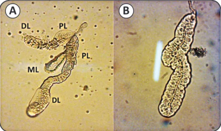
FIGURE 1 - Single salivary glands from adult Anopheles sundaicus mosquitoes. A) A female salivary gland. B) A male salivary gland. DL: distal region of lateral lobe; PL: proximal
region of the lateral lobe; ML: median lobe. (Nikon stereoscopic microscope at 4× magnifi cation).
Protein profi les of Anopheles sundaicus salivary glands
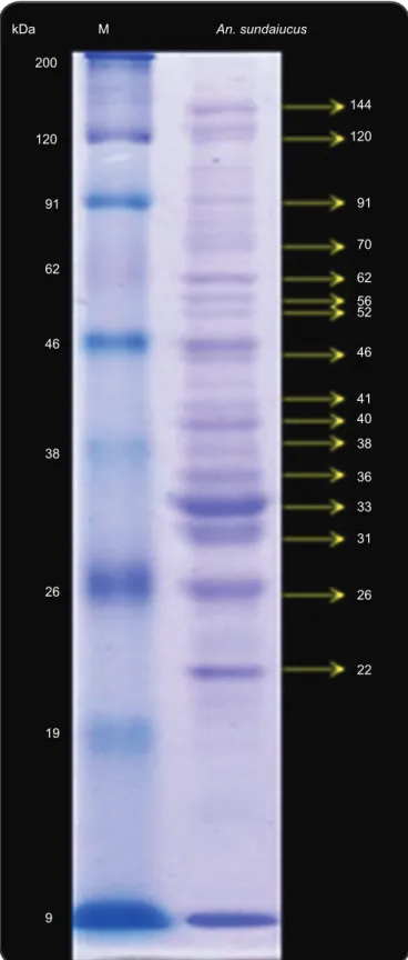
Salivary glands of female mosquitoes consist of three lobes: the two lateral lobes and the medial lobe, which is attached to the salivary duct (Figure 1). The lateral lobes are longer than medial lobe and are formed by the proximal, intermediate, and distal regions, whereas the median lobe is formed by a short neck region and distal region. Based on the results of SDS-PAGE, we found that the protein profi les of salivary glands from female
An. sundaicus consisted of 16 bands with molecular weights
ranging from 24 to 138kDa (Figure 2). Among these 16 bands, there were some major bands observed at estimated molecular weights of 144, 120, 91, 62, 46, 40, 36, 33, 31, 26, and 22kDa.
Levels of anti-salivary gland protein IgG antibodies
ELISA was used to detect and measure the levels of anti-salivary gland protein IgG antibodies. The levels of antibodies against salivary proteins in sera from healthy individuals living in Bangsring village were variable. These variations could be infl uenced by the intensity of exposure to mosquito bites and mosquito density(11). There were three serum samples with low
OD values (less than that of the negative control) among the 20 serum samples. Therefore, these three samples were not used for detection of immunogenic proteins by western blotting.
Immunogenic proteins found in Anopheles sundaicus salivary glands
The results of western blotting showed the presence of several immunogenic proteins with molecular masses of 56, 46, 41, 38, 36, 33, 31, and 26kDa (Figure 3). These results were obtained by using sera from individuals living in Bangsring village, and three repetitions were performed. Among the protein bands recognized by the anti-salivary protein antibody, the most immunogenic proteins had molecular masses of 46, 41, 33, and 31kDa (these protein bands were present in 10-12 serum samples). These immunogenics proteins were consistently recognized by individual responses from 17 serum samples and by pooled serum from individuals living in malaria-endemic area (Bangsring village) as a positive control (Figure 4A). The human antibody response to salivary proteins from female Anopheles mosquitoes is also specifi c to Anopheles mosquito bites(12) (17). Our result
kDa M An. sundaiucus
200
120
91
62
46
38
26
19
9
144
120
91
70
62
56 52
46
41
40
38
36
33
31
26
22
FIGURE 2 - Salivary gland proteins from female Anopheles sundaicus mosquitoes, separated using 12% SDS-PAGE (right lane) and stained with Coomassie Blue. Molecular weight markers are shown in the left lane. kDa: Kilodalton; M: Marker; An.: Anopheles; SDS-PAGE: sodium dodecyl sulfate polyacrylamide gel electrophoresis.
kDa M
200 120 91 62 46
38
26
19
9
46 41 33 31
FIGURE 3 - Western blotting using Anopheles sundaicus salivary gland proteins detected with human sera from individuals living in a malaria-endemic area. The most highly immunogenic proteins are shown by intense bands at 46, 41, 33, and 31kDa.
kDa: Kilodalton; M: Marker.
kDa M kDa M
200
120
91
62
46
38
26
19
9
46
41
31
91 200
120
62
46
38
26
19
9
A
B
The protein profiles of Anopheles sundaicus salivary glands have not been studied before; however, some studies have reported the protein profi les of other Anopheles species, i.e., Anopheles dirus, Anopheles gambiae, and Anopheles stephensi(12) (17) (18) (21). A previous study by Cornelie et al.(17) demonstrated the expression of 20 proteins from the salivary glands of An. gambiae, which were clearly detected in silver stained gels. Some of these proteins had the same molecular weights as proteins from salivary glands of
Anopheles sundaicus, i.e., 61-63, 52-54, 41-44, 38-39, 31-34, and 26kDa, similar to the major protein bands detected from the salivary glands of An. dirus(21). This result may be explained
by the presence of several proteins in the salivary glands of Anopheles that are conserved at the genus level(7). Some
proteins families are found in all Anopheles species; these are called genus-specifi c anopheline secreted proteins and include apyrase/5′ nucleotidase, antigen 5/gvag, GE-rich/30kDa, long and short form D7, mucin/13.5kDa, SG3, SG7, SG10, and hypothetical 6.2-kDa protein families(18). Potential proteins having molecular weights of 61-63kDa include apyrase/5′ nucleotidase, which is involved in the blood feeding process through degradation (hydrolysis) of adenosine diphosphate (ADP) and adenosine triphosphate (ATP) to adenosine monophosphate (AMP), a mediator of platelet aggregation, infl ammation, and neutrophil activation(5) (22 (23). Long form D7
protein families have molecular weights ranging from 33 to 34kDa, and GE-rich/anti-platelet family proteins have molecular weights around 30kDa; these proteins are also involved in the blood feeding process(22) (23).
The salivary gland proteins of An. sundaicus could be recognized by IgG antibodies in human sera from individuals living in Bangsring village, particularly proteins with molecular masses of 56, 46, 41, 38, 36, 33, 31, and 26kDa. Among these proteins, those with molecular masses of 46, 41, 33, and 31kDa were the most immunogenic. This result showed that exposure to mosquito bites in individuals living in malaria-endemic areas, could induce the immune response by producing salivary protein-specifi c IgG antibodies(10) (12). Indeed, the IgG antibody response against salivary gland proteins can be infl uenced by exposure to mosquito bites. In individuals with different levels of exposure to An. gambiae
bites, the levels of anti-salivary gland protein IgG antibodies are positively related to the intensity of exposure to Anopheles
mosquito bites. The IgG response increases signifi cantly with the increase in Anopheles exposure, as evaluated using conventional
entomological methods during the transmission season(10). The
results of a study conducted by Fontaine et al. also demonstrated a positive correlation between the average IgG response to SGEs of Aedes caspius and Aedes caspius density, which was affected by changes in the season and the ecological environment(18). The level of the IgG response increased signifi cantly during peak exposure to Ae. caspius in September and declined to baseline values within 4 months (January)(11). These results also showed that IgG
responses induced by mosquito saliva antigens persisted for only
a short time. Consistent with this, in a study in Senegal, children who experienced low and moderate exposure exhibited decreased antibody concentrations clearly after more than 3 months(10). Another study showed that the IgG antibody response to salivary gland proteins appearing after exposure to mosquito bites lasts 3-6 weeks(8). Although the duration of the IgG response to salivary gland
proteins may be brief (between 1 and 4 months), this response can detect specifi c proteins of the salivary gland, i.e., gSG6, in children exposed to very infrequent Anopheles bites. According to this study, immunogenic proteins from the salivary glands of Anopheles could be developed as immunoepidemiological markers to assess the risk to very infrequent Anopheles bites in the context of changes
in seasons, different environmental conditions, and travel(13).
The observed immunogenic proteins with molecular weights of 41 and 46kDa could be members of the SG1 family or TRIO proteins, which have molecular weights ranging from 40 to 48kDa(23) (24). TRIO is a multidomain protein that
binds the lymphocyte activating receptor transmembrane tyrosine phosphatase (PTPase) and contains a protein kinase domain; however, the function of this protein still needs to be determined(22). Our results also showed that proteins with
molecular weights of 41 and 46kDa were the most immunogenic because these proteins were recognized by anti-salivary gland protein antibodies from 12 serum samples. According to a previous study, TRIO protein is antigenic in four species
Anopheles, i.e., Anopheles gambiae, Anopheles arabiensis,
Anopheles stephensi, and Anopheles albimanus; the results of mass spectrometric analyses showed that this protein is conserved within the Cellia subgenus(18). TRIO protein is one of several antigenic proteins, in addition to gSG6, gSG1b, SG5, and long form D7, that are over expressed in salivary glands infected with Plasmodium falciparum. Therefore, TRIO proteins may be involved in malaria transmission and may represent a new candidate biomarker for infected Anopheles bites(25).
The observed immunogenic protein with a molecular weight of 33 kDa could be a long form D7 family protein; these proteins range in molecular weight from 33 to 36 kDa(23). D7 proteins
are also members of the odorant-binding protein superfamily (ODP) and are found in the salivary glands of blood-sucking insects, such as mosquitoes, sand flies, and Culicoides(26).
This protein has two types, i.e., the short form, which is only found in mosquitoes, and the long form, which can be found in mosquitoes and sand fl ies(23) (24). D7 proteins have functions
related to the binding of one or more agonists hemostasis and the blood feeding process(26). Some D7 proteins, i.e., D7r1,
D7r2, D7r3, D7r4, and long form D7, have been shown to bind with biogenic amines compounds, such as serotonin, histamine, and norepinephrine, thus becoming antagonistic to vasoconstrictors and affecting platelet aggregation and induction of pain(27).
According to a previous study(26), which analyzed the salivary
glands of An. darlingi by mass spectrometry, the 31-kDa protein identifi ed in our study could be a member of the 30-kDa allergen protein family or the GE-rich/anti-platelet family. Members of the 30-kDa allergen protein family from the salivary glands of
Ae. aegypti were shown to be associated with an allergic reaction to mosquito bites involving IgE and lymphocyte-mediated
hypersensitivity(28). This protein, also called aegyptin, has been
shown to inhibit platelet aggregation induced by collagen in humans and to impede granule secretion. Aegyptin recognizes the specifi c binding sites of glycoprotein IV, integrin α2β1, and von Willebrand factor and can the refore prevent the interaction between collagen and these major ligands(29). Anophelin
anti-platelet protein (AAPP) is a protein found in the salivary glands of An. stephensi that is homologous to the 30-kDa Aedes aegypti
allergen(23). This protein is also a member of the GE-rich family of
proteins. AAPP has been shown to block the adhesion of platelets to collagen via direct binding to collagen, subsequently inhibiting the increase in intracellular calcium ion concentration(30).
Therefore, this protein has an important role in blood feeding by inhibition of the hemostatic process.
This study is the first to show the protein profile and immunogenic proteins from the salivary glands of
An. sundaicus, one of the major malaria vectors in South Asian countries. Several studies have described anti-salivary protein antibody responses that recognize some proteins of Anopheles
salivary glands, particularly malaria vectors in Africa(17) (18).
However, few studies have investigated immunogenic proteins from salivary glands of Anopheles mosquitoes in Asia(12).
Therefore, this analysis herein provides important insights into immunogenic proteins from Anopheles mosquitoes in Asia.
Several immunogenic proteins from different Anopheles
species have been shown to have similar molecular weights, suggesting that these proteins could be conserved at the genus level(18). The same immunogenic proteins in different Anopheles mosquitoes, such as SG6, may trigger wide cross-reactivity between Anopheles species(31). This universal characteristic
of immunogenic proteins may improve their applicability as malaria vaccines in different regions. This fact also supports the use of this protein as an indicator of exposure to several
Anopheles mosquitoes in different areas. The results presented in this study represent the initial step for identifi cation and further characterization of immunogenic salivary proteins from
An. sundaicus, which is necessary to determine their role in malaria transmission and blood feeding processes.
In conclusion, the present study showed that there were four major immunogenic proteins, i.e., 46, 41, 33, and 31kDa, expressed in the salivary glands of An. sundaicus. These proteins exhibited a strong reaction in 10-12 out of 20 serum samples in western blotting results of human sera. The reaction was highly specifi c and generated anti-salivary gland protein IgG antibodies; thus, these proteins could be developed as new biomarkers of exposure to malaria vectors or new candidates for multivalent malaria vaccines containing different component of the malaria transmission cycle. Therefore, further studies are needed to determine the identities of these proteins and their biological functions in
REFERENCES ACKNOWLEDGMENTS
The authors would like to thank all the laboratory staff of the Biology Laboratory, Biology Department, Mathematic and Natural Sciences Faculty, Jember University. This study was funded by a Doctorate Research Grant of the Directorate General of Higher Education Indonesia.
The authors declare that there is no confl ict of interest. CONFLICT OF INTEREST
FINANCIAL SUPPORT
This study was funded by a Doctorate Research Grant of the Directorate General of Higher Education Indonesia.
1. World Health Organization (WHO). World Malaria Report 2013.
(Accessed 2013). Available at: http://www.who.int/en/
2. Dusfour I, Harbach RE, Manguin S. Bionomics and systematics of the oriental Anopheles sundaicus complex in relation to malaria transmission and vector control. Am J Trop Med Hyg 2004; 71:518-524.
3. Elyazar IR, Sinka ME, Gething PW, Tarmidzi SN, Surya A, Kusriastuti R, et al. The distribution and bionomic of Anopheles malaria vector mosquitoes in Indonesia. Adv Parasitol 2013; 83:173-266.
4. Billingsley PF, Snook LS, Johnston VJ. Malaria parasite growth is stimulated by mosquito probing. Biol Lett 2005; I:185-189. 5. Ribeiro JMC, Francischetti IMB. Role of Arthropod saliva in
blood feeding: Sialome and Post-sialome perspective. Annu Rev Entomol 2003; 48:73-88.
6. Titus RG, Bishop JV, Mejia ZS. The immunomodulatory factors of arthropod saliva and the potential for these factors to serve as vaccine targets to prevent pathogen transmission. Parasit Immunol 2006; 28:131-141.
7. Fontaine A, Diouf I, Bakkali N, Missé D, Pagès F, Fusai T, et al. Implication of haematophagous salivary proteins in host-vector interactions. Parasit Vectors 2011; 4:1-17.
8. King JG, Vernick KD, Hillyer JF. Members of the salivary gland surface protein family (SGS) are major immunogenic components of mosquito saliva. J Biol Chem 2011; 286:40824-40834.
9. Remoue F, Alix E, Cornelie S, Sokhna C, Cisse B, Doucoure S, et al. IgE and IgG4 antibody responses to Aedes saliva in African children. Acta Trop 2007; 104:108-115.
10. Remoue F, Cisse B, Ba F, Sokhna C, Herve JP, Boulanger D, et al. Evaluation of antibody respone to Anopheles salivary antigens as a potential marker of risk of malaria. Trans R Soc Trop Med Hyg 2006; 100:363-370.
12. Waitayakul A, Somsri S, Sattabongkot J, Looraesuwan S, Cui L, Udomsangpetch R. Natural human humoral response to salivary gland protein of Anopheles mosquitoes in Thailand. Acta Trop
2006; 98:66-73.
13. Poinsignon A, Cornelie S, Ba F, Boulanger D, Sow C, Rossignol M,
et al. Human IgG respon to a salivary peptide, gSG6-P1, as a new immuno-epidemiological tool for evaluating low-level exposure to
Anopheles bites. Malar J 2009; 8:198.
14. Stone W, Bousema T, Jones S, Gesase S, Hashim R, Gosling R,
et al. IgG responses to Anopheles gambiae salivary antigen gSG6
detect variation in exposure to malaria vectors and disease risk. PloS One 2012; 7:e40170.
15. Andrade BB, Rocha BC, Reis-Filho A, Camargo LMA, Barral A, Barral-Netto M. Anti-Anopheles darlingi saliva antibodies as marker of Plasmodium vivax infection and clinical immunity in the
Brazilian Amazon. Malar J 2009; 8:121.
16. Andrade BB, Barral-Netto M. Biomarkers for susceptibility to infection and disease severity in human malaria. Mem Inst
Oswaldo Cruz 2011; 106:70-78.
17. Cornelie S, Remoue F, Doucoure S, Ndiaye T, Sauvage FX, Boulanger D, et al. An insight into immunogenic salivary proteins
of Anopheles gambiae in African children. Malar J 2007; 6:75.
18. Fontaine A, Fusai T, Briolant S, Buffet S, Villard C, Baudelet E, et al. Anopheles salivary gland proteomes for major malaria
vectors. BMC Genomics 2012; 13:614.
19. Mardiana W, Suwaryono T. Aktifi tas Menggigit Anopheles
sundaicus di Kecamatan Wongsorejo, kabupaten Banyuwangi,
Jawa Timur. Media Litbang Kesehatan 2003; 13:26-30.
20. Lormeau VMC. Dengue viruses bindings protein from Ae. aegypti
and Ae. polynesiensis salivary glands. Virol J 2009; 6:1-4.
21. Jariyapan N, Choochote W, Jitpakdi A, Harnnoi T, Siriyasaten P,
Wilkinson MC, et al. Salivary gland proteins of the human malaria
vector, Anopheles dirus B (Diptera: Culicidae). Rev Inst Med Trop
Sao Paulo 2007; 49:5-10.
22. Francischetti IMB, Valenzuela JG, Pham VM, Garfi eld MK, Ribeiro JMC. Toward a catalog for the transcript and protein (sialome) from the salivary gland of the malaria vector Anopheles
gambiae. J Exp Biol 2002; 205:2429-2451.
23. Valenzuela JG, Francischetti, IMB, Pham VM, Garfi eld MK,
Ribeiro JMC. Exploring the salivary gland transcriptome and
proteome of the Anopheles stephensi mosquito. Insect Biochem
Mol Biol 2003; 33:717-732.
24. Arca B, Lombardo F, Valenzuela JG, Francischetti IMB, Marinotti O, Coluzzi M, et al. An update catalogue of salivary gland transcripts in the adult female mosquito, Anopheles gambiae.
J Exp Biol 2005; 208:3971-3986.
25. Marie A, Holzmuller P, Tchioffo MT, Rossignol M, Demettre E, Seveno M, et al. Anopheles gambiae salivary protein expression
modulated by wild Plasmodium falciparum infection: highlighting
of new antigenic peptides as candidates of An. gambaie bites.
Parasit Vectors 2014; 7599:1-13.
26. Calvo E, Pham VM, Marinotti O, Andersen JF, Ribeiro JMC. The salivary gland transcriptome of the neotropical malaria vector
Anopheles darlingi reveals accelerated evolution of genes relevant
to hemophagy. BMC Genomics 2009; 10:57.
27. Calvo E, Mans BJ, Andersen JF, Ribeiro JM. Function and evolution of a mosquito salivary protein family. J Biol Chem 2006;
281:1935-1942.
28. Simons FE, Peng Z. Mosquito allergy: recombinant mosquito salivary antigens for new diagnostic tests. Int Arch Allergy Immunol 2011;124:403-405.
29. Calvo E, Tokumasu F, Marinotti O, Villeval JC, Ribeiro JMC, Francischetti IMB. Aegyptin, a novel mosquito salivary gland
protein, specifi cally binds to collagen and prevents its interaction with platelet glycoprotein IV, integrin α2β1, and von Willebrand factor. J Biol Chem 2007; 282:26928-26938.
30. Yoshida S, Sudo T, Niimi M, Tao L, Sun B, Kambayashi J, et al. Inhibition of collagen-induced platelet aggregation by anopheline antiplatelet protein, a saliva protein from a malaria vector mosquito.
Blood Res 2008; 111:2007-2020.
31. Rizzo C, Ronca R, Fiorentino G, Mangano VD, Sirima SB, Nebie I, et al. Wide cross-reactivity between Anopheles gambiae and
Anopheles funestus SG6 salivary proteins support exploitation of
gSG6 as a marker of human exposure to major malaria vectors in