Bull Pan Am Health Organ 18(l), 1984
AN EPIDEMIOLOGIC
STUDY OF AMERICAN
CUTANEOUS
LEISHMANIASIS
IN MAIPUCO,
PERUi-*
Carmen Arzubiaga,s Jorge Huayanay,s
and Italo Biaggionis
Little is known about the reservoirs, vectors, transmission patterns, and dis- tribution of American leishmaniasis in the region of the Pemvian Amazon. The work reported here, which involved testing 67 villagem in an area where leish- maniasis cases had been occurring, was performed to help assess the extent of the leishmaninris problem in that region.
Introduction
Two forms of leishmaniasis occur in Peru. One, known as uta, occurs in the western valleys of the Andes and produces a cutaneous lesion of spontaneous remission that rarely causes contiguous mucosal damage. The other, called espundiu, occurs in the Amazon region and produces cutaneous lesions that in some cases are indistinguishable from those of utu. However, it can also produce destructive metastatic mucosal lesions, typically in the nasal septum or the oropharyngeal mucosa c-1,2).
Little is known about the epidemiology of the two forms, particularly in the Amazon region, or about the natural history, distri- bution, reservoirs, transmission mechanisms, and pathogenic agents of the disease in these areas. Furthermore, an indeterminate num- ber of cases go unreported every year in Peru, largely because of the sociocultural circum- stances of the affected populations. Never- theless, endemic areas are recognized within the country; and the focal behavior of the
‘Also appearing in Spanish in the Bofetin de la Ofiina Sanitaria Panamcricana 96(4), 1984.
2The work reported here was funded in part by the Department of Epidemiology of Peru’s Northeast Health Region, Loreto, Peru, and by the United Nations De- velopment Program/World Bank/World Health Orga- nization Special Program of Research and Training in Tropical Diseases, projects SWG 780235 and RSG 780488.
3Research Fellow, Instituto de Medicina Tropical “Alexander van Humboldt, Universidad Peruana Caye- tano Heredia, A. P. 5045, Lima, Peru.
disease-involving the reported presence of different clinical forms of leishmaniasis, pre- sumably owing to the existence of several Leishania strains-has created a need to elaborate upon the few studies performed in the affected regions (2,3,4,5).
For some time the value of the Montenegro intradermal test in assessing the prevalence of leishmania infections has been recognized (S), and significant progress has been made with regard to improving both the quality of the antigen and the methods of data analysis em- ployed (7,8). Nevertheless, some question has remained regarding the test’s specificity in ecological settings such as those found in Peru’s Amazon region.
Therefore, once an Amazon area that was producing leishmaniasis cases was identified, interest arose in defining the extent of the problem more clearly. The purpose of the work reported here was to determine the prev- alence of leishmaniasis infection by means of the Montenegro intradermal test, as a basis for determining the need for later studies. At the same time, the study was designed to evaluate both the intradermal test and the in- direct immunofluorescence test as field meth- ods applicable to the particular area and sit- uation involved.
Materials and Methods
The Study Population
The study was conducted in the village of Maipuco, a settlement located in Maipuco
20 PAHO BULLETIN + vol. 18, no. 1, 1984
District, Loreto Province, Loreto Depart- ment, Peru (Figure 1). The village, 240 km southwest of the city of Iquitos on the right bank of the MaraiGn River, is situated in a low jungle terrain subject to flooding. Its few inhabitants, numbering 380 in all, were dis- tributed by age and sex as shown in Table 1.
Figure 1. A map of Peru showing Iquitos, Lagunas, and the survey village of Maipuco.
‘:‘.:;:i, ..y::.:. 3
PACIFIC OCEAN
As indicated in the table, a survey sample con- sisting of 67 Maipuco residents was selected. Application of the Kolmogorov-Smirnov test indicated that this sample was representative of the village population at the 95 per cent confidence level (9).
The Intradermal and Indirect Immunojluorescence Tests
Montenegro’s intradermal test, exposing the test subject to leishmanin (6), was per- formed on all 67 members of the survey popu- lation. The leishmanin was prepared at the laboratory of the Alexander von Humboldt Institute of Tropical Medicine, using as an- tigen dead promastigotes of Leishmania brazi- liensis in a saline solution with 0.5 per cent phenol at a protein nitrogen concentration equivalent to 35 pg/mg. This solution (0.1 ml) was inoculated by the intradermal route into the upper inside portion of each subject’s left forearm. The subject’s response to this in- oculation was read at 48 hours by measuring the diameters of the induration at the inocu- lation site, a measurement of 5 mm or more being taken to indicate a positive response.
To obtain specimens for the indirect im- munofluorescence test, blood samples were obtained from the survey subjects and were centrifuged within six hours of their procure- ment in Maipuco. Sodium azide (0.01 per cent) was added to the resulting serum speci- mens as a preservative in Iquitos, and the
Table 1. The Maipuco population and survey sample, by age group and sex.
Age P‘OUP (in years)
M&s
Total population Survey population
Total Total
FCXW3leS NO. % Males Females NO. %
<l 25 22 47 (12.4) 3 1 4 (6.0)
l-4 43 37 80 (21.1) (11.9)
5-14 69 45 114 (30.0) 1: 1: 2: (35.8)
15-44 54 55 109 (28.7) 15 11 26 (38.8)
45-64 16 12 28 (7.4) 2 3 5 (7.5)
2 65 0 2 2 (0.5) 0 0 0 -
Arzubiaga et al. l LEISHMANIASIS IN PERU 21
specimens were transported under refrigera- tion to Lima. They were then maintained at -70% until tested for leishmania antibodies by the indirect immunofluorescence method within 15 days of their collection.
Promastigotes of the Yumare strain of Leishmania braziliensis isolated by C. F. Pifano in the Department of Yumare, Venezuela, were used as antigen for this test. The general guidelines of the method used by Guimaraes (9,IO) were followed, evaluating dilutions of 1:16, 1:32, 1:64, and 1:128. Serum samples known to contain leishmania antibodies served as positive controls, and a specimen processed only with diluent served as a negative control. Positive results at dilutions of 1:32, 1:64, and 1: 128 were to have been considered signifi- cant, as the method yields appropriately sen- sitive and specific results at these concentra- tions (10).
Results
A total of 18 subjects (26.8 per cent of the 67 tested) yielded positive responses to the Montenegro test. As indicated in Table 2, all were over 15 years of age, and nearly all (17 of the 18) were agricultural workers-including the “sanitarian,” who performed the duties of a primary health care worker. None of the 18 exhibited active leishmaniasis at the time the test was performed, but scars clinically com- patible with previous leishmaniasis were seen on four of the 18 subjects (22 per cent). No such scars were observed on any of the 49 Montenegro-negative subjects, and none of the 14 Montenegro-positive subjects without dermal scarring had any known history of the disease. The Montenegro-positive subjects in- cluded 12 males and six females, making the sex ratio two to one.
Table 2. Data on the 18 subjects responding positively to the Montenegro intradermal test-including each subject’s age, sex, occupation, place of infection (if known), presence or absence of cutaneous scarring
compatible with leishmaniasis, width of induration resulting from the Montenegro test, and serologic results of the indirect immunofluorescence test.
Subject Age
NO. (ye=) Sex Occupatlo”
Place of infection
Montenegro test Indirect Immunoiluores- Presence of response (width cence test responses at all
scarli”g of induration) serum dilutions testeda 1 2 10 11 12 13 14 15 16 17 18
39 M
51 F
36 M
39 M
15 M
18 M
37 F
28 F
36 M
53 M
40 F
49 M
19 M
42 M
38 F
15 M
16 M
18 F
Agricultural worker and
sanitarian Agricultural worker II I> ,> I, II Housewife Agricultural worker 0 1, I, I, I, 1, ,, 1, ,,
unknown No 6x5 mm Negative
Lagunasb Unknown Maipuco ,I ,, L agunasb Maipuco Unknown ,1 Maipuco Unknown Maipuco 0 I, I, NO
NO 16x20 mm
NO 5x5 mm
No 8x8 mm
Yes 6x6 mm
Yes 6x6 mm
Yes 5x7 mm
No 13x13 mm
Yes 14x14 mm
No 9x9 mm
NO 14x15 mm
No 6x5 mm
No 4x5 mm
No 6x6 mm
No 10x7 mm
No 5x5 mm
No 5x5 mm
10x11 mm 3,
I, 1, 0
Positive, 1: 16 only II ,, ,, Negative
1,
Positive, 1: 16 only Negative
,, Positive, 1: 16 only
Negative Positive, 1:16 only
9, ,1 1, Negative
,,
22 PAHO BULLETIN . vol. 18, no. 1, 1981
In the case of the four subjects with scars, all the scars were found on the lower extremi- ties. The initial lesions involved were said to have lasted anywhere from a month to a year, and the time elapsing between the appearance of the lesion and the survey examination was said to have ranged from one month to 17 years.
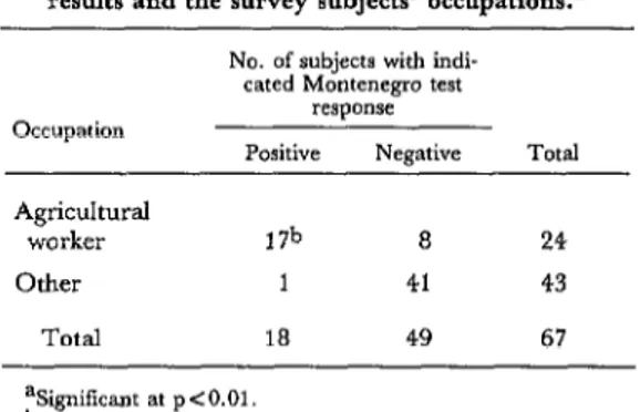
It is noteworthy that 58 per cent of the 31 survey subjects above 14 years of age re- sponded positively to the Montenegro test (Table 3), and that 17 of the 25 tested agri- cultural workers also responded positively. As Tables 4 through 6 indicate, these relation-
Table 5. Relationship between the Montenegro test results and the survey subjects’ occupations.a
No. of subjects with indi- cated Montenegro test
RSpO”X Occupation
Positive Negative Total Agricultural
worker 17b 8 24
Other 1 41 43
Total 18 49 67
%igniiicant at p<O.Ol.
bI”cluding the one agricultural worker also employed as a sanitarian.
Table 3. Responses to the Montenegro test of all the survey subjects categorized by sex, age group, and occupation.
Age group (in years)
Montenegro tea response:
Positive Negative
Agricultural Occupied Other Agricultural Occupied Other workers at home occupation workers at home occupation
Ma Fa M F M F M F M F M F Total
<l - _ - - - 3 1 - - 4
l-4 _ _ - - - 4 4 - - 8
5-14 - - - 2 - 12 10 - - 24
15-44 10 -b 4 - 1 - - 5 1 - 5 - - 26
45-64 *1---2--s
2 65 - - - 0
Total 12 5 0 1 0 0 7 1 19 22 0 0 67
aM - males, F - females.
bone of these 10 also worked as a sanitarian.
Table 4. Relationship between the Montenegro test results and the survey subjects’ age.a
Age (in years)
No. of Subjects with indi- cated Montenegro test
response
Positive Negative Total
Cl5 0 36 36
215 18 13 31
TOtal 18 49 67
Table 6. Relationship between the Montenegro test results and the presence of scarring compatible with
cutaneous leishmaniasis.a No. of subjects with indi-
cated Montenegm test response
Positive Negative Total Subjects with
scarring Subjects with-
out scarring Total
4 0 4
14 49 63
18 49 67
Ar..ubia,ga et al. l LEISHMANIASIS IN PERU 23
ships as well as the relationship between the presence of leishmaniasis-compatible scarring and the Montenegro test results were found to be statistically significant. No statistical signif- icance was found between the Montenegro and indirect immunofluorescence test results using the contingency coefficient4 (12), the results of the latter test being generally nega- tive.
Discussion
The value of the intradermal test for de- termining exposure to leishmaniasis is well established. Many years ago, Wagner dem- onstrated the existence of delayed hypersen- sitivity to leishmaniasis among guinea pigs previously inoculated with inactive forms of the infective agent (6). However, Montenegro was the first to use this cutaneous test for the diagnosis of human leishmaniasis, which is why the test bears his name (6). Later, in 1940, Pessoa and Pestana applied the intra- dermal reaction for clinical epidemiologic purposes, and many subsequent observations have since verified the test’s great sensitivity and worth (13).
It is also generally recognized that the intra- dermal reaction remains positive for years, even for life (14), following clinical remission of the disease. The test thus permits retro- spective diagnosis of leishmania infections and hence a fairly precise determination of the prevalence of the disease.
The leishmanin used in the present survey has been shown by work not yet published to be highly sensitive and specific, demonstrating the latter quality by inducing only one false positive response in a survey population of 300 apparently healthy schoolchildren (15).
In general, the findings obtained (a 27 per cent positive response in village residents with few scars or dermal lesions characteristic of leishmaniasis) are comparable to those re- ported by other studies of American leish-
4Employed to estimate the degree of association be- tween two nonparametric variables.
maniasis (13,16) and are similar to those ob- tained by Herrer in a highland jungle area (Tambopata, Puno, Peru) where it was found that 26 per cent of 1,613 residents studied were Montenegro-positive, of whom 21 per cent had no history of dermal lesions (4).
As various authors have suggested, the presence of reactors without any history or evidence of lesions may be explained by the occurrence of subclinical infections (4,14,17). In other countries, cases of positive intra- dermal reactions have been described that re- sulted from exposure to animal leishmanins (I&19), but such reactions have not been demonstrated in our area. Cross-reactions with Chagas’ disease (Trypanosoma cruzi) anti- bodies have also been described (20); how- ever, this matter is somewhat controversial, and in any event no cases of Chagas’ disease have been reported in the Maipuco area.
The fact that positive responses occurred only in people over 14 years old, most of whom were males and farmers, indicates that most exposure to the insect vector occurred far from the village-in the course of farming work or while hunting in river gorges. The absence of positive reactors among those under 14 suggests that transmission of the disease within the household is improbable, and that the leishmaniasis vector and/or the natural hosts of the disease are not present in the village proper. (There is no proof that peo- ple, apart from serving as accidental hosts, can become reservoirs of the disease agent.)
24 PAHO BULLETIN l vol. 18, no. 1, 1984
tor in that region (21). In discussing this vec- tor, Herrer describes it as having habits that keep it outside of settled areas, a behavior pattern consistent with what was found in our study.
Regarding the immunofluorescence test, Oddo and Cascio demonstrated in 1963 that sera from patients with Mediterranean vis- ceral leishmaniasis could be shown to react with cultures of Leishmania donovani by the in- direct immunofluorescence method (6). The value of this technique for diagnosing kala- azar (the disease caused by L. donovani) was demonstrated a year later by Duxbury, who also reported a cross-reaction with sera from patients with American leishmaniasis (22, 23). Finally, Camargo (24) showed that when such sera were treated with a heterologous antigen, then the cross-reactions could be eliminated, and sera monospecific against L. braziliensis (principal agent of American leishmaniasis) could be obtained.
Experience using the indirect immuno-
fluorescence test, especially in Brazil, has shown it to be particularly useful in diagnos- ing and monitoring patients with active American leishmaniasis, with the test results being positive in over 90 per cent of the cases (6). The antibody titers obtained are not cor- related with the degree of parasitism in the le- sions or with the intensity of the Montenegro reaction (2.5,26,27), although some authors have found sera from patients with delayed forms of the disease and mucosal lesions to yield higher antibody titers than sera from pa- tients with recent cutaneous lesions (27).
It has also been reported that the antibody titers obtained tend to decrease or disappear after treatment (25’, so that unlike the Mon- tenegro test the indirect immunofluorescence test is negative during the inactive (cicatricial) phase of the disease. These findings are con- sistent with those of the present study, in which no active disease cases were observed and the indirect immunofluorescence test de- tected no clearly significant antibody titers.
ACKNOWLEDGMENTS
The authors wish to thank Drs. Hugo Lumbreras and Humberto Guerra for reviewing the manuscript and offering suggestions, and Dr. Jorge Sibina, Director of Peru’s Northeast Health Region, and Dr. Eduardo Garcia, Head of the Department of Epidemiology, for their assistance.
SUMMARY
The study reported here tested residents at a Peruvian Amazon village in an area where Ameri- can leishmaniasis cases were occurring. To help assess the extent of the problem, as well as the specificity of the Montenegro intradermal test in the Peruvian Amazon, 67 residents at the lowland jungle village of Maypuco in Loreto Department
were selected. Each member of this representative population sample was given the Montenegro test, and each provided a blood specimen for later indi- rect immunofluorescence testing.
The immunofluorescence test results were neg- ative, but 18 of the 67 subjects (26.8 per cent) re- sponded positively to the Montenegro test. Four of the 18 positive subjects exhibited scars compatible with prior leishmaniasis. None of the other subjects
were found to have such scars, nor were any sub- jects found to have active leishmaniasis lesions.
Because all 18 Montenegro-positive subjects were above 14 years of age and most were men engaged in farmwork, it would appear that most ex- posure to the insect vector occurred outside the village proper, and that transmission of the disease
within the home was unlikely.
Arzubia,ga et al. l LEISHMANIASIS IN PERU 25
REFERENCES (1) Herrer, A., V. Hidalgo, and 0. Meneses. Leishmaniasis tegumentaria e insecticidas en el Peti: Reactivation de la uta durante 10s Gltimos aiios. Rev Inst Med Trap Sio Paul0 22~203-206, 1980. (2) Lainson, R., and J. J. Shaw. Leishmanias and Leishmaniasis of the New World, with partic- ular reference to Brazil. Bull Pan Am Health Organ 7:1-19, 1973.
(3) Tejada, A. Leishmaniasis tegumentaria en el Peri: Investigation epidemiologica-clinica de la leishmaniasis tegumentaria en 10s departamentos de Cuzco y Madre de Dios. Doctoral thesis, Uni- versidad National Mayor de San Marcos, Lima, Peru, 1973.
(4) Herrer, A. Report on the ecologic and epi- demiologic investigations conducted into American leishmaniasis in Upper Tambopata, Puno Depart- ment, in 1976. Personal communication.
(5) Mori, R. Leishmaniasis mucocutanea, Lo- reto, Peru: La exploration petrolifera y su proble- mitica en relation a la leishmaniasis tegumentaria americana. Tesis de Bachiller 3661, Universidad Peruana Cayetano Heredia, Lima, Peru, 1977.
(6) Furtado, T. Criterios o diagnostic0 da leish- maniose tegumentar americana. Annais Brasileiros de Dermetologie 55:81-86, 1980.
(7) Mayrink, W., M. N. Melo, C. A. da Costa, P. A. Magalhzes, M. Dias, M. V. Coelho, F. G. Araujo, P. Williams, and B. Figueiredo. Intrader- morreacIo de Montenegro na leishmaniose tegu- mentar americana ap6s terap&utica antimonial. Rev Inst Med Trap Go Paul0 18: 182-185, 1976.
(8) Restrepo Isaza, M. La reaction de Monte- negro en la epidemiologfa de la leishmaniasis sud- americana. Bol Of Sanit Panam 89:130-138, 1980.
(9) Armitage, P. Statistical Methodr in Medical Research. Blackwell Scientific Publications, Oxford,
1973.
(10) Guimarges, M. C. S., V. L. Giovannini, and M. E. Camargo. Antigenic standardization for mucocutaneous leishmaniasis immunofluorescence test. Rev Inst Med Trap Szo Paul0 16:145-148, 1974.
(11) Cantella, R., A. Colichon, L. Lopez, and C. La Terre. Curso de introduccidn a las tknicas de im- munojluorescencia. Departamento de Microbiologfa, Universidad Peruana Cayetano Heredia, Lima, 1973.
(1.2) Ostle, B., and R. Mensing. Statistics in Re- search (third edition). Iowa State University Press, Ames, Iowa, 1975.
(13) Pifano, F. La evaluation de la leishmaniasis tegumentaria americana en el Valle de Aroa, Esta- do de Yaracuy, mediante el indice al&gico (intra- dermoreaccion con antigen0 de L. braziliemis). Ar- chives Venezolanos de Medicina Tropical y Parasitologia Medica 4:25-35, 1962.
(14) Zuckerman, A. Current status of the im-
munology of blood and tissue Protozoa: 1. Leish- mania. Exp Parasitol38:370-400, 1975.
(15) Benavente, L. Andean cutaneous leish- maniasis: Epidemiologic and cultural aspects of an outbreak in the locality of Surco, Peru, in Septem- ber 1981. Personal communication.
(16) Melo, M. N., W. Mayrink, C. A. da Cos- ta, P. A. MagalhHes, M. Dias, P. Williams, F. G. Araujo, M. V. Coelho, and S. M. Batista. Padro- nizac5.o do antigen0 de Montenegro. Rev Inst Med Trop Sao Paul0 19: 161-164, 1977.
(17) Pessoa, S. B., and J. A. S. Lopez. SBbre a intradermorreac5,o de Montenegro em regiso end& mica de leishmaniose tegumentar e visceral. Rev Inst Med Trap Sio Paul0 5:170-175, 1963.
(18) Southgate, B. A. Studies in the epidemiolo- gy of East African leishmaniasis: 5. Leishmania adleri and natural immunity. J Trop Med Hyg 70:33-36, 1967.
(19) Imperato, P. J., 0. Sow, and B. Fofana. Positive leishmanin skin sensitivity in the absence of clinical leishmaniasis. J Trap Med Hyg 76:132- 134, 1973.
(20) Cohen, S. Immunology of Parasitic Infections.
Blackwell Scientific Publications, Oxford, 1976. (21) Herrer, A. Lutzomyia peruensis Shannon, 1929: Posible vector natural de la uta (leishmaniasis tegumentaria). Rev Inst Med Trap Scio Paul0 24: 168- 172, 1982.
(22) Duxbury, R. E., and E. H. Sadun. Fluo- rescent antibody test for the serodiagnosis of vis- ceral leishmaniasis. Am J Trop Med Hyg 13:525-529, 1964.
(23) Bray, R. S., and R. Lainson. The im- munology and serology of leishmaniasis: The fluo- rescent antibody staining technique. Trans R Sot Trap Med Hyg 59:535-544, 1965.
(24) Camargo, M. E., and C. Rebonato. Cross- reactivity in fluorescence tests for Trypanosoma and Leishmania antibodies. Am J Trop Med Hyg 18: 500-505, 1969.
(25) Chiari, C. de A., W. Mayrink, and P. Ma- galhzes. ReacBo de imunofluoresc&ncia indireta no controle de tratamento da leishmaniose tegumentar americana. Rev Inst Med Trap &io Paul0 15:298-303, 1973.
(26) Bittencourt, A. C., A. Sod&, and Z. A. Andrade. Pesquisa de anticorpos circulantes pelo mttodo de imunofluoresctncia na leishmaniose te- gumentar. Rev Inst Med Trop Szo Paul0 10:247-252,
1968.