1 Interunit Graduate Program in Bioengineering, São Carlos School of Engineering (EESC/FMRP/IQSC), Universidade de São Paulo (USP),
São Carlos, SP, Brazil
2 Cardiopulmonary Physical Therapy Laboratory, Exercise Research Center, Physical Therapy Department, Universidade Federal de São Carlos
(UFSCar), São Carlos, SP, Brazil
Received: 11/11/2012 Revised: 05/20/2013 Accepted: 07/29/2013
a r t i c l e
Cardiac autonomic responses during upper versus lower
limb resistance exercise in healthy elderly men
Heloisa G. Machado-Vidotti1, Renata G. Mendes2, Rodrigo P. Simões2,
Viviane Castello-Simões2, Aparecida M. Catai2, Audrey Borghi-Silva1,2
ABSTRACT | Objective: To investigate the cardiac autonomic responses during upper versus lower limb discontinuous resistance exercise (RE) at different loads in healthy older men. Method: Ten volunteers (65±1.2 years) underwent the one-repetition maximum (1RM) test to determine the maximum load for the bench press and the leg press. Discontinuous RE was initiated at a load of 10%1RM with subsequent increases of 10% until 30%1RM, followed by increases of 5%1RM until exhaustion. Heart rate (HR) and R-R interval were recorded at rest and for 4 minutes at each load applied. Heart rate variability (HRV) was analyzed in 5-min segments at rest and at each load in the most stable 2-min signal. Results: Parasympathetic indices decreased signiicantly in both exercises from 30%1RM compared to rest (rMSSD: 20±2 to 11±3 and 29±5 to 12±2 ms; SD1: 15±2 to 8±1 and 23±4 to 7±1 ms, for upper and lower limb exercise respectively) and HR increased (69±4 to 90±4 bpm for upper and 66±2 to 89±1 bpm for lower). RMSM increased for upper limb exercise, but decreased for lower limb exercise (28±3 to 45±9 and 34±5 to 14±3 ms, respectively). In the frequency domain, the sympathetic (LF) and sympathovagal balance (LF/HF) indices were higher and the parasympathetic index (HF) was lower for upper limb exercise than for lower limb exercise from 35% of 1RM. Conclusions: Cardiac autonomic change occurred from 30% of 1RM regardless of RE limb. However, there was more pronounced sympathetic increase and vagal decrease for upper limb exercise than for lower limb exercise. These results provide a basis for more effective prescription of RE to promote health in this population.
Keywords: physical therapy; resistance exercise; autonomic nervous system; elderly; upper limbs; lower limbs.
HOW TO CITE THIS ARTICLE
Machado-Vidotti HG, Mendes RG, Simões RP, Castello-Simões V, Catai AM, Borghi-Silva A. Cardiac autonomic responses during upper versus lower limb resistance exercise in healthy elderly men. Braz J Phys Ther. 2014 Jan-Feb; 18(1):9-18. http:// dx.doi.org/10.1590/S1413-35552012005000140
Introduction
Resistance exercise (RE) training has become an important component of physical exercise programs for older people1. It is well established that RE
training combined with endurance training is the
most effective way to achieve the expected beneicial
results of the physical training program1,2.
Several studies have shown that strength (resistance)
training can counteract age-related impairments3,4.
Aging is associated with decreases in muscle mass,
strength5,6, and cardiovascular fitness and with
increased cardiovascular risk factors. Some age-related
chronic diseases include cardiovascular disease, type 2 diabetes and obesity7. Impaired heart rate variability (HRV) with increased sympathetic modulation and decreased parasympathetic modulation is also related to aging8,9.
Considering all age-related physiological changes, an RE program may be able to enhance muscle strength, power, and local muscular endurance10,11,
increase HRV and, consequently, decrease the risk of mortality8,12 and co-morbidities associated with advancing age9,13. Therefore, RE is a valuable
tool for physical therapists and understanding the cardiovascular adjustments involved in it can provide the theoretical basis for better prescription of these exercises to effectively promote health in this population.
Cardiovascular responses to RE depend on exercise intensity and muscle mass1. In this context, upper
limb RE generates different cardiovascular responses
compared to lower limb exercise. It is well-established
that upper limb exercise leads to higher blood pressure
to a higher work component and elevated peripheral resistance caused by reduced active muscle mass14-16.
In addition, upper limb exercise also induces greater
perceived exertion compared to leg exercise at the same relative workload17.
Regarding autonomic cardiac responses, Tulppo et al.18 found a more rapid vagal withdrawal in arm cycle ergometer exercise than leg cycle ergometer exercise in young healthy men. This condition leads to higher HR and hemodynamic changes in arm exercise and it may be explained by a more powerful central command by vagal withdrawal and a stronger muscle afferent input in upper limb exercise.
However, the autonomic responses of upper versus lower limbs in RE are still unclear. Consequently, the aim of this study was to investigate the cardiac autonomic responses during upper versus lower limb discontinuous RE at different loads in healthy older men. The hypothesis was that the cardiac responses would be different.
Method
Subjects
Ten healthy elderly men were included in this study after clinical evaluation. The inclusion criteria
were: age between 60 and 80 years; no participation
in exercise training programs such as resistance or aerobic training in the previous 6 months; no obesity; and apparent good health. The exclusion criteria were: current smoking habit, current use of
any medication, musculoskeletal pain or dificulty in
understanding the RE protocol.
Screening of subjects was performed with clinical
evaluation, physical evaluation, and laboratory tests, including analysis of hemoglobin, triglycerides, total cholesterol, low-density lipoprotein (LDL) and high-density lipoprotein (HDL), fasting glucose, and uric acid. For clinical safety, a physician-supervised exercise test with gas analysis (CPX-D, Medical
Graphics, St. Paul, MN, USA) was performed for all
volunteers before participation in our experimental protocol. The volunteers were considered able to perform the protocol when the cardiopulmonary, metabolic, and electrocardiographic variables showed normal physiological behavior during testing.
This study was approved by the Human Ethics
Committee of Universidade Federal de São Carlos (196/2006), São Carlos, SP, Brazil, and followed the principles of the declaration of Helsinki. All subjects
gave written informed consent for participation.
Procedures
After clinical evaluation, all volunteers underwent
a physical and anthropometric evaluation. Height (m) and weight (kg) were measured and body
mass index (BMI) (kg/m2) was calculated using
a scale and stadiometer (Welmy, São Paulo, SP, Brazil). The subsequent evaluations (maximum load
determination and incremental RE) were performed with a one-week interval. The tests (upper or lower
limb) were performed in a random order deined by numbered envelopes. All tests were performed within
a three-week period.
Experiments were conducted at approximately the same time of day (morning) to minimize subject-to-subject variation due to circadian rhythms at a controlled room temperature of 22-24 °C and relative air humidity of 50-60%. Each volunteer was instructed to wear comfortable clothes for the test; to avoid alcoholic and caffeinated beverages 24 hours before the test; to avoid heavy meals at least 2 hours before the test; and to abstain from exhaustive training on the day prior to the test.
Immediately prior to data collection, subjects conirmed a normal night’s sleep and HR and blood
pressure were checked to ensure they were within the normal range. The subjects were instructed to avoid unnecessary talking during the protocol in order to minimize interference in the HR and eletrocardiogram (ECG) signal and to take note of symptoms before, during, and after the protocols.
One-repetition maximum (1RM) test – 45° Incline Bench Press and Leg Press
To determine the protocol loads for RE, the subjects were tested for maximal strength performing a 1RM test for upper limb using the incline bench
press set at 45° (Vitally Convergent, São Paulo, Brazil) and for lower limbs using the leg press at 45° (Pro-Fitness, São Paulo, Brazil) with a one-week
interval.
During the 1RM test on the incline bench press, the volunteers remained seated with trunk inclined at 45° from horizontal and feet on the ground. They were instructed to hold the free bar which supported the load. During exercise, the elbow and shoulder
were lexed and then returned to extension as the
initial position, avoiding isometric contraction and the Valsalva maneuver19. Based on a previous study20,
the 1RM load was estimated (1RM-E) by multiplying the body weight by 0.6.
For the 1RM test on the leg press at 45°, the volunteer remained seated with trunk inclined at
45° from horizontal and knees and hips lexed at
extended and returned to the initial position after
the lexion position avoiding isometric contraction
and the Valsalva maneuver19. Based on a previous
study21, the 1RM-E was estimated by multiplying
the body weight by 4.
For both exercises, 80% was applied to the 1RM-E value to start the test. If the estimated load was not the
maximum, an increase of 10% was achieved after 5 minutes or the time required for vital signs to return
to baseline levels. It was expected that 1RM would
be determined in up to six attempts22.
Discontinuous incremental resistance exercise protocol
One week after the 1RM test, a discontinuous incremental RE protocol was performed with the same equipment and movements described for the maximum test. The volunteer remained at rest seated
on the equipment for 10 min. After this, the protocol
began at a load of 10%1RM and subsequent increases
of 10% until 30%1RM. After that, the incremental
adjustments were 5%1RM until exhaustion or
SBP>200 mmHg, HR≥85% of maximum (HR (220 - age × 0.85)) and the development of any
potential arrhythmias. The volunteers performed 4 min of exercise at a cadence of 12 repetitions per minute in each load combined with respiratory rhythm of 2 seconds of inspiration and 3 of expiration. Respiratory standardization was determined to reduce
the inluence of the respiratory rhythm on autonomic modulation, more speciically on high frequency bands. A recovery period of approximately 15 min was observed in order to return to base HR and BP
values. The incremental tests (supine and leg) were also separated by a one-week interval.
Safety and ECG monitoring during protocols
During evaluation, HR in real time was veriied and
recorded by an HR monitor (Polar Vantage, Kempele,
Finland) linked to an interface (Polar Advantage, Kempele, Finland) and a microcomputer (Notebook Soyo - PW 9800, Taiwan, China). In addition, an ECG monitor (Ecaix TC 500, São Paulo, Brazil)
was used to detect any potential arrhythmias or
ischemia. Blood pressure was veriied by auscultation (sphygmomanometer BD, São Paulo, Brazil) before
and after each test and pain and muscle fatigue were
evaluated using the Borg scale at end of each stage23.
Heart rate and R-R interval data acquisition, signal processing and HRV analysis
HR and R-R interval (R-Ri) were recorded using an HR monitor (Polar Electro™, Kempele, Finland).
Measurements were subsequently obtained at rest before starting the exercise on the respective bench press or leg press at 45° for 10 min and during incremental RE exercise protocol for 4 minutes.
After acquisition, the signals were transferred to the Polar Precision Performance Software and
visually inspected and corrected for ectopic beats (premature, supraventricular, and ventricular). Times series data were processed using the Kubios
HRV Analysis software (MATLAB, version 2 beta,
Kuopio, Finland).
For HRV analysis, we used the HR and R-Ri signals acquired in the pre-exercise rest period and during exercise. The segments selected were 5 minutes of rest and visually the most stable exercise section containing 2 min free of noise at each load effort, excluding the initial 40 seconds of the exercise (period during which a rapid withdrawal of vagal
activity occurred) and the inal 30 seconds to avoid inluences of blood pressure measurement.
The HRV indices obtained were rMSSD, RMSM, SD1, LF, and HF. The rMSSD consists of the square
root of the mean of the sum of the squares of the differences between the R-Ri registered, divided by
the number of R-Ri in a speciied time period minus
one, which provided information on parasympathetic cardiac modulation24. The RMSM index corresponds
to the square root of the sum of the squares of the differences of the individual values in relation to the mean value, divided by the number of R-Ri in a
speciied time period, characterized as a marker of
the total HRV25.
Quantitative Poincaré plot analysis consisted of plotting each R-Ri as a function of the preceding interval and in this type of analysis the stationary
signal is not required. The SD1 index was obtained
in milliseconds (ms), which provides information on the standard deviation of instantaneous beat-to-beat variability, characterized as a parasympathetic cardiac modulation marker26.
Frequency-domain analysis of HRV was performed using Fast Fourier Transformation27. Two spectral components were obtained: low frequency (LF), from 0.04 to 0.15 Hz, and high frequency (HF), from 0.15 to 0.4 Hz. The spectral components were expressed in normalized units (nu) and also in absolute units (ms2) to HF. Normalization is obtained by dividing
Statistical analysis
The sample size was calculated using the
GraphPad StatMate software, version 1.01. To reach statistical signiicance (p<0.05) at a power of 80%, a sample of 8subjects was required to demonstrate a mean difference between upper and lower limbs,
based on the rMSSD index in a previous study21.
Data normality was verified by the
Shapiro-Wilk test, and the data were expressed in mean and
standard error of the mean (SEM). Student’s t-test was used to compare the 1RM test between upper and lower limb at rest and at the end of exercise.
Repeated measures analysis of variance (ANOVA)
was used to compare the variables at different loads of 1RM. Tukey-Kramer post-hoc was used to identify
differences. Analysis was performed using the software STATISTICA for Windows (Stat Soft Inc., 2000). A p value less than 0.05 was taken to denote statistical signiicance.
Results
Ten healthy, previously sedentary older adults successfully completed the proposed protocol. Descriptive characteristics of participants are: 10
males, age 65.2±1.2 years, weight 70.0±2.2 kg, height 1.66±0.01 m, BMI 25.0±0.8 kg/m2. There were no
adverse events experienced due to either exercise. The exercise interruption criterion reported by all volunteers was muscle fatigue.
Cardiovascular variables and HRV indices in the upper and lower limb exercises (1RM test and incremental resistance exercise protocol) are shown
in Table 1. For the 1RM test, there was a signiicant
increase in HR when comparing rest and the peak of exercise, however, no difference was found between the different exercise modalities.
In relation to the incremental resistance exercise protocol, there was a signiicantly higher value in SBP
and HR when comparing rest and peak of exercise
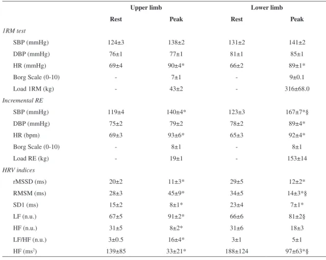
Table 1. Cardiovascular variables and HRV in upper and lower limb exercises at rest and peak of 1RM test and incremental resistance exercise (RE) protocol in healthy older men.
Upper limb Lower limb
Rest Peak Rest Peak
1RM test
SBP (mmHg) 124±3 138±2 131±2 141±2
DBP (mmHg) 76±1 77±1 81±1 85±1
HR (mmHg) 69±4 90±4* 66±2 89±1*
Borg Scale (0-10) - 7±1 - 9±0.1
Load 1RM (kg) - 43±2 - 316±68.0
Incremental RE
SBP (mmHg) 119±4 140±4* 123±3 167±7*§
DBP (mmHg) 75±2 79±2 78±2 89±4*
HR (bpm) 69±3 93±6* 65±3 92±4*
Borg Scale (0-10) - 8±1 - 8±1
Load RE (kg) - 19±1 - 153±14
HRV indices
rMSSD (ms) 20±2 11±3* 29±5 12±2*
RMSM (ms) 28±3 45±9* 34±5 14±3*§
SD1 (ms) 15±2 8±1* 23±4 7±1*
LF (n.u.) 67±5 91±2* 66±6 81±2§
HF (n.u.) 31±5 8±2* 31±6 18±3
LF/HF (n.u.) 3±0.5 16±4* 3±1 5±1
HF (ms2) 139±85 33±21* 188±124 97±63*§
in both upper and lower limb exercises, while for
diastolic blood pressure (DBP), this difference was
observed only for exercise performed on the leg press.
In addition, a signiicantly higher SBP was found at
the end of lower limb exercise compared to upper limb exercise.
Regarding cardiac autonomic responses, Table 1 shows the HRV indices obtained at rest and peak of discontinuous incremental exercise protocol. For both
groups, there was a signiicant decrease in rMSSD, SD1, and HF in absolute units (ms2) comparing rest
and peak of exercise. For the RMSM index, however,
there was an increase for the incline bench press and a decrease for the leg press at 45°.
For upper limb exercise, there was a signiicant
increase in LF and LF/HF and a decrease in HF also comparing rest and peak of exercise for these frequency-domain indices assessed in normalized
units. Additionally, at the peak of incremental load,
intergroup differences were also observed with
signiicantly higher values for RMSM and LF (nu)
and lower values for HF (ms2) in upper limb exercises
compared to lower limb exercises.
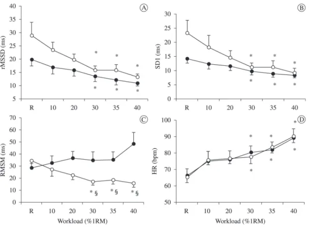
Considering the results on different % loads during
the incremental protocol, we observed a signiicant
decrease in the rMSSD index (Figure 1A) and SD1 index (Figure 1B) from 30%1RM compared to rest,
while the HR values increased from this percentage
(Figure 1D). In contrast, the RMSM index decreased
in lower limb exercise and increased in upper limb
from 30%1RM. From this load, there was a signiicant
difference between exercise modalities, with higher values for the incline bench press (Figure 1C).
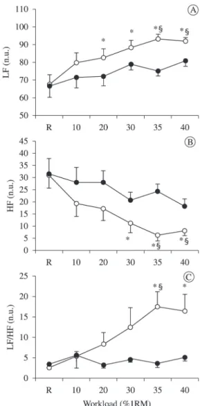
The behavior of the frequency-domain indices of HRV are shown in Figure 2. The LF component
(Figure 2A) increased and HF (Figure 2B) decreased signiicantly from 30%1RM in the incline bench
press. Regarding the LF/HF ratio (Figure 2C), there
was a signiicant increase from 35% of 1RM in the bench press. In addition, we found differences
between exercises at 35% and 40% of 1RM, with higher values of LF and lower HF in the upper limb exercise. For LF/HF, this difference was observed only at 35%1RM, with higher values for the bench
press exercise. It is worth noting that the results at
different % loads during the incremental protocol for HF values analyzed in absolute units (data not shown) were the same as for HF values analyzed in normalized units, with lower values in arm exercise from 30%1RM compared to rest and also lower
values in bench press compared to leg press exercise at 35% and 40% of 1RM.
Discussion
The present study investigated the autonomic cardiac responses during upper versus lower limb resistance exercise at different loads in healthy older
men. The main indings of this study were: 1) a signiicant reduction in the rMSSD and SD1 indices,
representative of parasympathetic modulation, from 30% of 1RM in both exercises, but more pronounced
in upper limb exercise; 2) a signiicant decrease in the RMSM index, representative of sympathovagal
balance, from 30% of 1RM in the 45° leg press, and in contrast, an increase in the incline bench press;
3) a signiicant increase in HR from 30% of 1RM in
both exercises.
To our knowledge this is the irst study to evaluate and compare HRV (rMSSD, RMSM, and SD1)
in acute upper and lower limb resistance exercise
on the bench press and leg press at 45°. Several
studies2,17,21,29-31 evaluated HRV in leg resistance exercise but none compared the HRV response during a discontinuous incremental exercise in upper and lower limbs. Our results showed that, despite the different percentages of load applied, there was a predominance of sympathetic modulation in the upper limb resistance exercises. These results may contribute to understanding cardiovascular responses
during this speciic type of exercise in older people. In the cardiovascular system, the behavior of
cardiac function is mediated by neuro-regulatory activity from the autonomic nervous system. The electrical stimuli reaching the heart through these reflexive pathways are responsible for beat-to-beat variations and reflect sympathetic and parasympathetic modulation27. At rest, the parasympathetic modulation remains predominant,
leading to lower values of HR and BP. However,
during exercise or stressor stimuli, the sympathetic modulation on the HR becomes predominant and
consequently leads to an increase in HR and BP32.
In our study, the rMSSD and SD1 indices decreased as load increased. These indices relect
the vagal modulation18,27,33 therefore, from 30% of 1RM, we can assume that there is a decrease in parasympathetic activity. However, it is not possible to determine the participation of the sympathetic branch by evaluating only these indices.
In this context, the RMSM index relects both
the sympathetic and parasympathetic modulation of HR27,33. In our study, the RMSM on the leg press presented lower values compared to the bench press
from 30%1RM. In the inclined bench press, this
index was stable with a slight increase throughout the load progression, while on the leg press there was
a signiicant decrease in this index from 30%1RM.
From this result, we noted that total HRV differentiates between the two types of exercise, however, with this index it is not possible to reach conclusions about the autonomic branches separately.
Although sympathetic and parasympathetic
autonomic responses are not always reciprocal, in both exercises we observed a decrease in parasympathetic indices from 30% of 1RM without
difference between them. So, we can only suggest
Figure 2. Frequency-domain indices of heart rate variability.
( ) upper limb exercise; ( ) lower limb exercise.
a possibly higher sympathetic modulation in upper limb exercise compared with lower limb exercise.
At the same time, autonomic changes have been
related to lactate accumulation at higher intensities, which can modify the autonomic modulation toward sympathetic predominance21. Simões et al.21 showed
that 30% of 1RM corresponded to the point at which aerobic metabolism is supplemented by anaerobic metabolism related to lactate accumulation in leg press exercise. This study, similarly to ours, also
showed a decrease in SD1 and rMSSD from the
same %RM, however, we did not evaluate lactate
concentration to allow consistent inferences. Another
study34 involving an incremental exercise test on a
bicycle ergometer demonstrated that all measures of vagal modulation decrease progressively until the ventilatory threshold level is reached, when sympathetic modulation begins to predominate.
In a previous study35, there was a prevalence
of sympathetic modulation on the HRafter the
anaerobic threshold point in dynamic exercises. The same behavior was found in the inclined bench press in the present study. However, even without a
signiicant difference between exercises, the values
for the arm exercise were lower than the leg exercise, suggesting stronger response in the arm compared to the leg exercise17. In a review, Helge36 suggested
that in arm exercise there is greater release of blood lactate compared to leg exercise, leading to a greater response of cardiovascular variables.
HRV spectral analysis was included to reinforce
the results in the time domain. Similarly to previous
studies that analyzed the frequency domain in short time series (2 and 3 min) we used 2 min segments during exercise37-40. Additionally, for HRV comparative analysis (rest and exercise), different time series were applied (5 min rest and 2 min of exercise), which has been shown previously38.
In the frequency domain, we observed a decrease
in HF and an increase in LF and LF/HF in the bench
press when comparing rest and exercise. Some
studies suggested that the HF spectral component is a marker of vagal modulation41, LF is a marker of
predominance of sympathetic modulation42, and LF/
HF may suggest sympathovagal balance43. Thus, our
results suggest that, in the incline bench press from 30% 1RM, there was a shift toward sympathetic modulation.
The segments selected were 5 minutes of rest and visually the most stable exercise section containing 2 min free of noise at each load effort, excluding the initial 40 seconds of the exercise (period during which a rapid withdrawal of vagal activity occurred)
and the inal 30 seconds to avoid inluences of blood
pressure measurement.
In a previous study comparing arm and leg exercise,
Tulppo et al.18 veriied faster vagal withdrawal during arm compared to leg incremental dynamic exercise performed on a cycle ergometer. The explanation can be the more powerful central command by vagal withdrawal and a stronger muscle afferent input
during arm exercise compared to leg exercise. In our
study, the vagal indices in the time domain did not
present signiicant differences between exercises, although rMSSD and SD1 values were lower in the
upper limb exercise than in the lower. However the HF index (in nu and ms2) was also lower in the upper
limb exercise compared to the lower limb exercise. Considering the maximal load exercise, some authors18 observed a higher level of sympathetic nervous activity during maximal lower limb exercise compared to upper, contrasting with our results. They support the idea of higher mass discharge of the sympathetic nervous system during lower rather than upper body exercise. These contradictory results
may be explained by the type of protocol chosen. In
our study, RE was performed discontinuously on resistance equipment (bench press and leg press) with different loads of 1RM, while the aforementioned study used continuous incremental exercise until exhaustion on a bicycle ergometer for legs and arms. Thus, the stimulus generated by the two types of protocols was different and can result in a distinct response.
Some differences in arm and leg exercise should
be taken into account to better understand the
results of the present study. In upper limb exercise,
there is greater input possibly caused by the higher number and/or sensitivity of afferent receptors in this musculature44. Central command activation
during RE depends on the exercise intensity and, consequently, the muscle tension, especially on the concentric phase of contraction, which can mechanically compress the peripheral arterial system
causing an obstruction of blood low. The mechanical
and metabolic changes caused by exercise and
muscle contraction can stimulate afferent ibers in type III and IV musculatures32, with greater intensity
in arm musculature. The metabolic changes in active musculature combined with reduced blood
low can stimulate mainly the type IV ibers which
send information to the central nervous system, triggering an increase in sympathetic discharge to the cardiovascular system45. In arm exercise, the
and this is another aspect that may explain the higher sympathetic modulation in upper limb exercise.
The present study has certain limitations that need
to be addressed, the irst being the progressive exercise protocol design. Although it was a discontinuous
incremental protocol, all loads were assessed in a single session, with an interval of 15 min between sets. This may have generated blood lactate accumulation and increased circulating catecholamine, thus
inluencing the HR responses mainly at higher loads
and inducing volunteers to reach the peak load at a
relatively low percentage of 1RM. Secondly, the RE
protocol had a relatively long duration, however this was necessary to obtain a viable record for analysis. Lastly, the visual assessment of stationarity in the short-term HR recordings may be unreliable and can affect primarily the frequency-domain indices of HRV.
In summary, we can observe that significant
cardiac autonomic changes occur from 30% of 1RM regardless of RE limb. However, there was more pronounced sympathetic increase and vagal decrease for upper limb exercises than for lower limb exercises. Therefore, this study provides a physiological understanding of HRV analysis comparing upper and lower limb exercise that can assist the physical therapist in prescribing RE more effectively to promote health in the elderly.
Additionally, knowledge of autonomic responses
in healthy older adults can serve as a benchmark for future studies involving the elderly population affected by chronic diseases such as heart disease.
Acknowledgements
The Fundação de Amparo à Pesquisa do Estado de São Paulo (FAPESP- 07/53202-9, 06/53414-3 and
09/01842-0), São Paulo, SP, Brazil and Coordenação de Aperfeiçoamento de Pessoal de Nível Superior
(CAPES), Brasília, DF, Brazil, for providing inancial support. The authors would like to thank
all colleagues at the Cardiopulmonary Physical
Therapy Laboratory and the School of Health for their
friendly collaboration. More importantly, however, we are indebted to the patients for their effort and enthusiastic cooperation throughout the study.
References
1. Pollock ML, Franklin BA, Balady GJ, Chaitman BL, Fleg JL, Fletcher B, et al. AHA Science Advisory. Resistance
exercise in individuals with and without cardiovascular
disease: beneits, rationale, safety, and prescription: An
advisory from the Committee on Exercise, Rehabilitation,
and Prevention, Council on Clinical Cardiology, American Heart Association; Position paper endorsed by the American College of Sports Medicine. Circulation. 2000;101(7):828-33. http://dx.doi.org/10.1161/01.
CIR.101.7.828
2. Williams MA, Haskell WL, Ades PA, Amsterdam EA, Bittner V, Franklin BA, et al. Resistance exercise in
individuals with and without cardiovascular disease:
2007 update: a scientiic statement from the American Heart Association Council on Clinical Cardiology and Council on Nutrition, Physical Activity, and Metabolism. Circulation. 2007;116:572-84. http://dx.doi.org/10.1161/
CIRCULATIONAHA.107.185214
3. Aagaard P, Suetta C, Caserotti P, Magnusson SP, Kjaer
M. Role of the nervous system in sarcopenia and muscle atrophy with aging: strength training as a countermeasure.
Scand J Med Sci Sports. 2010;20:49-64. http://dx.doi.
org/10.1111/j.1600-0838.2009.01084.x
4. Mayer F, Scharhag-Rosenberger F, Carlsohn A, Cassel M, Müller S, Scharhag J. The intensity and effects of strength training in the elderly. Dtsch Arztebl Int. 2011;108(21):359-64. PMid:21691559 PMCid:PMC3117172. http://dx.doi. org/10.3238/arztebl.2011.0359
5. Cruz-Jentoft AJ, Baeyens JP, Bauer JM, Boirie Y, Cederholm T, Landi F, et al. Sarcopenia: European
consensus on definition and diagnosis: report of the
European Working Group on Sarcopenia in Older People. Age Ageing. 2010;39:412-23. http://dx.doi.org/10.1093/ ageing/afq034
6. Fried LP, Hadley EC, Walston JD, Newman AB, Guralnik JM, Studenski S, et al. From bedside to bench: research agenda for frailty. Sci Aging Knowledge
Environ. 2005;2005(31):pe24 http://dx.doi.org/10.1126/ sageke.2005.31.pe24
7. Chodzko-Zajko WJ, Proctor DN, Fiatarone Singh MA, Minson CT, Nigg CR, Salem GJ, et al. American College of Sports Medicine position stand. Exercise and physical activity for older adults. Med Sci Sports Exerc. 2009;41(7):1510-30. http://dx.doi.org/10.1249/
MSS.0b013e3181a0c95c
8. Forte R, De Vito G, Figura F. Effects of dynamic resistance training on heart rate variability in healthy older women.
Eur J Appl Physiol. 2003;89(1):85-9. http://dx.doi.
org/10.1007/s00421-002-0775-1
9. Hu M, Finni T, Zou L, Perhonen M, Sedliak M, Alen
M, et al. Effects of strength training on work capacity and parasympathetic heart rate modulation during exercise in
physically inactive men. Int J Sports Med.
2009;30(10):719-24. http://dx.doi.org/10.1055/s-0029-1225329
10. Ades PA, Savage PD, Cress ME, Brochu M, Lee NM,
Poehlman ET. Resistance training on physical performance
in disabled older female cardiac patients. Med Sci Sports Exerc. 2003;35(8):1265-70. http://dx.doi.org/10.1249/01.
MSS.0000079044.21828.0E
11. Deschenes MR, Kraemer WJ. Performance and physiologic adaptations to resistance training. Am J Phys Med Rehabil. 2002;81(11 Suppl):S3-16. http://dx.doi.
org/10.1097/00002060-200211001-00003
training on coronary artery disease risk factors. Exp Biol Med (Maywood). 2003;228(4):434-40. PMid:12671188.
13. Braith RW, Beck DT. Resistance exercise: training
adaptations and developing a safe exercise prescription.
Heart Fail Rev. 2008;13(1):69-79. http://dx.doi.
org/10.1007/s10741-007-9055-9
14. Kang J, Chaloupka EC, Mastrangelo MA, Angelucci J. Physiological responses to upper body exercise on an arm and a modified leg ergometer. Med Sci Sports Exerc. 1999;31(10):1453-9. http://dx.doi.
org/10.1097/00005768-199910000-00015
15. Volianitis S, Secher NH. Arm blood low and metabolism
during arm and combined arm and leg exercise in
humans. J Physiol. 2002;544(Pt 3):977-84. http://dx.doi. org/10.1113/jphysiol.2002.023556
16. Volianitis S, Yoshiga CC, Nissen P, Secher NH. Effect of itness on arm vascular and metabolic responses to upper body exercise. Am J Physiol Heart Circ Physiol. 2004;286(5):H1736-41. http://dx.doi.org/10.1152/ ajpheart.01001.2003
17. Okamoto T, Masuhara M, Ikuta K. Upper but not lower
limb resistance training increases arterial stiffness in
humans. Eur J Appl Physiol. 2009;107(2):127-34. http://
dx.doi.org/10.1007/s00421-009-1110-x
18. Tulppo MP, Mäkikallio TH, Laukkanen RT, Huikuri HV. Differences in autonomic modulation of heart rate during arm and leg exercise. Clin Physiol. 1999;19(4):294-9.
http://dx.doi.org/10.1046/j.1365-2281.1999.00180.x
19. Wilborn C, Greenwood M, Wyatt F, Bowden R, Grose
D. The effects of exercise intensity and body position on
cardiovascular variables during resistance exercise. Journal of Exercise Physiology Online. 2004;7(4):29-36.
20. Machado HG, Simoes RP, Mendes RG, Castello V, Di Thommazo L, Almeida LB, et al. Cardiac autonomic
modulation during progressive upper limb exercise by
patients with coronary artery disease. Braz J Med Biol Res. 2011;44(12):1276-84. http://dx.doi.org/10.1590/
S0100-879X2011007500134
21. Simões RP, Mendes RG, Castello V, Machado HG, Almeida LB, Baldissera V, et al. Heart-rate variability
and blood-lactate threshold interaction during progressive
resistance exercise in healthy older men. J Strength Cond
Res. 2010;24(5):1313-20. http://dx.doi.org/10.1519/
JSC.0b013e3181d2c0fe
22. Kraemer WJ, Fry AC. Strength testing: development and
evaluation of methodology: physiological assessment of
human itness champaign. Human Kinetics: 1995;115-138.
23. Borg G. Ratings of perceived exertion and heart rates
during short-term cycle exercise and their use in a new
cycling strength test. Int J Sports Med. 1982;3(3):153-8. http://dx.doi.org/10.1055/s-2008-1026080
24. Antila K. Quantitative characterization of heart rate during exercise. Scand J Clin Lab Invest Suppl. 1979;(153):3-68. PMid:293886.
25. Zuttin RS, Moreno MA, César MC, Martins LEB, Catai AM, Silva E. Evaluation of autonomic heart rate
modulation among sedentary young men, in seated and
supine postures. Rev Bras Fisioter. 2008;12(1):7-12. http:// dx.doi.org/10.1590/S1413-35552008000100003
26. Mourot L, Bouhaddi M, Perrey S, Rouillon JD, Regnard J. Quantitative Poincaré plot analysis of heart rate variability: effect of endurance training. Eur J Appl Physiol. 2004;91(1):79-87. http://dx.doi.org/10.1007/ s00421-003-0917-0
27. Heart rate variability. Standards of measurement,
physiological interpretation, and clinical use. Task Force
of the European Society of Cardiology and the North American Society of Pacing and Electrophysiology. Eur Heart J. 1996;17(3):354-81. http://dx.doi.org/10.1093/
oxfordjournals.eurheartj.a014868
28. Malliani A, Pagani M, Lombardi F, Cerutti S.
Cardiovascular neural regulation explored in the frequency
domain. Circulation. 1991;84(2):482-92. http://dx.doi.
org/10.1161/01.CIR.84.2.482
29. Billman GE. Cardiac autonomic neural remodeling
and susceptibility to sudden cardiac death: effect of
endurance exercise training. Am J Physiol Heart Circ Physiol. 2009;297(4):H1171-93 http://dx.doi.org/10.1152/ ajpheart.00534.2009
30. Motohiro M, Yuasa F, Hattori T, Sumimoto T, Takeuchi
M, Kaida M, et al. Cardiovascular adaptations to exercise training after uncomplicated acute myocardial infarction.
Am J Phys Med Rehabil. 2005;84(9):684-91. http://dx.doi.
org/10.1097/01.phm.0000171167.31010.f4
31. Takahashi AC, Melo RC, Quitério RJ, Silva E, Catai AM.
The effect of eccentric strength training on heart rate and on its variability during isometric exercise in healthy older
men. Eur J Appl Physiol. 2009;105(2):315-23. http://
dx.doi.org/10.1007/s00421-008-0905-5
32. Mitchell JH. J.B. Wolffe memorial lecture. Neural control of the circulation during exercise. Med Sci Sports
Exerc. 1990;22(2):141-54. PMid:2192221. http://dx.doi.
org/10.1113/expphysiol.2011.058156
33. Malliani A, Montano N. Heart rate variability as a clinical tool. Ital Heart J. 2002;3(8):439-45. PMid:12407819.
34. Tulppo MP, Makikallio TH, Takala TE, Seppanen T,
Huikuri HV. Quantitative beat-to-beat analysis of heart
rate dynamics during exercise. Am J Physiol. 1996;271(1 Pt 2):H244-52. PMid:8760181.
35. Sinski M, Lewandowski J, Abramczyk P, Narkiewicz
K, Gaciong Z. Why study sympathetic nervous
system? J Physiol Pharmacol. 2006;57 Suppl 11:79-92. PMid:17244940.
36. Helge JW. Arm and leg substrate utilization and muscle adaptation after prolonged low-intensity training. Acta Physiol (Oxf). 2010;199(4):519-28. http://dx.doi.
org/10.1111/j.1748-1716.2010.02123.x
37. Vardas PE, Kochiadakis GE, Manios EG, Kanoupakis
EM, Zouridakis EG, Chlouverakis GI. Spectral analysis
of heart rate variability before and during episodes of nocturnal ischaemia in patients with extensive coronary
artery disease. Eur Heart J. 1996;17(3):388-93. http://
dx.doi.org/10.1093/oxfordjournals.eurheartj.a014870 38. Mendonca GV, Fernhall B, Heffernan KS, Pereira FD.
39. Cottin F, Médigue C, Leprêtre PM, Papelier Y, Koralsztein JP, Billat V. Heart rate variability during exercise
performed below and above ventilatory threshold. Med
Sci Sports Exerc. 2004;36(4):594-600. http://dx.doi.
org/10.1249/01.MSS.0000121982.14718.2A
40. Weippert M, Kumar M, Kreuzfeld S, Arndt D, Rieger A, Stoll R. Comparison of three mobile devices for measuring R-R intervals and heart rate variability: Polar S810i, Suunto t6 and an ambulatory ECG system. Eur J Appl Physiol. 2010;109(4):779-86. http://dx.doi.org/10.1007/
s00421-010-1415-9
41. Malliani A. Principles of cardiovascular neural regulation in health and disease. Boston: Kluwer Academic Publishers;
2000. http://dx.doi.org/10.1007/978-1-4615-4383-1
42. Montano N, Porta A, Cogliati C, Costantino G, Tobaldini
E, Casali KR, et al. Heart rate variability explored in the frequency domain: a tool to investigate the
link between heart and behavior. Neurosci Biobehav Rev. 2009;33(2):71-80. http://dx.doi.org/10.1016/j.
neubiorev.2008.07.006
43. Sandercock GR, Brodie DA. The use of heart rate
variability measures to assess autonomic control during
exercise. Scand J Med Sci Sports. 2006;16:302-13. http://
dx.doi.org/10.1111/j.1600-0838.2006.00556.x
44. Ishida K, Takaishi T, Miyamura M. Ventilatory responses
at the onset of passive movement and voluntary exercise
with arms and legs. Acta Physiol Scand.
1994;151(3):343-52. http://dx.doi.org/10.1111/j.1748-1716.1994.tb09753.x
45. Iwamoto GA, Kaufman MP. Caudal ventrolateral medullary cells responsive to muscular contraction. J Appl Physiol. 1987;62(1):149-57. PMid:3558174.
Correspondence
Audrey Borghi-Silva
Universidade Federal de São Carlos (UFSCar)
Departamento de Fisioterapia
Laboratório de Fisioterapia Cardiopulmonar Rod. Washington Luis, Km 235, Monjolinho
CEP 13565-905, São Carlos, SP, Brazil