Introduction
The speciic mechanisms that inluence the central nervous system (CNS) changes during or after different types of exer -cises are controversial1,2. Distinct modes of exercise may result
in differential afferent feedback signals to CNS and/or direct central effects via intrinsic brain modulations and conscious perceived of effort3,4. However, great variations among exercise
modalities in the amount of muscle work, and the consequent cardiorespiratory demands and other factors such as cerebral oxygenation consumption rate may also explain the differential corticospinal responses after dynamic vs. static exercise5,7.
There have been some reports in which transcranialmagnetic stimulation (TMS) has been used to test the effects of physical exercise on corticospinalexcitability8,9.Its effects on the responsive
-ness of corticospinal cells appear to be dependent on the type of exercise (dynamic vs static)5,8. Some of dynamic exercises (exercises
involving multiple limb muscles) do not change the amplitude of motor evoked potentials (MEPs)5,10. However, reduction in the
amplitude of MEPs for at least 20 min after fatiguing exercise has also been documented11,12. Decrease in the amplitudes of MEPs
after fatiguing exercise, a phenomenon called post-exercise MEP depression, and an increase in MEP amplitudes after non-fatiguing exercise, a phenomenon called post-exercise MEP facilitation are shown in static exercise, but are not observed in some dynamic
exercise10,12. Other studies suggested some differences in cortical
and spinal responses to dynamic exercise (e.g. locomotor) versus static exercise (e.g. single-joint)3,5,8,13.
There is little evidence about whether the effect of exercise on corticospinal excitability is also dependent on the exercise intensity. To the best of our knowledge, Höllge, Kunkel, Ziemann, Tergau, Geese, Reimers14 were the irst who evaluated the inlu
-ence of different dynamic exercise with different intensities on the TMS-induced MEP amplitude. They demonstrated distinct responses on excitability mainly between exhaustive (high inten -sity) and non-exhaustive exercise (low inten-sity). Furthermore, few researches have investigated corticospinal excitability after dynamic non-exhaustive exercise with different intensities. Therefore, the present study was designed to test the hypothesis that differences in MEPs amplitude would occur after dynamic non-exhaustive cycling exercise with different intensities.
Experimental Procedures
Subjects
Eighteen healthy subjects participated in this crossover study. The level of physical activity of subjects were categorised into very active (2 women and 3 men), active (8 women) and irregularly
Original Article (short paper)
Intensity-dependent effects of cycling exercise on
corticospinal excitability in healthy humans: a pilot study
Isis Suruagy Adriana Baltar
Universidade Federal de Pernambuco, Recife, PE, Brasil
Luis Paulo Gomes
Universidade Tiradentes, Aracajú, SE, Brasil
Marina Berenguer Armele Dornelas Kátia Monte-Silva
Universidade Federal de Pernambuco, Recife, PE, Brasil
Abstract — Aims: the aim of this study was to verify the effects of different intensities of locomotor exercise on
corticospinal excitability. Methods: 18 healthy subjects (27.6 ± 6.5 years,) participated in a design study of three different
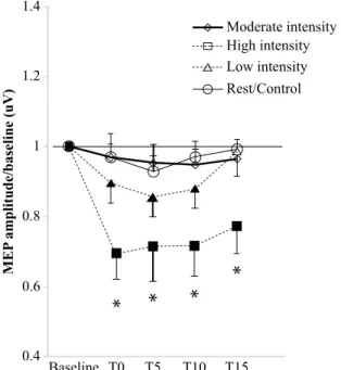
exercise protocols on a cycle ergometer: (i) 10 min at 75% Wmax (high intensity); (ii) 15min at 60% Wmax (moderate intensity) or (iii) 30 min at 45% Wmax (low intensity). The protocols of lower body cycling were assigned in random order in separate sessions. A control session was done with subjects at rest. Corticospinal excitability was assessed before (baseline) and every 5 min for 15min after the end of exercise/rest (time: 0, 5, 10 and 15) by measurement of the motor evoked potential (MEP) elicited by transcranial magnetic stimulation in the relaxed irst-dorsal interosseus muscle. Results: Compared to the resting session, a signiicant decrease (64%) in the motor evoked potential amplitudes
was found only in the session of exercise of high intensity. This result seems depend on the level of physical activity of subject. No change was found after rest, low and moderate exercises. Conclusions: These indings suggest that changes in the corticospinal excitability depend on exercise intensity and level of physical activity of subjects.
active (4 women and 1 men) by International Physical Activity Questionnaire (IPAQ)15,16. All participants were informed of the procedures and risks before giving written informed consent to participate in the research. Approval for study procedures was obtained from the Institutional Research Ethics Committee and wasperformed in accordance with the Declaration of Helsinki.
Maximal incremental exercise test
Before the experimental sessions (session 1), the subjects were submitted to maximal incremental exercise test to ind out their maximal tolerated power (Wmax). For the test, participants sat on the cycle ergometer (ERGO-FIT® model 167 cycle, Pirmansens, Germany) with similar riding position (saddle and handlebar height and position) and were asked to begin cycling at 70 rpm during a warm-up phase (3 min) at a work load of 15W. The work rate was then increased by 25W per minute until exhaustion when participants could not keep a cadence of 70 rpm for 5 seconds or when the subjects voluntarily terminated the test. During the test, the subjects were verbally encouraged to continue for as long as possible. Wmax was deined as the highest work load sustained by the subject for longer than 1 min.
Experimental design
The protocol included three experimental sessions (sessions 2, 3 and 4) which were conducted in a pseudo-randomized (counterbal -anced sequence) crossover design. In each session, one of three different exercise intensities on a cycle ergometer was tested: (i) 10min at 75% Wmax (high intensity); (ii) 15min at 60% Wmax (moderate intensity) or (iii) 30 min at 45% Wmax (low intensity). A control session (session 5) was done with subjects at rest, the subjects were asked to stay sit without move the hands. The ses -sions were separated by at least two days.
In all experimental sessions, during the exercise, the heart rate (HR) was continuously registered using an HR recorder (Polar RS800, Polar Electro, Kempele, Finland). In addition, the Borg Rating of Perceived Exertion (RPE)17 was used to estimate
whole-body perceived exertion. Every three minutes during exercise, the RPE was displayed in front of the subjects who were asked to point the number that the best described their perceived exertion (number 7 represents unloaded cycling while number 19 indicates an exertion similar to exhaustive cycling)17.
The volunteers were instructed to refrain from vigorous activities and to avoid the consumption of caffeine, alcohol and tobacco for 24 h prior to each session. For all exercise time, the laboratory temperature (19 ± 1°C) remained constant in order to minimize environmental inluence.
Monitoring of motor corticospinal excitability
Before and after each session, the corticospinal excitability was assessed by MEPs elicited by TMS. For this, the subjects were seated in a comfortable chair with head and arm rests. Single-pulse
TMS was applied using a igure-of-eight magnetic coil (7 cm diam -eter) connected to a magnetic stimulator (Neurosoft Ltd., Russia; peak magnetic ield=2.2 tesla). The coil was held tangentially to the skull, with the handle pointing backwards and laterally at an angle of 45° from midline in the right hemisphere. The optimal position was deined as the site where stimulation resulted in the largest MEPs. Surface EMG was recorded from the left irst dorsal interosseous (FDI) with AgCl electrodes in a belly-tendon montage. The signals were ampliied and iltered with a time constant of 80 ms and a low-pass ilter of 5.0 Hz, then digitized at an analogue-to-digital rate of 20 kHz and further relayed into a laboratory computer using the Neuro-MEP-Micro software (Neurosoft Company, Russia). The intensity was adjusted to elicit, on average, baseline MEPs of 1 mV peak-to-peak amplitude and was kept constant for the post-stimulation assessments. During the registers, EMG was recorded online in order to ensure the rest of FDI. Twenty MEPs were recorded at frequency of 0.25 Hz at baseline and every 5, 10 and 15 minutes.
The design of the experiment is shown in igure 1.
Statistical analysis
Individual MEP amplitude means were calculated for each time bin, including baseline and post-exercise/rest time points. The post – exercise/rest MEPs were normalized intra-individually and are given as baseline ratios. In order to verify any difference in the conditions before each session one – way ANOVA were employed.
A repeated measure ANOVA was calculated using the within-subject factors “time course” (baseline, 0,5,10 and 15min) and “exercise protocols” (low, moderate, high intensity and rest) and the dependent variable MEP amplitude. Also by repeated measure ANOVA, the behaviour of excitability after physical exercise was also analysed considering the level of physical activity of the subjects. Post hoc analyses were performed with a student paired-samples t test when appropriate. The Mauchly test of sphericity was checked and the Greenhouse-Geisser correction was performed, if necessary. The data were analyzed using the program SPSS (version 18.0). A p value of <0.05 was considered signiicant for all statistical analysis.
Results
Table 1 shows the characteristics of sample and averaged values for HR and RPE obtained during each exercise protocols and work load correspondent are given in the table 2.
The results of the one-way ANOVA showed that MEP am -plitudes did not differ signiicantly in baseline measurements among the sessions. The repeated measures ANOVA revealed
Table 1.Participants characteristics
Variable
Age, in years mean (±SD) 27.6 (±6.5)
Gender n(%) Male Female
4 (22.2%) 14 (77.7%)
BMI, in Kg/cm² mean (±SD) 21.5 (±1.82)
Physical activity1 n(%) Irregularly active Active
Very active
5 (28%) 8 (44%) 5 (28%)
Wmax2 mean (±SD) Irregularly active Active
Very active
197.7 (±60.2) 153 (±48.3) 178 (±14.5) 265 (±62.7)
1Determined by International physical actitvity questionnaire – IPAQ; 2
De-termined by the maximal incremental exercise test. W – Power output. BMI – body mass index
Table 2. Mean (± standard deviations) of heart rate (HR) 1, Borg rat -ing of perceived exertion (RPE) 2 and power (watts) dur-ing exercise protocols on a cycle ergometer.
Exercise Protocols HR¹ RPE² Power
High intensity (10min at 75% Wmax) (±10.6)163.3 (±2.0)15.5 (±45.1)148.3
Moderate intensity (15min at 60%
Wmax) (±9.2)149.4 (±1.4)13.0 (±36.1)118.6
Low intensity (30 min at 45% Wmax) (±2.9)135.0 (±1.0)10.2 (±27.1)89.0
1 Bpm, beat per minute; 2 score of Borg scale
RPE - rating perception effort e HR - heart rate S2
S3
S4
S5
RANDOMIZADE
S1
MEP
MAXIMAL INCREMENTAL EXERCISE TEST (Wmax)
RPE/HR HIGH-INTENSITY MODERATE-INTENSITY
LOW-INTENSITY REST/CONTROL
MEP MEP MEP MEP
T0 T5 T10 T15Figure 1.Experimental course. In session 1, subjects were submitted to maximal incremental exercise test to ind out their maximal tolerated power (Wmax). In sessions 2,3 and 4, the effect of different exercise protocols on motor cortex excitability, as monitored via motor evoked potential (MEP – 20 stimulus) elicited by single pulse transcranial magnetic stimulation (TMS), was explored. Speciically, high (10min at 75% Wmax); moderate (15min at 60% Wmax) and low intensity (30 min at 45% Wmax) were compared with control session (session 5) with subjects at rest. The excitability was monitored every 5 min for one quarter of an hour (T0, T5, T10 and T15) after the end of exercise/rest. RPE - rating perception effort, HR - heart rate, MEP - motor evoked potential.
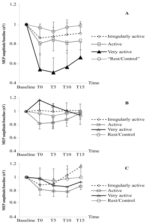
Figure 3 plots the mean (±SE) of baseline-standardized MEP amplitude change over the course of the session of high-intensity (3A), moderate-intensity (3B) and low-intensity exercise (3C) for groups of subjects classiied as very active, active or irregu -larly active, according to IPAQ. The post hoc test revealed a signiicant corticospinal excitability reduction only after session with exercise of high – intensity in very active subjects when compared to baseline condition and to the resting/control session.
*
*
*
*
Moderate intensity High intensity Low intensity Rest/Control
Baseline T0 T5 T10 T15 1.4
1.2
1
0.8
0.6
0.4
MEP
amplitude/baseline (uV)
Discussion
The present study evaluated the effect of a single session of dynamic exercise with three different intensities modiies in the corticospi -nal excitability. The results demonstrate a signiicant decrease in the MEP amplitude after high intensity exercise for 10 min. No changed was found after low and moderate-intensity exercise.
This pattern of change after high intensity exercise is similar to that observed after sustained single-joint contrac -tion18,19 fatiguing running exercise20, strength exercises14 and
after maximal incremental treadmill exercise21. Most studies
that found a reduction of the MEP amplitude are those which used fatiguing exercise12, 14,20. Despite the intense exercise (75%
Wmax) used in our study not be considered fatiguing, the in -tensity and duration that were used probably were enough to promote accumulation of fatigue metabolites and corresponding increased iring of muscle metabosensitive receptors, or other acute exercise-induced responses that are common to fatiguing exercise5. Although the precise cellular mechanisms underlying
post-exercise MEP depression are unclear, Sammi, Wassermann, Hallett22 hypothesize that exercise may modify synaptic trans
-mission within the motor cortex for several minutes in a way
Irregularly active Active
Very active “Rest/Control”
Time
T0 T5 T10 T15
Baseline 1.2
1
0.8
0.6
0.4
MEP
amplitude/baseline (uV)
MEP
amplitude/baseline (uV)
1.2
1
0.8
0.6
0.4
Irregularly active Active
Very active Rest/Control
B A
Time
T0 T5 T10 T15
Baseline
MEP
amplitude/baseline (uV)
1.2
1
0.8
0.6
0.4 Time
T0 T5 T10 T15
Baseline
C
Irregularly active Active
Very active Rest/Control
similar to that in which high-frequency microstimulation of a synaptic pathway leads to depressed transmission.
In contrast to our results, previous TMS studies23-24 that used
cycling exercise of similar high intensity (80% Wmax) and lon -ger duration (~45min) reported no changes in MEP responses measured either in vastuslateralis23or rectus femoris muscles24.
One reason for this discrepancy might be due the place where the corticospinal excitability was evaluated, in the cortical rep -resentation area of non-exercised muscle (our study) in contrast to exercised muscle (previous studies)23-24. However, contrary to
this hypothesis, Takahashi et al.25 showed that intense exercise of
leg muscles leads to pronounced effects on the corticospinal and corticocortical excitability of two non-exercised arm muscles (FID and biceps brachi). Therefore, exhaustive exercise of large muscle groups might cause a widespread cerebral deoxygenation and affects the excitability of circuits in the non-exercised area of motor cortex26.
In contrast to the effects of high-intensity exercise, no change on corticospinal excitability was found after low – and moderate-intensity exercise in our study. Similar indings have been demonstrated by previous studies27-28. It is likely that
non-fatiguing dynamic exercises of low and moderate intensity are unable to inluence non-exercised muscles cortical area. However, a recent study reported that even when measured in the muscles directly involved in the exercise, a period of sustained cycling did not signiicantly inluence motor cortex excitability10. This
response pattern in corticospinal responsiveness differs from that observed after non-fatiguing single-joint contraction, which corticospinal excitability increases markedly22,29. For example,
Samii, Wassermann, Ikoma, Mercuri, Hallett29 observed the
amplitude of MEPs induced by TMS from the resting muscle after 30-second periods of non-fatiguing isometric exercises of the extensor carpi radialis was on average more than twice the pre-exercise value. Given that the amount of muscle work and associated oxygen demands are considerable different to the two modes of exercise (single-joint vs. locomotor exercises), these and other differences in their associated systemic physiological con -sequences may explain the contrasting corticospinal responses10.
The differences among the systemic physiological conse -quences, such as hyperthermia, arterial hypoxemia, decreases in cerebral oxygenation, respiratory muscle loading, and brain catecholamines, occasioned by different exercise intensities may also help to explain the variable responses of central nervous system drive to high and low/moderate levels of exercise intensity in our study. This seems to happen because different modes of exercise may result in different afferent feedback signals to the central nervous system and/or direct central effects via intrinsic brain modulations and conscious perceptions of effort3,4. The
signiicant reduction of MEPs amplitudes when the subjects were submitted to high intensity protocol of exercise in our study may be related to intrinsic brain regulation mechanisms related to an increased internal sense of effort and/or mental stress and support the concept of central fatigue as a protective mechanism3. Alternatively, the lack of changes in excitability
of the cortical cells after low/moderate exercises may be due to these modes of exercises have been unable to trigger the intrinsic brain regulation mechanisms.
The central response to the level of exercise intensity seems also depend on the levels of physical activity of subject. We demonstrated that behavior of the corticospinal excitability after dynamic exercise differs among the volunteers with different levels of physical activity according to IPAQ. Highly active individuals had signiicant depression in of excitability only after exercise of high-intensity. This inding demonstrates that in addition to the type, duration and intensity of exercise per -formed, the level of physical activity of the subjects may have inluence the modulation of brain and spinal cord projections to exercising lower limb muscles during dynamic tasks like cycling. Cirilo, Lavender, Ridding, Semmler30 found that regular
physical activity, primarily involving lower limb muscles, was accompanied by increased motor cortex plasticity in a small hand muscle compared with sedentary reinforcing that there are differences in behavior of brain and spinal cord according to level of physical activity.
In a previous study, it was showed that, the physically very active subjectshad an increased neuroplastic response to a non-invasive brain stimulation protocol (Paired associate stimulation) when compared with sedentary individuals30. This result provides
evidence that high levels of physical activity maintained over an extended period of time can enhanced the capacity for cortical plasticity. This supposed neuroplastic capacity enhancement as -sociated to level of physical activity of individual may, in part, explain why physical activity has a positive effect on memory and executive function31. Another point to be considered is the
production of lactate during exercise, studies have found that blood lactate concentration can inluence the level of cortical excitability, mainly in the motor cortex32. It is suggested that during vigorous
exercise, the accumulation of lactate in the blood relects that the oxygen supply was inadequate to meet the energy require -ments of the working muscle during the exercise33. Coco et al.32
showed that after acute and exhaustive exercise, the increases in blood lactate concentration are accompanied by decreased motor threshold, indicating an increase in the excitability of the motor cortex proportional concentrations of this metabolite. It is proven that individuals with better itness level produce a smaller amount of blood lactate than irregular active subjects33, it is proven that
individuals with better itness level produce a smaller amount of blood lactate, this may be one explanation of the decrease in corticospinal excitability only in very active subject during high-intensity exercise. However, these possibilities are speculative and more research is needed to conirm these indings.
in subjects with different levels of physical activity, and differ -ent repres-entations of motor cortex (i.e., exercised muscle) are need to elucidate the mechanisms responsible for this effect.
In conclusion, our result highlights that changes in the corti -cospinal excitability depend on exercise intensity, duration and level of physical activity of subjects.
References
1. Amann M, Dempsey JA. Locomotor muscle fatigue modiies central motor drive in healthy humans and imposes a limitation to exercise performance. J. Physiol. 2008;586(1):161-73. 2. Brink-Elfegoun T, Kaijser L, Gustafsson T, Ekblom B. Maximal
oxygen uptake is not limited by a central nervous system governor. J Appl Physiol. 2007;102(2):781-6.
3. Gandevia S. Spinal and supraspinal factors in human muscle fatigue. Physiol. Rev. 2001;81(4):1725-89.
4. Marcora S. Perception of effort during exercise is independent of afferent feedback from skeletal muscles, heart, and lungs. J Appl Physiol. 2009;106(6):2060-2.
5. Sidhu SK, Cresswell AG, Carroll TJ. Corticospinal responses to sustained locomotor exercises: moving beyond single-joint studies of central fatigue. Sports Medicine. 2013;43(6):437-49. 6. Périard JD, Caillaud C, Thompson MW. Central and peripheral
fatigue during passive and exercise-induced hyperthermia. Med Sci Sports Exerc. 2011;43(9):1657-65.
7. Dempsey JA, Amann M, Romer LM, Miller JD. Respiratory system determinants of peripheral fatigue and endurance perfor -mance. Med Sci Sports Exerc. 2008;40(3):457-61.
8. Goodall S, Howatson G, Romer L, Ross E. Transcranial magnetic stimulation in sport science: A commentary. Eur J Sport Sci. 2014;14(sup1):S332-S40.
9. Kidgell DJ, Stokes MA, Castricum TJ, Pearce AJ. Neurophysiological responses after short-term strength training of the biceps brachii muscle. J Strength Cond Res. 2010;24(11):3123-32.
10. Sidhu SK, Cresswell AG, Carroll TJ. Motor cortex excitability does not increase during sustained cycling exercise to volitional exhaustion. J. Appl. Physiol. 2012;113(3):401-9.
11. Racinais S, Girard O, Micallef J-P, Perrey S. Failed excitability of spinal motoneurons induced by prolonged running exercise. Journal of neurophysiology. 2007;97(1):596-603.
12. Brasil-Neto JP CL, Hallett M. . Central fatigue as revealed by post-exercise decrement of motor evoked potentials. Muscle Nerve. 1994;17:713-19.
13. Taylor J, Gandevia S. Noninvasive stimulation of the human corticospinal tract. J. Appl. Physiol. 2004;96(4):1496-503. 14. Höllge J, Kunkel M, Ziemann U, Tergau F, Geese R, Reimers C.
Central fatigue in sports and daily exercises. A magnetic stimula -tion study. Int J Sports Med. 1997;18(08):614-7.
15. Booth ML, Ainsworth BE, Pratt M, Ekelund U, Yngve A, Sallis JF, et al. International physical activity questionnaire: 12-country reliability and validity. Med Sci Sports Exerc. 2003;195(9131/03):3508-1381.
16. Matsudo S, Araújo T, Marsudo V, Andrade D, Andrade E, Braggion G. Questinário internacional de atividade f1sica (IPAQ):
estudo de validade e reprodutibilidade no Brasil. Rev bras ativ fís saúde. 2001;6(2):05-18.
17. Borg GA. Psychophysical bases of perceived exertion. Med Sci Sports Exerc. 1982;14(5):377-81.
18. Hoffman BW, Oya T, Carroll TJ, Cresswell AG. Increases in cor -ticospinal responsiveness during a sustained submaximal plantar lexion. J. Appl. Physiol. 2009;107(1):112-20.
19. McNeil CJ, Giesebrecht S, Gandevia SC, Taylor JL. Behaviour of the motoneurone pool in a fatiguing submaximal contraction. J. Physiol. 2011;589(14):3533-44.
20. Ross EZ, Middleton N, Shave R, George K, Nowicky A. Corticomotor excitability contributes to neuromuscular fatigue fol -lowing marathon running in man. Exp. Physiol. 2007;92(2):417-26. 21. Verin E, Ross E, Demoule A, Hopkinson N, Nickol A, Fauroux
B, et al. Effects of exhaustive incremental treadmill exercise on diaphragm and quadriceps motor potentials evoked by transcranial magnetic stimulation. J Appl Physiol. 2004;96(1):253-9. 22. Samii A, Wassermann EM, Hallett M. Post-exercise depression
of motor evoked potentials as a function of exercise duration. Electroencephalogr Clin Neurophysiol /Electromyogr Motor C. 1997;105(5):352-6.
23. Goodall S, González-Alonso J, Ali L, Ross EZ, Romer LM. Supraspinal fatigue after normoxic and hypoxic exercise in hu -mans. J. Physiol. 2012;590(11):2767-82.
24. Sidhu SK, Bentley DJ, Carroll TJ. Locomotor exercise induces long-lasting impairments in the capacity of the human motor cor -tex to voluntarily activate knee extensor muscles. J Appl Physiol. 2009;106(2):556-65.
25. Takahashi K, Maruyama A, Hirakoba K, Maeda M, Etoh S, Kawahira K, et al. Fatiguing intermittent lower limb exercise inluences corticospinal and corticocortical excitability in the nonexercised upper limb. Brain Stimul. 2011;4(2):90-6. 26. Rasmussen P, Nielsen J, Overgaard M, Krogh-Madsen R, Gjedde
A, Secher NH, et al. Reduced muscle activation during exercise related to brain oxygenation and metabolism in humans. J Physiol. 2010;588(11):1985-95.
27. McDonnell MN, Buckley JD, Opie GM, Ridding MC, Semmler JG. A single bout of aerobic exercise promotes motor cortical neuroplasticity. J Appl Physiol . 2013;114(9):1174-82.
28. Smith AE, Goldsworthy MR, Garside T, Wood FM, Ridding MC. The inluence of a single bout of aerobic exercise on short-interval intracortical excitability. Exp. Brain Res. 2014;232(6):1875-82. 29. Samii A, Wassermann E, Ikoma K, Mercuri B, Hallett M.
Characterization of postexercise facilitation and depression of motor evoked potentials to transcranial magnetic stimulation. Neurology. 1996;46(5):1376-.
30. Cirillo J, Lavender AP, Ridding MC, Semmler JG. Motor cortex plasticity induced by paired associative stimulation is enhanced in physically active individuals. J. Physiol. 2009;587(24):5831-42. 31. Cotman CW, Berchtold NC. Exercise: a behavioral interven -tion to enhance brain health and plasticity. Trends Neurosci. 2002;25(6):295-301.
33. Myers J, Ashley E. Dangerous curves a perspective on ex -ercise, lactate, and the anaerobic threshold. CHEST Journal. 1997;111(3):787-95.
Corresponding author
Katia Monte-Silva
Applied Neuroscience Laboratory, Universidade Federal de Pernambuco, Depart
-ment of Physical Therapy. Av. Prof. Moraes Rego s/n, Recife, Brazil. Email: [email protected]
Manuscript received on August 25, 2016 Manuscript accepted on November 15, 2016