Quim. Nova, Vol. 39, No. 7, 832-835, 2016
Artigo
http://dx.doi.org/10.5935/0100-4042.20160091
*e-mail: [email protected]
SPECTROSCOPIC STUDY OF EFFECTS OF TETRAALKYLAMMONIUM CATIONS ON F−-SENSING
PROPERTIES OF CALIX[4]PYRROLE BORADIAZAINDACENE DYE
Yongjun Lv*
College of Material and Chemical Engineering, Sichuan University of Science and Engineering, Zigong, 643000, China
Recebido em 08/01/2016; aceito em 17/03/2016; publicado na web em 25/05/2016
A novel meso-tetracyclohexylcalix[4]pyrrole-based boradiazaindacene dye 3 was synthesized and characterized. F−-binding properties of the dye in the presence of tetrabutylammonium (TBA+), tetraethylammonium (TEA+), and tetramethylammonium (TMA+) counter ions were investigated by UV–Vis, fluorescence, and NMR spectroscopies. Dye 3 displayed various degrees of absorption red shift, fluorescence quenching, and downfield shifts of NH signals for the three fluoride salts. The association constants of these salts mainly depend on cation size effects and ion-pairing effects and were in the order KTMA+ > KTEA+ > KTBA+. Thus, we speculate that both F− and tetraalkylammonium cations are concomitantly located above and below a bowl-shaped calix[4]pyrrole cup in an ion-paired complex, respectively.
Keywords:calix[4]pyrrole; sensing; spectroscopy; cation size effect.
INTRODUCTION
Calix[4]pyrrole, as a pyrrole-derived macrocycle, was first reported as an efficient halide anion receptor by Sessler’s group in 1996.1 Since then, calix[4]pyrrole-based receptors have attracted
an increasing attention to modulate the anion-binding affinity and selectivity of the parent calix[4]pyrrole.2-6 In general, calix[4]pyrrole
derivatives are prepared to sense halide anions through hydrogen bonds7,8 or the ion-pairing effect.9,10 In an anion-sensing process,
tetrabutylammonium (TBA+) cation salts are readily utilized, yet
the effects of various tetraalkylammonium cations have been largely ignored. Sessler and co-workers elucidated that cations of different sizes can affect the anion-binding affinity of calixpyrroles in the solid state.11 However, this effect in solution has not been studied in detail.
As a result, we herein report the F−
-binding properties of calix[4] pyrrole boradiazaindacene (BODIPY) in CH3CN solution using
te-trabutylammonium fluoride (TBA-F), tetraethylammonium fluoride (TEA-F), and tetramethylammonium fluoride (TMA-F) salts. Their sensing properties were investigated by UV–Vis, fluorescence, and NMR spectroscopies. With the introduction of a BODIPY fluorophore on a calix[4]pyrrole, F−-recognition processes can be monitored by
colorimetric and fluorometric dual-channel patterns.12-17 EXPERIMENTAL
Materials and methods
All reagents were commercially obtained and used without fur-ther purification. In titration experiments, F−
was added in the form of TBA+, TEA+, and TMA+ salts. The fluoride salts were purchased
from Alfa Aesar and were stored in a desiccator under vacuum. Chromatography-grade CH3CN was used.
1H NMR and 13C NMR
spectra were obtained using a Varian 400 MHz INOVA spectrom-eter with the samples dissolved in CDCl3. ESI-MS spectra were
obtained using a Waters Micromass ZQ-4000 mass spectrometer. C, H, N elemental analyses were performed using Vario-EL. UV–Vis spectra were obtained using a PerkinElmer Lambda 35 spectrom-eter. Fluorescence spectra were obtained using a PerkinElmer LS55
spectrometer. All experiments were conducted at room temperature (25 °C). A solution of BODIPY dye 3 (1.1 × 10⁻³ mol L−1) in CH
3CN
was prepared and diluted to a concentration appropriate for UV–Vis and fluorescence spectroscopies; the appropriate F− salt (TBA-F,
TEA-F, or TMA-F) dissolved in CH3CN was added to attain the
F− concentration of 1.1 × 10⁻² mol L−1, and the absorption/emission
spectra of the resulting solution were immediately obtained. Nonlinear curves were fit to the absorption and fluorescence data and were expressed as equations (1) and (2), respectively.18
, (1)
, (2)
where A and A0 are the absorbance of dye 3 in the presence and
absence of F−
, respectively, FI and FI0 are their corresponding
fluo-rescence intensities, CF and CD are the concentrations of F
− and dye
3, respectively, and K is the association constant.
Synthesis of 3-[2-(β -tetracyclohexylcalix[4]pyrrolyl)ethenyl]- 4,4-difluoro-8-(4-bromo)phenyl-1,5,7-trimethyl-3a,4a-diaza-4-bora-s-indacene (3)
Targeted dye 3 was prepared as shown in Scheme 1.Compound
119 (0.91 g, 2.00 mmol) and formylcalix[4]pyrrole 2 (1.23 g, 2 mmol)
were refluxed in a mixture of dry toluene (50 mL), piperidine (0.5 mL), and AcOH (0.5 mL). The mixture was heated for 10 h in a Dean–Stark apparatus to azeotropically remove any water formed. The solvents were removed under reduced pressure, and the product was purified by column chromatography (CH2Cl2/hexane, 1:1) to
obtain 3 as a purple solid (0.70 g). Yield: 35%. M.p. > 300 °C. ESI-MS: m/z = 1001.3 (M + H+). 1H NMR (400 MHz, CDCl
3) δ: 1.42 (s,
6H, CH3), 1.49 (m, 30H, CH2), 2.00 (m, 10H, CH2), 2.60 (s, 3H, CH3),
Spectroscopic study of effects of tetraalkylammonium cations on F−-sensing 833 Vol. 39, No. 7
CH), 7.63–7.67 (m, 3H, 2H for ArH, 1H for vinylic). 13C NMR (100
MHz, CDCl3) δ: 14.7, 14.7, 15.0, 115.8, 116.9, 117.8, 121,4, 123.3,
129.3, 129.4, 130.1, 132.4, 134.1, 136.5, 138.3, 142.1, 142.4, 142.5, 153.8, 153.8, 155.1, 156.7. Anal. calcd. for 3 (C60H68BBrF2N6): C,
71.93; H, 6.84; N, 8.39. Found: C, 71.96; H, 6.90; N, 8.34.
RESULTS AND DISCUSSION
UV-Vis spectral studies
Figure 1 shows changes in the UV–Vis spectra of 3 (1.1 × 10⁻⁵ mol
L−1) upon titration with the CH
3CN solution of TBA-F. Dye 3 exhibits
a typical absorption band at 590 nm; this band is red shifted by 90 nm compared to the corresponding band of parent BODIPY 1 (λab = 499 nm). This absorption shift is attributed to the extension of π-electron
conjugation systems with the modification of the calixpyrrole unit on the BODIPY core.20 With the stepwise addition of TBA-F, the
absorbance intensity at 590 nm decreased and a new band gradually appeared at 609 nm. The color correspondingly changed from fuchsia to blue (Figure 1 inset). These observed changes were attributed to the activated intramolecular charge transfer (ICT) process by F−
binding.21 Interestingly, adding TEA-F and TMA-F caused similar
changes in the UV–Vis spectra and color of the solution, as evident in Figures 1S and 2S (Supplementary Material). However, on a per molar basis, the addition of TMA-F caused the greatest absorption band red shift, followed by the red shifts caused by the additions of TEA-F and TMA-F. On the basis of the UV–Vis data, we determined the association constants K (Figure 1 inset);22 the results are shown in
Table 1. The F−-binding order is K
TMA+ > KTEA+ > KTBA+, opposite the
order of their size. Specifically, TMA+, as a relatively small cation,
is prone to associate with an electron-rich bowl-shaped cavity of the calix[4]pyrrole unit. This good match between the cation size and cavity would promote strong hydrogen bonding interactions between four pyrrolic hydrogen atoms and fluoride ions.23
Fluorescence studies
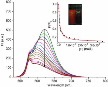
The fluorescence spectrum of dye 3 exhibits a typical emission
band at 623 nm, as shown in Figure 2. The quantum yield was determined to be 0.04 using rhodamine B (Φ = 0.49 in EtOH) as a
standard.24 Compared to the emission wavelength of parent BODIPY 1 (λem = 510 nm), that of 3 exhibits a red shift of approximately 110 nm, which reveals the ICT nature of the excited states of 3. The addi-tion of TBA-F resulted in obvious fluorescence quenching at 623 nm and a 20-nm blue shift; these sensing events instantaneously occurred upon the addition of F−
(Figure 2 inset). The disappearance of red fluorescence is ascribed to the occurrence of an ICT process from an electron-rich calix[4]pyrrole unit upon binding F− to an
electron-deficient BODIPY group. The blue shift may be due to a slight twist of the conjugated pyrrole BODIPY plane after the coordination of F−,
which affects the co-planarity of the entire structure of molecule 3.25
The additions of TEA-F and TMA-F induced similar fluorescence changes (Figures 3S and 4S, Supplementary Material). However,
Scheme 1. Synthetic procedure for dye 3
Figure 1. UV–Vis spectra of 3 (1.1 × 10⁻⁵ mol L−1) in CH
3CN upon the
addi-tion of TBA-F from 0 to 2.1 × 10⁻⁵ mol L−1. Inset: nonlinear curve fitting as
a function of [F−] monitored at 591 nm
Table 1. Association constants and detection limits for 3 with F− as different tetraalkylammonium salts in CH3CN
Fluoride salt K (L
−1 mol) determined by UV-Vis spectra at 590 nm
K (L−1 mol) determined by
fluorescence at 623 nm Detection limit (mol L
−1)
TBA-F 8.00 × 10³ 9.29 × 10³ 3.34 × 10⁻⁵
TEA-F 1.32 × 10⁴ 1.12 × 10⁴ 2.76 × 10⁻⁵
Lv
834 Quim. Nova
quenching exhibited the trend TBA+ < TEA+ < TMA+ on a per molar
basis. This order is related to the structure of tetraalkylammonium cations and to the affinity of 3 for fluoride. Similarly, association constants were also determined using equation (2), and lower de-tection limits were determined as three times the signal-to-noise ratio; the results are reported in Table 1.26 As reported in the table,
good agreement is observed between K values and those obtained from UV–Vis analyses. Fluorescence titrations indicated that 3 de-monstrates a stronger affinity for TMA-F compared with the other two fluoride salts.
1H NMR spectral studies
The interactions of 3 with F− were further validated by 1H NMR
experiments. Figure 3 shows partial spectra of 3 upon the addition of 2.0 equivalents of F− with different cations. The signals of NH
protons at 6.40, 6.49, and 7.16 ppm disappeared, and downfield shifted to various degrees, which suggests the formation of a 3-F−
hydrogen-bonding complex. The signals of the NH proton shifted to 12.42 and 12.92 ppm for TBA-F, 12.50 and 13.02 ppm for TEA-F, and 12.54 and 13.08 ppm for TMA-F. The downfield shifts also followed this order. As a result, we can reasonably speculate about the proposed interaction model of 3 and tetraalkylammonium
fluoride salts (Figure 4). The F− and tetraalkylammonium cations
were concomitantly located above and below the bowl-shaped ca-lix[4]pyrrole cup. The formation of an ion-pairing complex may be based on a stepwise binding mechanism, whereby a calix[4]pyrrole subunit binds the F− anion initially to form a cone-conformation
anionic complex with an electron-rich cavity opposed to the bound F−. The short distance between the F− ion and N atom in the cation
would afford a close-contact ion-pairing arrangement. The hydro-gen bonding interaction and affinity of 3 for F− would increase.27-29
Therefore, TMA+, as the smallest quaternary ammonium cation
among those tested, was found to induce substantial spectroscopic responses in the F− recognition process of dye 3.
CONCLUSIONS
We investigated the F−-binding properties of a calix[4]pyrrole
BODIPY 3 in the presence of TBA+, TEA+, and TMA+ as counter
ions. Upon the addition of F−, the UV–Vis wavelength of the
ma-ximum absorption of 3 exhibited a 90-nm red shift, the color of the solution changed from fuchsia to purple, fluorescence was quenched, and pyrrole NH signals in the 1H NMR spectrum shifted downfield.
Sensing responses were attributed to the ICT process from the electron-rich calix[4]pyrrole to electron-withdrawing BODIPY core. Moreover, the trend of association constants was KTMA+ > KTEA+ >
KTBA+, which is a consequence of the magnitude of cation size effects
and ion-pairing effects. Specifically, ion-pairing interactions can cooperatively strengthen hydrogen bonds, which would contribute to enhancing the affinity of 3 toward F− anions. Studies of the effects
of tetraalkylammonium cations combined with other anions such as Cl−, Br−, I−, H
2PO4
−, and AcO− are currently underway.
ACKNOWLEDGMENTS
This work was supported by the Foundation of Sichuan Educational Committee (No. 16ZA0260), the Scientific Research Fund of Sichuan University of Science and Engineering (No. 2012RC02), and the Sichuan Province Science and Technology Innovation Talent Project (No. 2015026).
SUPPLEMENTARY MATERIAL
UV–Vis spectra and fluorescence spectra of 3 in CH3CN upon the
addition of TEA-F and TMA-F are available. Supplementary material associated with this article can be found, in the online version, at http://www.quimicanova.sbq.org.br/.
REFERENCES
1. Gale, P. A.; Sessler, J. L.; Lynch, K. V.; J. Am. Chem. Soc. 1996, 118, 5140.
2. Kim, D. S.; Sessler, J. L.; Chem. Soc. Rev.2015, 44, 532.
Figure 2. Fluorescence spectra of 3 (1.1 × 10⁻⁶ mol L−1) in CH
3CN upon
addition of TBA-F at concentrations from 0 to 3.6 × 10⁻⁶ mol L−1. Inset:
nonlinear curve fitting as a function of [F−] monitored at 623 nm. Excitation
was at 580 nm
Figure 3. Partial 1H NMR spectra of 3 (1.1 × 10⁻² mol L−1) in CDCl 3 and in
the presence of 2.0 equivalents of F− as TBA+ , TEA+
, and TMA+ salts
Spectroscopic study of effects of tetraalkylammonium cations on F−-sensing 835 Vol. 39, No. 7
3. Chang, K. C.; Minami, T.; Koutnik, P.; Savechenkov, P. Y.; Liu, Y.; Anzenbacher, P. J.; J. Am. Chem. Soc. 2014, 136, 1520.
4. Liu, Y.; Minami, T.; Nishiyabu, R.; Wang, Z.; Anzenbacher, P. J.; J. Am. Chem. Soc.2013, 135, 7705.
5. Kim, S. K.; Gross, D. E.; Cho, D. G.; Lynch, V. M.; Sessler, J. L.; J. Org. Chem.2011, 76, 1005.
6. Nielsen, K. A.; Cho, W. S.; Lyskawa, J.; Levillain, E.; Lynch, V. M.; Sessler, J. L.; Jeppesen, J. O.; J. Am. Chem. Soc. 2006, 128, 2444. 7. Choi, J. H.; Cho, D. G.; Tetrahedron. Lett.2013, 54, 6928.
8. Yoon, J.; Kim, M. S.; Hong, S. J.; Sessler, J. L.; Lee, C. H.; J. Org. Chem.2009, 74, 1065.
9. Kim, S. K.; Sessler, J. L.; Acc. Chem. Res.2014, 47, 2525. 10. Kim, S. K.; Sessler, J. L.; Chem. Soc. Rev. 2010, 39, 3784.
11. Sessler, J. L.; Gross, D. E.; Cho, W. S.; Lynch, V. M.; Schmidtchen, F. P.; Bates, G. W.; Light, M. E.; Gale, P. A.; J. Am. Chem. Soc.2006, 128, 12281.
12. Lv, Y.; Xu, J.; Guo, Y.; Shao, S.; Chem. Pap.2011, 65, 553.
13. Lv, Y.; Xu, J.; Guo, Y.; Shao, S.; J. Inclusion Phenom. Macrocyclic Chem.2012, 72, 95.
14. Gotor, R.; Costero, A. M.; Gacina, P.; Gil, S.; Parra, M.; Eur. J. Org. Chem.2013, 2013, 1515.
15. Taner, B.; Kursunlu, A. N.; Güler, E.; Spectrochim. Acta, Part A2014, 118, 903.
16. Kursunlu, A. N.; Sahin, E.; Güler, E.; RSC Adv. 2015, 5, 5951.
17. Kursunlu, A. N.; RSC Adv. 2015, 5, 41025.
18. Liu, W.; Xu, L.; Sheng, R.; Wang, P.; Li, H.; Wu, S.; Org. Lett.2007, 9, 3829.
19. Zhang, X.; Xiao, Y.; Qian, X.; Org. Lett.2008, 10, 29. 20. Louder, A.; Burgess, K.; Chem. Rev. 2007, 107, 4891.
21. Baruah, M.; Qin, W.; Flors, C.; Hofkens, J.; Vallee, R. A.; Beljonne, D.; Auweraer, M. V.; Borggraeve, W. M.; Boens, N.; J. Phys. Chem. A 2006, 110, 5998.
22. Connors, K. A.; Binding Constants: The Measurement of Molecular Complex Stability, Wiley-VCH: New York, 1987.
23. Bates, G. W.; Gale, P. A.; Light, M. E.; CrystEngComm.2006, 8, 300. 24. Almonasy, N.; Neoras, M.; Hykova, S.; Lycka, A.; Cermak, J.; Dvorak,
M.; Michl, M.; Dyes Pigm.2009, 82, 164.
25. Valeur, B.; Molecular Fluorescence, Principles and Applications, Wiley-VCH: New York, 2002.
26. Shortreed, M.; Kopelman, R.; Kuhn, M.; Hoyland, B.; Anal. Chem.
1996, 68, 1414.
27. Park, I. W.; Yoo, J.; Kim, B.; Adhikari, S.; Kim, S. K.; Yeon, Y.; Haynes, C. J. E.; Sutton, J. L.; Tong, C. C.; Lynch, V. M.; Sessler, J. L.; Gale, P. A.; Lee, C. H.; Chem.-Eur. J.2012, 18, 2514.
28. Park, I. W.; Yoo, J.; Adhikari, S.; Parl, J. S.; Sessler, J. L.; Lee, C. H.; Chem.-Eur. J.2012, 18, 15073.