Comparison of sweat rate during graded
exercise and the local rate induced by
pilocarpine
Laboratório de Fisiologia do Exercício, Escola de Educação Física,
Fisioterapia e Terapia Ocupacional, Universidade Federal de Minas Gerais, Belo Horizonte, MG, Brasil
A.C. Vimieiro-Gomes, F.C. Magalhães, F.T. Amorim, C.A. Machado-Moreira, M.S. Rosa, N.R.V. Lima and L.O.C. Rodrigues
Abstract
Centrally stimulated sweat rate produced by graded exercise until exhaustion was compared to the local sweat rate induced by pilo-carpine, often used as a sweating index for healthy individuals. Nine young male volunteers (22 ± 4 years) were studied in temperate environment in two situations: at rest and during progressive exercise with 25 W increases every 2 min until exhaustion, on a cycle ergom-eter. In both situations, sweating was induced on the right forearm with 5 ml 0.5% pilocarpine hydrochloride applied by iontophoresis (1.5 mA, 5 min), with left forearm used as control. Local sweat rate was measured for 15 min at rest. During exercise, whole-body sweat rate was calculated from the body weight variation. Local sweat rate was measured from the time when heart rate reached 150 bpm until exhaustion and was collected using absorbent filter paper. Pharmaco-logically induced local sweat rate at rest (0.4 ± 0.2 mg cm-2 min-1) and mean exercise-induced whole-body sweat rate (0.4 ± 0.1 mg cm-2 min-1) were the same (P > 0.05) but were about five times smaller than local exercise-induced sweat rate (control = 2.1 ± 1.4; pilocarpine = 2.7 ± 1.2 mg cm-2 min-1), indicating different sudorific mechanisms. Both exercise-induced whole-body sweat rate (P < 0.05) and local sweat rate (P < 0.05) on control forearm correlated positively with pilocarpine-induced local sweat rate at rest. Assuming that exercise-induced sweating was a result of integrated physiological mechan-isms, we suggest that local and whole-body sweat rate measured during graded exercise could be a better sweating index than pilo-carpine.
Correspondence
L.O.C. Rodrigues Av. Antônio Carlos, 6627 31310-250 Belo Horizonte, MG Brasil
Fax: +55-31-3499-2325 E-mail: [email protected] Research supported by CNPq, FAPEMIG, CAPES, and Pro-Reitoria de Pós-Graduação da UFMG.
Received September 22, 2004 Accepted March 31, 2005
Key words •Sweat rate
•Graded exercise test •Pilocarpine •Thermoregulation
Introduction
The ability of humans to dissipate heat during exercise and in hot environments de-pends mainly on the evaporation of sweat
The local sweat rate has been measured during pharmacological stimulation of the eccrine sweat glands with cholinergic ago-nists (methacholine, acetylcholine, pilo-carpine) injected subcutaneously or admistered by iontophoresis. The sweat rate in-duced by cholinergic agents is considered to reflect the maximal rate (2), and the sweat rate induced by pilocarpine at rest is re-garded to be an objective index of sweat gland output and to reflect sweat production during continuous exercise (3-6).
Sweat production stimulated by the sym-pathetic system has been measured during prolonged exercise and/or exposure to dif-ferent thermal environments to evaluate the acclimation status and to establish rehydra-tion protocols according to water losses caused by sweating (7-9).
However, no studies are available as-sessing the sweat rate during graded exer-cise tests until exhaustion (GXT) despite the technical simplicity of its measurement. Physiological variables are determined in protocols habitually adopted for the meas-urement of aerobic power (V. O2max):
ventila-tory volume, oxygen uptake, carbon dioxide extraction, respiratory exchange ratio, heart rate (HR), arterial pressure, lactate concen-tration, rate of perceived exertion (RPE), and body temperature. In addition, we are unaware of studies that compared sweat rate stimulated by graded exercise to sweat rate induced by pharmacological agents.
We hypothesized that high sympathetic nerve activity, which is observed during graded exercise intensity that elicits an HR above 150 bpm (10), should be related to sweat production. Furthermore, we assumed that exercise sweating stimulation (central) would be different in nature than the pilo-carpine stimulation (local) (6).
The purpose of the present study was to compare the centrally stimulated sweat rate (local and whole-body) produced by graded exercise until exhaustion with the local sweat rate induced by pilocarpine iontophoresis.
Material and Methods
The study was approved by the Research Ethics Committee of the Federal University of Minas Gerais (No. 238-02). All proce-dures were performed according to the norms of Resolution 196 of the National Health Council (1996) on scientific research in-volving humans, and written informed con-sent was obtained from each volunteer.
The experiments were carried out in a tropical region in the southern hemisphere between April and June 2003, a period com-prising the end of autumn and beginning of winter, always from 7:30 to 10:00 am.
Subjects
Nine young male volunteers, considered to be healthy after medical examination, were submitted to anthropometric evaluation and determination of the following variables: body weight (Filizola® MF-100 scale,
preci-sion of 0.02 kg, São Paulo, SP, Brazil), height, the sum of biceps, triceps, subscapu-lar, pectoral, midaxillary, suprailiac, abdomi-nal, midthigh, and medial calf skinfold thick-ness (Skinfold Caliper, Lange, Santa Cruz, CA, USA), and body surface area (AD) (11).
The physical characteristics of the volun-teers were: age, 22 ± 4 years; body weight, 69.56 ± 7.60 kg; height, 176.5 ± 6.0 cm; AD,
1.8 ± 0.1 m2; sum of skinfolds, 68 ± 23 mm.
Procedures
The study was conducted in two situa-tions: a) measurement of local sweat rate at rest (only the pilocarpine iontophoresis test as the “gold standard”; REST), and b) meas-urement of local (control and pilocarpine) and whole-body sweat rate during progres-sive exercise until exhaustion on a cycle ergometer (GXT). The order of the experi-mental situations was randomized and crossed.
At REST and during GXT, sweating was induced locally in the right forearm with 5 ml 0.5% (50 mg/ml) pilocarpine hydrochlo-ride (Galena Química e Farmacêutica, Cam-pinas, SP, Brazil) applied by iontophoresis (1.5 mA, 60 µA cm-2, 5 min) as described by
Gibson and Cooke (12) and Buono and Sjoholm (3). In the resting condition, after induction with pilocarpine, the volunteers remained in the sitting position for 15 min for the collection of sweat from the right forearm.
Immediately before GXT, induction of sweating with pilocarpine was repeated in the right forearm, with the left forearm being used as control. Symmetry between the right and left forearm was assumed. Initial body weight was measured immediately after in-duction. Next, the volunteers pedaled on a mechanically braked cycle ergometer (Monark, 824E, Varberg, Sweden) accord-ing to the ACSM (13) protocol used for the assessment of aerobic power. The exercise was progressive, starting with a 50-W load, followed by 25-W increases every 2 min until the individual reached one of the ex-haustion criteria, i.e., the volunteer asked to stop the exercise (subjective exhaustion), stabilization of HR or V. O2 occurred despite
an increase in the ergometer workload, and a respiratory exchange ratio higher than 1. At this point, total exercise time was measured. After exhaustion, the volunteers continued pedaling for 2 min at a 50-W load, and then remained sitting for 3 min. Final body weight
was then measured and whole-body sweat rate (mg cm-2 min-1) was calculated by
divid-ing body weight variation by time between measurements (min) and by AD (cm2),
un-corrected for respiratory water loss and meta-bolic weight loss.
The following variables were measured during GXT: ventilation (l min-1), oxygen
uptake (V.O2, l min-1), carbon dioxide
ex-traction ( V.CO2, l min-1) and respiratory
ex-change ratio, which were determined with an open-circuit spirometer (Medigraphics VO-2000, Ann Arbor, MI, USA), and HR (Polar Vantage NV, Kempele, Finland). RPE was determined every 2 min using the Borg scale. In addition, rectal temperature (Tre)
was measured with a disposable rectal probe (YSI, 4400 series - 4491-E, Yellow Springs, OH, USA) inserted 10 cm beyond the anal sphincter. Mean skin temperature (T-sk) was
determined with skin thermosensors (YSI, 400 series) positioned on the chest, arm, thigh and leg, and calculated by the follow-ing equation: T-sk = 0.3 (chest + arm) + 0.2
(thigh + leg) (14). The mean body tempera-ture (T-b) was calculated using the T
-b = 0.67
(Tre) + 0.33 (T
-sk) equation.
During GXT, pilocarpine-stimulated lo-cal sweat production on the right forearm was measured from the moment when HR reached 150 bpm until exhaustion and sweat production on the left forearm was used as control. This HR was chosen to permit the observation of sweating during a phase of the exercise when the sympathetic nervous system is activated (10). Local sweat pro-duction during GXT and at REST was deter-mined using a 16-cm2 piece of absorbent
filter paper (J Prolab, S.J. dos Pinhais, PR, Brazil) positioned in the medial region close to the elbow joint of the right and left fore-arms. To prevent sweat evaporation, the ab-sorbent filter papers were covered with a 64-cm2 piece of plastic fixed to the skin with
(Consul AirMaster 7500, São Paulo, SP, Brazil).
Statistical analysis
One-way ANOVA and the post hoc Stu-dent-Newman-Keuls test were used to com-pare mean local sweat rate and mean body temperatures at rest and during GXT. Pearson’s correlation was calculated between the pilocarpine-induced sweat rate for the right forearm at rest (independent variable) and whole-body sweat rate and local sweat rate for the left forearm (control) during GXT (dependent variables). Data are re-ported as means ± SD and the level of signif-icance was set at 5%.
Results
Table 1 shows the variables measured during GXT. RPE was higher than 18, HR reached 190 bpm and the respiratory ex-change ratio was higher than 1, showing that exercise was performed until exhaustion. In addition, the subjects reached a maximum power of 292 W corresponding to a V. O2max
of 56.9 ml kg-1 min-1.
Table 2 shows the variations in body temperature during REST and GXT. The variations in rectal temperature, mean skin temperature and mean body temperature were greater during GXT than at REST (P < 0.05). Figure 1 shows the whole-body sweat rate obtained during GXT, the pilocarpine-induced local sweat rate at rest and the local sweat rate on the pilocarpine-induced and control forearms during GXT. The local sweat rate of both forearms was about five times higher in the GXT situation than the pilocarpine-induced local sweat rate at rest (P < 0.05). However, during GXT, the local sweat rate on the pilocarpine-induced fore-arm did not differ from the sweat rate on the control forearm (P > 0.05).
Figure 2 shows that a positive and sig-nificant correlation was observed between (Mettler Toledo AB 204 analytical scale,
precision of 0.1 mg, Columbus, OH, USA) divided by the area of the absorbent filter paper and time of sweat collection (mg cm-2
min-1).
The procedures were performed in a room (40 m3) with the following environmental
thermal conditions: REST = 21 ± 0.4ºC and relative humidity = 65 ± 4%; GXT = 21 ± 0.5ºC and relative humidity = 67 ± 5%, kept constant with a commercial air conditioner
Table 2. Variation of body temperatures at rest and during graded exercise tests until exhaustion.
Temperature (ºC) REST GXT
∆-Tre 0.1 ± 0.1 0.4 ± 0.2*
∆-Tsk 0.1 ± 0.1 2.6 ± 0.6*
∆-Tb 0.1 ± 0.1 1.1 ± 0.3*
Variables are reported as mean ± SD for 9 male volunteers. ∆-Tre = rectal temperature variation;
∆T
-sk = -skin temperature variation; ∆T
-b = -body temperature variation. GXT = graded exercise tests until exhaustion. *Body temperature varia-tions were greater at GXT than at REST (P < 0.05, Student Newman-Keuls test).
Table 1. Variables measured during the graded exercise test.
Variable
Wmax (W) 292 ± 45
RPE 19 ± 1
Total exercise time (min) 21.6 ± 3.3
HRmax (bpm) 190 ± 7
V.O2max (ml kg-1 min-1) 56.9 ± 11.8
V.Emax (l min-1) 94.3 ± 10.9
V.CO2max (l min-1) 4.1 ± 0.7
Rmax 1.1 ± 0.1
Data are reported as means ± SD for 9 male volunteers. Wmax = maximal power; RPE = final rate of perceived exertion; HRmax = maximal heart rate; V.O2max = maximal oxygen uptake; V
. Emax = maximal ventilation; V.CO2max = maximal carbon dioxide extraction; Rmax = maximal respiratory exchange ratio. The values reported for V.O2max, V.Emax, V
.
Figure 1. Whole-body and local sweat rates at rest (REST) and during graded exercise tests until exhaus-tion (GXT). Data are reported as mean ± SD for 9 male volunteers. Local pilocarpine REST = sweat rate on the right forearm after 15 min of pilocarpine iontophoresis. Whole-body GXT = whole-body sweat rate during GXT. Local control GXT = sweat rate on the left forearm produced by exercise. Local pilocarpine GXT = sweat rate on the right forearm induced by exercise in combi-nation with pilocarpine. *Local control GXT and local pilocarpine GXT greater than local pilocarpine REST (P < 0.05, Student Newman-Keuls test).
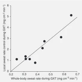
Figure 2. Correlation between whole-body sweat rate and local sweat rate on the left forearm (control) during the graded exercise test until exhaustion (GXT). The Pearson correlation test was used to obtain r = 0.92 and P = 0.001.
the control local sweat rate (left forearm) and whole-body sweat rate during GXT, with a correlation coefficient (r) of 0.92, P = 0.001.
As shown in Figure 3, positive and sig-nificant correlations were observed between A) the pilocarpine-induced local sweat rate at rest and whole-body sweat rate (r = 0.70, P = 0.03), and B) the pilocarpine-induced local sweat rate at rest (right forearm) and the control local sweat rate (left forearm) during GXT (r = 0.73, P = 0.02).
Discussion
The most important result of the present study was that the exercise stimuli induced a higher local sweat rate than pilocarpine stim-ulation (Figure 1). In addition, there was a positive and significant correlation between the pilocarpine-induced local sweat rate at rest and whole-body and local sweat rate during GXT (Figure 3), indicating that both variables could be used as an index of sweat gland output. However, GXT is a more widely used procedure for the measurement of aerobic power, for clinical diagnosis and for exercise prescription than pilocarpine iontophoresis, a method practically restricted to the diagnosis of cystic fibrosis and to scientific studies.
In the present study, the mean whole-body sweat rate during GXT was found to be similar to the pilocarpine-induced local sweat rate at rest. On the other hand, the higher local sweat rate observed during exercise (Figure 1) suggests that the mechanisms un-derlying the two sweat rates are different: the neural central stimuli of GXT seem to be greater than the local pilocarpine stimulus directly applied to the eccrine sweat glands. Thus, exercise-induced sweating is triggered by an integrated central-peripheral physi-ological mechanism. Moreover, the similar local sweat rate observed in pilocarpine-induced and control forearms during GXT indicates that pilocarpine did not act as an
additional stimulus besides the central stimuli from exercise on the eccrine sweat glands.
Pilocarpine is a cholinergic agonist able to directly stimulate the eccrine sweat gland
by activating acetylcholine receptors, with-out the need for stimulation of periglandular nerve endings. GXT is a physiological situ-ation corresponding to heat-producing physi-cal activities, which depends on the secre-tion of sweat by the eccrine sweat glands for the maintenance of homeostasis. Thus, the measurement of sweat gland output based on GXT seems to be more adequate to ex-press real physiological adjustments than measurement by pilocarpine induction.
Sweat production stimulated by the sym-pathetic autonomic nervous system (cholin-ergic) is known to depend on thermal and non-thermal mechanisms. Hypothalamic stimuli increase sweat production, which is proportional to the internal body tempera-ture (as a result of body metabolism) and skin temperature (as a result of thermal envi-ronmental stress) (15,16). Non-thermal stimuli such as certain emotional states and mental activities, the level of mental alert-ness and exercise-related factors (activation of central drive and muscle mechano- and metaboreceptors) also trigger sudorific re-sponses immediately after stimulation, and these responses are dissociated from the variation in body temperature (17-19). It is possible that the sweat production observed in the present study during GXT was the result of neural stimuli originating from ther-mal and non-therther-mal mechanisms and that
Figure 3. A, Correlation between the local sweat rate on the right forearm induced by pilocarpine at rest (REST) and whole-body sweat rate during the graded exercise test until exhaustion (GXT; r = 0.70, P = 0.03, Pear-son correlation test). B, Correla-tion between the local sweat rate on the right forearm induced by pilocarpine at rest and the local sweat rate on the left forearm (control) during GXT (r = 0.73, P = 0.02, Pearson correlation test).
the pilocarpine-induced sweat rate was only a consequence of local stimulation of the eccrine sweat glands.
Furthermore, exercise is known to in-duce higher concentrations of noradrena-line, adrenaline and vasoactive peptides (10) which might represent co-factors in the su-dorific response (1), variations in plasma osmolality (20), greater local blood flow (21), and increased temperature in the periglandular region (16). Thus, it could be hypothesized that these physiological fac-tors may play a role in the response of the eccrine sweat gland during exercise, con-tributing to the differences in sweating be-tween the two situations (REST and GXT). We observed wider intra-subject varia-bility in the sweat rate induced by the phar-macological agent at rest than in the sweat rate induced by exercise (Figure 3). This finding suggests a broader range of glandu-lar sensitivity to pilocarpine iontophoresis than to the central stimuli, related to biologi-cal variations (gland size, skin temperature, acclimation status, and training effect).
Another important finding was the posi-tive and significant correlation observed be-tween whole-body sweat rate and the local sweat rate on the left forearm (control), both produced by GXT (Figure 2), indicating that the forearm responded to exercise with a proportional sweat production. This finding
corroborates the known principle of univer-sal sweating in response to central thermal stimuli (17) and may indicate that the fore-arm is an area effectively participating in sweat production during exercise, as reported in the literature (22-24). Thus, this result shows that the measurement of forearm sweat rate reflects the whole-body sweat rate pro-duced during GXT.
In conclusion, the integrated central-pe-ripheral physiological mechanism during graded exercise elicited a greater sweating response than local stimulation with pilo-carpine. Thus, we suggest that local and whole-body sweat rate measured during graded exercise is more adequate to express physiological sudorific adjustments, being a better sweating index than pilocarpine.
References
1. Sato K, Kang WH, Saga K & Sato KT (1989). Biology of sweat glands and their disorders. I. Normal sweat gland function. Journal of the American Academy of Dermatology, 20: 537-563.
2. Sato K & Sato F (1990). Methods for studying eccrine sweat gland function in vivo and in vitro. Methods in Enzymology, 192: 583-599. 3. Buono MJ & Sjoholm NT (1988). Effect of physical training on peripheral sweat production. Journal of Applied Physiology, 65: 811-814.
4. Davis SL, Wilson TE, Vener JM, Crandall CG, Petajan JH & White AT (2005). Pilocarpine-induced sweat gland function in individuals with multiple sclerosis. Journal of Applied Physiology (in press). 5. Buono MJ, White C & Connolly KP (1991). Pilocarpine-induced
sweat rate at rest versus whole body sweat rate during exercise.
Journal of Applied Sport Science Research, 5: 82-86.
6. Hjortskov N, Jepsen LT, Nielsen B, Juul A & Skakkebaek NE (1995). Pilocarpine iontophoresis test: an index of physiological sweat se-cretion? Clinical Physiology, 15: 409-414.
7. Nielsen B, Strange S, Christensen NJ, Warberg J & Saltin B (1997). Acute and adaptive responses in humans to exercise in a warm, humid environment. Pflügers Archiv. European Journal of Physiolo-gy, 434: 49-56.
8. Armstrong LE, Maresh CM, Gabaree CV, Hoffman JR, Kavouras SA, Kenefick RW, Castellani JW & Ahlquist LE (1997). Thermal and circulatory responses during exercise: effects of hypohydration, de-hydration, and water intake. Journal of Applied Physiology, 82: 2028-2035.
9. Vimieiro-Gomes AC & Rodrigues LOC (2001). Avaliação do estado de hidratação dos atletas, estresse térmico do ambiente e custo calórico do exercício durante sessões de treinamento em voleibol de alto nível. Revista Paulista de Educação Física, 15: 201-211. 10. Rowell LB & O’Leary DS (1990). Reflex control of the circulation
during exercise: chemoreflexes and mechanoreflexes. Journal of Applied Physiology,69: 407-418.
11. Dubois D & Dubois EF (1916). A formula to estimate the approxi-mate surface area if height and weight be known. Archives of Internal Medicine, 17: 831-836.
12. Gibson LE & Cooke RE (1959). A test for concentration of electro-lytes in sweat in cystic fibrosis of the pancreas utilizing pilocarpine
by iontophoresis. Pediatrics, 23: 545-549.
13. American College of Sports Medicine (2000). Guidelines for Exer-cise Testing and Prescription. 6th edn. Lippincott Williams & Wilkins, Philadelphia, PA, USA.
14. Ramanathan NL (1964). A new weighting system for mean surface temperature of the human body. Journal of Applied Physiology, 19: 531-533.
15. Nadel ER, Bullard RW & Stolwijk JAJ (1971). Importance of skin temperature in the regulation of sweating. Journal of Applied Physi-ology,31: 80-87.
16. Nadel ER, Mitchell JW, Saltin B & Stolwijk JAJ (1971). Peripheral modifications to the central drive for sweating. Journal of Applied Physiology,31: 828-833.
17. Kuno Y (1956). Human Perspiration. Charles C. Thomas Publisher, Springfield, IL, USA.
18. Yamazaki F, Sone R & Ikegami H (1994). Responses of sweating and body temperature to sinusoidal exercise. Journal of Applied Physiology,76: 2541-2545.
19. Shibasaki M, Kondo N & Crandall CG (2003). Non-thermoregulatory modulation of sweating in humans. Exercise and Sport Sciences Reviews, 31: 34-39.
20. Fortney SM, Wenger CB, Bove JR & Nadel ER (1984). Effect of hyperosmolality on control of blood flow and sweating. Journal of Applied Physiology, 56: 1688-1695.
21. Sugenoya J, Iwase S, Mano T, Sugiyama Y, Ogawa T, Nishiyama T, Nishimura N & Kimura T (1998). Vasodilator component in sympa-thetic nerve activity destined for the skin of the dorsal foot of mildly heated humans. Journal of Physiology, 507: 603-610.
22. Cotter JD, Patterson MJ & Taylor NA (1995). The topography of eccrine sweating in humans during exercise. European Journal of Applied Physiology and Occupational Physiology, 71: 549-554. 23. Kondo N, Takano S, Aoki K, Shibasaki M, Tominaga H & Inoue Y
(1998). Regional differences in the effect of exercise intensity on thermoregulatory sweating and cutaneous vasodilation. Acta Physi-ologica Scandinavica, 164: 71-79.