Endogenous vasopressin and the
central control of heart rate during
dynamic exercise
Departamento de Fisiologia e Biofísica, Instituto de Ciências Biomédicas, Universidade de São Paulo, São Paulo, SP, Brasil
L.C. Michelini
Abstract
The present article contains a brief review on the role of vasopressinergic projections to the nucleus tractus solitarii in the genesis of reflex bradycardia and in the modulation of heart rate control during exer-cise. The effects of vasopressin on exercise tachycardia are discussed on the basis of both the endogenous peptide content changes and the heart rate response changes observed during running in sedentary and trained rats. Dynamic exercise caused a specific vasopressin content increase in dorsal and ventral brainstem areas. In accordance, rats pretreated with the peptide or the V1 blocker into the nucleus tractus
solitarii showed a significant potentiation or a marked blunting of the exercise tachycardia, respectively, without any change in the pressure response to exercise. It is proposed that the long-descending vasopressinergic pathway to the nucleus tractus solitarii serves as one link between the two main neural controllers of circulation, i.e., the central command and feedback control mechanisms driven by the peripheral receptors. Therefore, vasopressinergic input could contri-bute to the adjustment of heart rate response (and cardiac output) to the circulatory demand during exercise.
Correspondence
L.C. Michelini
Departamento de Fisiologia e Biofísica, ICB, USP Av. Prof. Lineu Prestes, 1524 05508-900 São Paulo, SP Brasil
Fax: 55 (011) 813-0845/818-7285 E-mail: [email protected]
Presented at the II International Symposium on Vasoactive Peptides, Ouro Preto, MG, Brasil,
October 6-8, 1997.
Publication supported by FAPESP.
Received January 29, 1998 Accepted March 4, 1998
Key words
•Exercise tachycardia •V1 receptors
•Brainstem areas •Peptides •Blood pressure •Training • Rats
Introduction
Mechanisms governing the reflex control of the circulation and their integration at the bulbar level have been extensively studied (see reviews 1-4). Today it is well known that cardiac output, total peripheral resis-tance and venous capaciresis-tance are regulated on a moment-to-moment basis to keep blood pressure (and volume) within a narrow range, maintaining adequate blood supply to differ-ent territories in any behavioral condition.
To maintain blood pressure and adjust flow efficiently, the central nervous system (CNS) processes peripheral information about pressure levels, blood gases, pH, blood
volume, temperature, etc. conveyed by dif-ferent sets of receptors such as barorecep-tors, chemorecepbarorecep-tors, cardiopulmonary re-ceptors, and others. This information is inte-grated in different areas and at different levels of the CNS to provide adequate changes of sympathetic and parasympathetic tone to the heart and blood vessels, the main effectors of the circulatory system.
Importance of baroreceptors and the genesis of reflex bradycardia
arterial mechanoreceptors are tonically ac-tive at baseline pressure since they are stim-ulated by the systolic stretching of the vessel during each cardiac cycle. The firing rate of afferents can be increased or decreased in-stantaneously according to the larger or smaller stretch of the wall, proportional to the transient increase or decrease in pressure from control levels.
Figure 1 shows both the pathways and main bulbar areas (with the respective exci-tatory and inhibitory amino acid neurotrans-mitters) involved in the primary circuitry of blood pressure control by baroreceptors. Si-nus and aortic nerve afferents (that join the IX and X cranial nerves, respectively) con-vey the encoded information on blood pres-sure levels to the nucleus tractus solitarii (NTS), the first synaptic relay in the brain-stem. Upon activation by a pressure increase, second-order neurons in the NTS excite: 1) the parasympathetic preganglionic neurons
located in the nucleus ambiguus (AMB) and dorsal motor nucleus of the vagus (DMV, not shown) determining an increase in vagal outflow to the heart and bradycardia, and 2) the inhibitory gabaergic neurons in the cau-dal ventrolateral medulla (CVLM) that project to the rostral ventrolateral medulla (RVLM). The RVLM is a major source of sympathetic premotor neurons projecting to the sympathetic pre-ganglionic neurons lo-cated in the intermediolateral cell column (IML) of the spinal cord. Inhibition of RVLM triggered by a pressure increase causes re-duction of sympathetic tone to blood vessels and heart, determining an increase in venous capacitance and a decrease in both total pe-ripheral resistance and cardiac output, with a marked bradycardic response. Opposite re-sponses, i.e., a decrease in venous capaci-tance (with an increase in venous return) and an increase in both total peripheral resis-tance and cardiac output, are observed
dur-EAA Baroreceptors
IX, X
Inhibitory inputs
Excitatory inputs
EAA
NTS
IML EAA
EAA
GABA
AMB
CVLM
RVLM
Heart and blood vessels Figure 1 - Schematic representation of afferents,
brainstem and spinal cord pathways and efferents that subserve cardiac and vasomotor components of the baroreceptor reflex. Proposed pathways that utilize excitatory (EAA) and inhibitory (Y-aminobu-tyric acid - GABA) amino acids as neurotransmitters are indicated. AMB = nucleus ambiguus, CVLM = caudal ventrolateral medulla, IML = intermediolat-eral cell column, NTS = nucleus tractus solitarii, RVLM = rostral ventrolateral medulla. Reproduced from Ref. 4, with permission.
ing a transient pressure decrease. In this case, the reduced or abolished baroreceptor discharge does not stimulate the parasympa-thetic preganglionic or the inhibitory gaba-ergic neurons.
It is important to note that activation of baroreceptors by a transient increase in pres-sure leads to a marked increase in vagal tone and withdrawal of sympathetic tone to the heart, causing strong reflex bradycardia.
Is reflex bradycardia always
observed during a pressure increase?
The answer is no. During dynamic exer-cise, for instance (see Figure 2), there is a moderate (10-20 mmHg) and abrupt increase in pressure, maintained throughout the exer-cise, that is not accompanied by bradycardia but by a marked tachycardia (increase of 100 beats/min or more). Tachycardia is essential to maintain increased cardiac output (and pressure) that provides appropriate flow for exercising muscles. Flow to skeletal muscles exhibits a large and prompt increase, even at
mild exercise intensity (0.4 km/h for rats, see Figure 2), an additional increase being ob-served at the transition from mild to moder-ate exercise intensity when the renal circula-tion (and other parts of the circulacircula-tion with flow in excess of metabolic activity) starts presenting vasoconstriction, contributing to an increase in flow to active territories.
It has been shown that normal blood pressure, heart rate, cardiac output and sys-temic vascular resistance responses to exer-cise require functional arterial baroreceptors (6-9). There is also some experimental evi-dence demonstrating that baroreceptors are working properly during exercise. In rats, loading of baroreceptors during running on a treadmill produced reflex bradycardia (10) and the sensitivity of the bradycardic re-sponse after an acute bout of exercise was of similar magnitude as that seen during the resting period (11). It has been proposed (8,12) that reflex sensitivity is maintained during exercise because the operating point of the arterial baroreflex is reset to higher pressures. However, the mechanism(s) that
1000
0 40
0
HR (bpm)
BF (ml/min)
250
0 150
50
0 20
0 0.4 km/h 0.8 km/h
0 2 4
Rest Exercise Recovery
0.4 km/h 0.8 km/h
0 2 4
Rest Exercise Recovery
Iliac probe Renal probe
1 min
AP (mmHg)
250
0
MAP (mmHg)
150
50 1000
would permit the coexistence of marked ta-chycardia with a moderate increase in blood pressure during exercise without changing the bradycardic protective response in the case of a further pressure challenge is (are) not known.
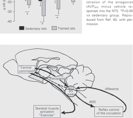
According to current theory, the circula-tory control during exercise is governed by two main neural mechanisms: a “central com-mand” and a feedback control mechanism driven by the receptors from cardiovascular areas and active muscles (9,12,13). As shown schematically in Figure 3, the “central com-mand” is a feed-forward control to set the basic pattern of motor activity for the skel-etal muscles. Skelskel-etal muscle activation stimulates receptors located within the muscles themselves and causes, directly or indirectly, changes in total peripheral resis-tance, leading to an increase in pressure which is accompanied by an increase in oxygen consumption, a reduction of venous capacitance with an increase in venous re-turn, and by heat production. These changes are continuously monitored by the
barore-ceptors, chemorebarore-ceptors, cardiopulmonary and thermosensitive receptors, respectively, which, together with the receptors located in active muscles, regulate the circulation in a reflex manner, determining appropriate changes in the autonomic control to the heart and vessels. Although it is likely that both feed-forward and feedback controllers of circulation should interact to regulate the arterial pressure and heart rate response dur-ing exercise, very little is known about this possible interaction. An attractive hypoth-esis, under investigation in our laboratory, is that central projections from integrative cen-ters, such as those from the hypothalamus to the cardiovascular relay areas in the brain-stem would serve as links between the “cen-tral command” and the feedback control driven by peripheral receptors (see dashed arrow in Figure 3).
Among the modulatory centers in the hypothalamus, the supraoptic (SON) and particularly the paraventricular nucleus (PVN) are of great importance in cardiovas-cular control. The PVN is a complex nucleus
”Central command“
NTS BS areas
ANS
Reflex control of the circulation
Afferents
Skeletal muscle activation
“Exercise”
∆ TPR ⇒ ⇑ BP ⇒ ⊕ baroreceptors ⇑ O2 consumption ⇒ ⊕ chemoreceptors
⇑ VR, ⇑ SV, ⇑ CO ⇒ ⊕ cardiopulmonary receptors ⇑ heat ⇒ ⊕ thermosensitive receptors
⇒ ⊕ skeletal muscle receptors Figure 3 - Schematic
with two distinct regions (14,15): the mag-nocellular region synthesizes vasopressin and oxytocin which are released into the blood via the neurohypophysis (16), while the neu-rons of the parvocellular region project to the brainstem areas (NTS, DMV, RVLM, IML) involved in the autonomic control of heart and vessels (17-22).
On the other hand, the NTS is a heteroge-neous cell group containing different types of neurons in the brainstem. It appears to be an important candidate site for the proposed interaction between forward and feed-back controllers of the circulation during exercise because: 1) it is the first synaptic relay of all peripheral afferents in the CNS (4,22,23); 2) it projects to brainstem areas controlling parasympathetic and sympathetic outflow (2,4); 3) it projects directly (and indirectly via bulbar, pontine and midbrain groups) to PVN and SON nuclei, amygdala and cortex (4,24-26), and 4) interestingly, it receives monosynaptic projections from the PVN (17-20). The reciprocal direct NTS → PVN, PVN → NTS innervation provides a prompt feedback control loop through which the PVN, an important hypothalamic inte-grative center involved in autonomic and neuroendocrine control, could also modu-late afferent cardiovascular inputs (level, type, distribution, etc.) coming from the pe-riphery to the NTS (26).
The long-descending monosynaptic pro-jections from the PVN to the NTS have been shown to contain vasopressin, oxytocin, en-kephalins and somatostatin (20-22). Most importantly, the PVN has been shown to be the only source of vasopressinergic projec-tions to the NTS (19,20); inside the NTS vasopressin is contained only in fibers which make axo-somatic and axo-dendritic con-nections with NTS neurons (27), an arrange-ment characteristic of a modulatory circuitry. Based on these neuroanatomical/immu-nohistochemical data, on the demonstration that vasopressin receptors are present at high
density in the NTS (28-31), on the knowl-edge that vasopressin has important cardio-vascular effects (32,33), and on our previous observations that peripheral and central va-sopressin administration produced hyperten-sion but quite different heart rate responses (34-36), our next question was:
Is vasopressin in the NTS involved in baroreceptor reflex control of heart rate?
To answer this question, some years ago we developed a technique to chronically cannulate the dorsal brainstem and to ad-minister peptides in the NTS of freely mov-ing rats (37). Administration of a suppressor dose of vasopressin restricted to the NTS (mimicking stimulation of the long-descend-ing vasopressinergic projections to this area) did not change the sensitivity of the barore-ceptor reflex control of heart rate, but dis-placed the set point of the reflex toward higher heart rate values (37). This response contrasts with both the potentiation of the bradycardic response after intravenous va-sopressin (38,39) and the blunting of reflex bradycardia following injection of vaso-pressin into the cerebrospinal fluid of con-scious rats (26,39, see also Table 1). The peripheral hormone vasopressin sensitizes
Table 1 -Vasopressin effects on baroreceptor reflex (BRS) control of heart rate in conscious rats.
NTS, Nucleus tractus solitarii.
Site of BRS effect Receptor involved References
administration
iv ↑ gain V2 38,39
Lateral ventricle ↓ gain V1 39
4thventricle ↓ gain not 26
determined
NTS gain unchanged V1 37
the baroreceptor reflex through V2 receptors
accessible from the blood, while neuronal V1 receptors show highly specific responses
such as inhibition of the reflex bradycardia by the neurohormone released into the cere-brospinal fluid, but displacement of the op-erating point of the reflex by the neurotrans-mitter vasopressin released directly in the NTS upon stimulation of projections origi-nating in the PVN. In addition, the lack of sensitivity for the bradycardic response dur-ing a pressure increase (gain not different from zero) observed after pretreatment of the NTS with a V1 blocker (37) indicates that
tonic activity of these projections is essential for the proper function of baroreceptor re-flex control of heart rate.
Therefore, the reply to this question is yes. Although neurohormonal vasopressin (as well as the peripheral hormone) could also influence the reflex control of the heart, it appears that vasopressinergic projections to the NTS are of major importance because they constitute a pathway by which the PVN could directly modulate baroreceptor func-tion (26). In addifunc-tion, by shifting the operat-ing point of the baroreflex control of the heart toward higher heart rate values, the
vasopressinergic input modulates the brady-cardic response to pressure challenges: dur-ing small pressure increases within a range of 10-20 mmHg (similar to that observed during dynamic exercise, see Figure 2) there was no bradycardic response, i.e., pressure increased without changes in basal heart rate (26,37). This effect is consistent with and not opposite to the appearance of exercise tachycardia during 10-20 mmHg pressure increase, as observed in Figure 2. On the other hand, in the same range of pressure change reflex bradycardia was significantly increased after endogenous blockade of V1
receptors in the NTS (26,37).
Based on these observations, one may speculate that an increased discharge of vasopressinergic neurons in the NTS during exercise may be relevant to the problem of the genesis of centrally mediated exercise tachycardia.
Are there changes in the
vasopressinergic input to the NTS during exercise?
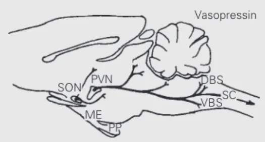
To deal with this query we quantitated the vasopressin content in different areas of the CNS and plasma of sedentary and trained rats sacrificed at rest or after acute exercise tests on a treadmill (40). The results obtained are summarized in Figure 4. Immediately after the acute exercise tests, which resulted in maximal HR in both the sedentary and trained groups (increases of 150-170 beats/min over baseline values), we observed specific in-creases in vasopressin content only in the dorsal (DBS) and ventral brainstem (VBS) areas, corresponding to the solitarii-vagal com-plex and ventrolateral medulla, respectively. There were no changes in the spinal cord, PVN, SON, median eminence, posterior pitu-itary and plasma vasopressin levels after exer-cise in the sedentary and trained groups. Exer-cise-induced changes in DBS and VBS vaso-pressin content during exercise were observed in both sedentary and trained groups, but
SON PVN
ME PP
DBS SC VBS Vasopressin
VP content immediately after acute exercise
S rats T rats
PVN ns ns
SON ns ns
ME ns ns
PP ns ns
DBS ns (+1.2 x) ⇑ 43 x*+ VBS ns (+3.2 x) ⇑ 6 x*+
SC ns ns
Plasma ns ns
were significantly higher only in trained rats. These results clearly demonstrate that vasopressinergic projections to the NTS are activated during dynamic exercise. Further-more, the specific increases of vasopressin content in DBS and VBS, with no detectable changes in the biosynthetic areas or in the magnocellular pathways to neurohypophy-sis or plasma levels (40), emphasize that the central vasopressin system exerts a differen-tial and specific control in special situations. Exercise has been shown to affect mostly the parvicellular vasopressinergic pathways from PVN to brainstem (40).
Interactions between vasopressin content and baroreceptor function were already docu-mented in a previous study (41) in which afferent input to the NTS region was inter-rupted via surgical denervation. Sinoaortic denervation produced differential changes in hypothalamic and brainstem vasopressin levels, with an increase in the brainstem and a decrease in the PVN and SON. It is not known whether alterations in peptide con-tent are associated with changes in the local (DBS/VBS) secretion of vasopressin, or whether the DBS and VBS changes in vaso-pressin content observed during exercise are associated with cardiovascular responses to acute exercise. Since our previous results showed that exogenous administration and endogenous blockade of arginine peptide (AVP) in the NTS were able to shift the operating point of the reflex during a tran-sient pressure increase, resulting in a smaller bradycardic response and an increased brady-cardic response, respectively, we next inves-tigated the role of AVP in exercise tachycar-dia.
Vasopressin in the NTS and its modulatory effect on exercise tachycardia
To determine whether changes in vaso-pressin content in the NTS are associated with cardiovascular responses to acute
exer-cise we studied the blood pressure and heart rate response to exercise of chronically in-strumented freely moving trained and seden-tary rats after pretreatment of the NTS with vasopressin or a V1 blocker (26,40).
Admin-istration of exogenous vasopressin in the NTS (mimicking an increased release into this area) specifically potentiated the tachycardic response without any change in blood pressure response during exercise. This effect was observed in both sedentary and trained rats. Most importantly (see Figure 5), in rats pretreated with a V1 blocker the
exer-cise tachycardia was significantly blunted without any change in the pressure response. Blunting of the tachycardic response was observed in both sedentary and trained rats but was significantly larger in the trained rats (40; see Figure 6). Therefore, the functional results are consistent with vasopressin con-tent, since the peptide was significantly in-creased in the DBS of trained rats immedi-ately after acute exercise and endogenous blockade of vasopressin at this level caused a significantly larger decrease in the exercise tachycardia of trained rats.
It was also shown (40) that the effect of vasopressin in the NTS is mediated by V1
receptors and is specific for the tachycardic response during exercise because V1
recep-tor blockade did not change control levels of heart rate or mean arterial pressure (observed during the rest period) nor did it change the pressor response to exercise. During dynam-ic exercise, specifdynam-ic adaptive responses of the heart without any change in the pressure response were also observed in trained spon-taneously hypertensive rats (SHR) when com-pared with sedentary control rats (42).
least 5 times higher than any other outflow tested (43). This is a clear demonstration of the importance of the central control of the heart (heart rate, contractility, cardiac out-put) during exercise. In addition, our results concerning the changes in brainstem vaso-pressin content and specific potentiation or blunting of the heart rate response after va-sopressin or V1 blocker treatment in the NTS
(40) suggest that vasopressinergic projec-tions from the PVN to the NTS are part of the central mechanism that specifically modu-lates heart rate control during exercise.
It should be noted that immediately after acute exercise vasopressin content was also increased in the VBS. Although it is possible that suprabulbar vasopressinergic pathways could modulate heart rate control by a com-bined action on dorsal (afferent input) and ventral (efferent output) brainstem areas, the functional effects of vasopressin in the ven-tral brainstem remain to be determined.
∆
HR (bpm)
200
160
24 min 120
80
40
0
40
20
0
0.4 0.8 1.1 1.4 km/h
-4 -2 0 2 4 6 8 10 12 14 16 18 20 22
Recovery Exercise
Rest
T rats
VEH AVPant
∆
MAP (mmHg)
Figure 5 - Heart rate (HR) (upper panel) and mean arterial pres-sure (MAP) responses (lower panel) during progressive exer-cise (0.4 up to 1.4 km/h) in 7 trained rats pretreated with ve-hicle (VEH) or a V1 blocker (AVPant) into the NTS. Repro-duced from Ref. 40, with per-mission.
Conclusions
In summary, we observed that vasopressin is released in the NTS during acute exercise (40) and that increased content of vasopressin in the NTS by acting on V1 receptors caused
both a shift of the operating point of the arterial baroreflex toward higher heart rate values (determining a smaller bradycardic response during pressure increases without changing baroreflex sensitivity; 37) and a larger tachycardic response during exercise (26,40). It is not known whether these re-sponses are caused by subtraction of the vagal output and/or by an increase in sympa-thetic output. Preliminary results (Michelini LC, unpublished observations) on loading of baroreceptors in the presence of sympathetic or vagal blockade (propranolol or atropine iv
higher heart rate values only in rats blocked with atropine (intact sympathetic outflow), suggesting that vasopressin in the NTS fa-cilitates the sympathetic pathway or favors the inhibition of sympathetic withdrawal that usually occurs during transient blood pres-sure increases. It was also shown that tachy-cardia during dynamic exercise was due to instantaneous withdrawal of parasympathetic tone followed by a maintained increase of sympathetic tone (44-47). Possible contribu-tions of vagal and/or sympathetic tone to the modulation of exercise tachycardia by cen-tral vasopressinergic input remain to be de-termined.
We may clarify that the vasopressinergic system of the NTS is not the only central mechanism involved in the genesis of exer-cise tachycardia (and/or central adaptations to training) because an AVP antagonist (AVPant) in this area did not block the heart
rate response but only caused a partial blunt-ing (Figure 5). Several peptides have been identified in the solitarii-vagal complex (22) and some of them were shown to be present in the projections from the hypothalamus to this area (20). It is likely that other peptidergic systems may be involved in the genesis of exercise tachycardia. Kregel et al. (48) showed that corticotropin-releasing factor is important since a partial blunting of the tachycardic response was observed after intracerebroventricular administration of its receptor antagonist. The oxytocinergic sys-tem has also been shown to be involved in stress-induced tachycardia (49) and to inter-act with the central vasopressinergic system in cardiovascular control (50,51). Prelimi-nary results with oxytocin (52) administered into the solitarii-vagal complex showed that the peptide caused a significant blunting of the tachycardic response during dynamic exercise. Although the V1 blocker used in
our experiments is 100 times more potent as an antivasopressor than as an antioxytocic, we could not exclude possible effects due to a partial blockade of oxytocin in the
brain-∆
HR (bpm)
0
-10
-20
-30
-40
0.4 0.8 1.1 1.4 km/h
Sedentary rats Trained rats *
*
*
Figure 6 - Comparison of maxi-mal heart rate (HR) reduction during exercise in sedentary and trained rats pretreated with the V1 blocker. Bars represent the differential effect of admin-istration of the antagonist (AVPant minus vehicle re-sponse) into the NTS. *P<0.05 vs sedentary group. Repro-duced from Ref. 40, with per-mission.
”Central command“
NTS BS areas
ANS
Reflex control of the circulation
Afferents
Skeletal muscle activation “Exercise”
Vasopressin
Figure 7 - Proposed vasopressinergic link to adjust the feed-forward (“central command”) and feedback controllers of the circulation during exercise. The dashed line represents the peripheral encoded information (from afferents) from cardiovascular effectors conveyed by the nucleus tractus solitarii (NTS) and other brainstem areas (BS) to suprabulbar integrative centers. ANS, Autonomic nervous system.
stem. However, stimulation of oxytocin re-ceptors did not seem to be involved in the potentiation of exercise tachycardia since oxytocin administration into the solitarii-vagal complex (52) caused a significant blunt-ing of the tachycardia durblunt-ing dynamic exer-cise, a response opposite to that observed with AVP. Therefore, during dynamic exer-cise oxytocinergic projections to the NTS should be less activated or not activated at all and the mechanism underlying exercise ta-chycardia seems to be different from that determining stress tachycardia.
biochemi-cal and functional data summarized in this review indicate that the long-descending pro-jections from the PVN to the NTS in the brainstem are part of the central mechanism modulating baroreceptor reflex control of heart rate during exercise. As illustrated in Figure 7, we propose that the
vasopressiner-gic pathway to the nucleus tractus solitarii serves as one link between the “central com-mand” and the reflex control of the heart and of the circulation, contributing to the adjust-ment of the heart rate response (and cardiac output) to the circulatory demand during dynamic exercise.
References
1. Kirchheim HR (1976). Systemic arterial baroreceptor reflexes. Physiological Re-views, 56: 100-176.
2. Palkovits M (1980). The anatomy of cen-tral cardiovascular neurons. In: Fuke K, Goldstein M, Hökfelt B & Hökfelt T (Edi-tors), Central Adrenalin Neurons: Basic Aspects and their Role in Cardiovascular Functions. Pergamon Press, Oxford, 3-17.
3. Chalmers J & Pilowsky P (1991). Brain-stem and bulbospinal neurotransmitter systems in the control of blood pressure. Journal of Hypertension, 9: 675-694. 4. Dampney RAL (1994). Functional
organi-zation of central pathways regulating the cardiovascular system. Physiological Re-views, 74: 323-364.
5. Amaral SL & Michelini LC (1997). Valida-tion of transit-time flowmetry for chronic measurements of regional blood flow in resting and exercising rats. Brazilian Jour-nal of Medical and Biological Research, 30: 897-908.
6. Ludbrook J (1983). Reflex control of blood pressure during exercise. Annual Review of Physiology, 45: 155-168.
7. DiCarlo E & Bishop VS (1990). Exercise training enhances cardiac afferent inhibi-tion of baroreflex funcinhibi-tion. American Jour-nal of Physiology, 258 (Heart and Circula-tory Physiology, 27): H212-H220. 8. DiCarlo E & Bishop VS (1992). Onset of
exercise shifts operating point of arterial baroreflex to higher pressures. American Journal of Physiology, 262 (Heart and Cir-culatory Physiology, 31): H303-H307. 9. Rowell LB (1992). Reflex control of the
circulation during exercise. International Journal of Sports Medicine, 13 (Suppl I): S25-S27.
10. Krieger EM, Brum PC & Negrão CE (1998). Role of arterial baroreceptors during acute and chronic exercise. Biological Research (in press).
11. Silva GJ, Brum PC, Negrão EC & Krieger EM (1997). Acute and chronic effects of
exercise on baroreflexes in spontaneously hypertensive rats. Hypertension, 30 (Part 2): 714-719.
12. Rowell LB & O’Leary DS (1990). Reflex control of the circulation during exercise: chemoreflexes and mechanoreflexes. Journal of Applied Physiology, 69: 407-418.
13. Mitchell JH (1990). Neural control of the circulation during exercise. Medicine and Science in Sports and Exercise, 22: 141-154.
14. Swanson LW & Kuypers HGJM (1980). The paraventricular nucleus of the hypo-thalamus: Cytoarchitectonic subdivisions and organization of projections to the pi-tuitary, dorsal vagal complex and spinal cord as demonstrated by retrograde fluo-rescence double-labeling methods. Jour-nal of Comparative Neurology, 194: 555-570.
15. Swanson LW & Sawchenko PE (1980). Paraventricular nucleus: A site for the in-tegration of neuroendocrine and auto-nomic mechanisms. Neuroendocrinology, 31: 410-417.
16. Morris JF, Chapman DB & Sokol HW (1987). Anatomy and function of the clas-sic vasopressin secreting hypothalamus-neurohypophyseal system. In: Gash DM & Boer GI (Editors), Vasopressin. Prin-ciples and Properties. Plenum, New York, 1-89.
17. Buijs RM, Swaab DF, Dogterom J & van Leeuwen FW (1978). Intra and extrahypo-thalamic vasopressin and oxytocin path-ways in the rat. Cell and Tissue Research, 186: 423-433.
18. Nilaver G, Zimmerman EA, Witkins J, Michaels J, Hoffman D & Silverman AJ (1980). Magnocellular hypothalamic pro-jection to the lower brain stem and spinal cord of the rat. Immunocytochemical evi-dence for predominance of the oxytocin-neurophysin system compared to the va-sopressin-neurophysin system. Neuroen-docrinology, 30: 150-158.
19. Sofroniew MV & Schrell U (1981). Evi-dence for a direct projection from oxyto-cin and vasopressin neurons in the hypo-thalamic paraventricular nucleus to the medulla oblongata: immunohistochemical visualization of both the horseradish per-oxidase transported and the peptide pro-duced by the same neurons. Neurosci-ence Letters, 22: 211-217.
20. Sawchenko PE & Swanson LW (1982). Immunohistochemical identification of neurons in the paraventricular nucleus of the hypothalamus that project to the me-dulla or to the spinal cord in the rat. Jour-nal of Comparative Neurology, 205: 260-272.
21. Palkovits M (1984). Distribution of neu-ropeptides in the central nervous system: A review of biochemical mapping studies. Progress in Neurobiology, 23: 115-189. 22. Van Giersbergen PLM, Palkovits M & De
Jong W (1992). Involvement of neuro-transmitters in the nucleus tractus solitarii in cardiovascular regulation. Physiological Reviews, 72: 789-824.
23. Miura M & Reis DJ (1969). Termination and secondary projections of carotid si-nus nerve in the cat brain stem. American Journal of Physiology, 217: 142-153. 24. Sawchenko PE & Swanson LW (1981).
Central noradrenergic pathways for the integration of hypothalamic neuroendo-crine and autonomic responses. Science, 214: 685-687.
25. Palkovits M (1988). Neuronal circuits in central baroreceptor mechanism. In: Saito H, Parvez H, Parvez S & Nagatsu T (Edi-tors), Progress in Hypertension. Vol. 1. VSP, Utretch, 387-409.
26. Michelini LC (1994). Vasopressin in the nucleus tractus solitarius: a modulator of baroreceptor reflex control of heart rate. Brazilian Journal of Medical and Biological Research, 27: 1017-1032.
Neurobiology of Vasopressin. Springer-Verlag, Berlin, 137-195.
28. Dogterom J, Snijdewint FGM & Buijs RM (1978). The distribution of vasopressin and oxytocin in the rat brain. Neurosci-ence Letters, 9: 341-346.
29. Van Leeuwen FW, Van der Beek EM, Van Heerikhuize JJ, Wolters P, Van der Meulen G & Wan Y-P (1987). Quantitative light microscopic autoradiographic local-ization of binding sites labelled with [3H] vasopressin antagonist d(CH2)5Tyr(Me)VP in the rat brain, pituitary and kidney. Neu-roscience Letters, 80: 121-126. 30. Tribollet E, Barberis C, Jard S,
Dubois-Dauphin M & Dreifuss JJ (1988). Localiza-tion and pharmacological characterizaLocaliza-tion of high affinity binding sites for vaso-pressin and oxytocin in the rat brain by light microscopic autoradiography. Brain Research, 442: 105-118.
31. Phillips PA, Abrahams JM, Kelly J, Paxinos G, Grzonka Z, Mendelsonhn FAO & Johnston CI (1988). Localization of vaso-pressin binding sites in rat brain by in vitro autoradiography using a radioiodinated V1 receptor antagonist. Neuroscience, 27: 749-761.
32. Cowley Jr AW, Liard JF, Skelton MM, Quinlen Jr EW, Osborn Jr JW & Webb RL (1985). Vasopressin-neural interactions in the control of cardiovascular function. In: Schrier RW (Editor), Vasopressin. Raven Press, New York, 1-10.
33. Cowley Jr AW & Liard JF (1987). Cardio-vascular actions of vasopressin. In: Gash DM & Boer GJ (Editors), Vasopressin: Principles and Properties. Plenum, New York, 389-433.
34. Michelini LC, Barnes KL & Ferrario CM (1983). Arginine vasopressin modulates the central action of angiotensin II in the dog. Hypertension, 11: I-75-I-79. 35. Ferrario CM, Mikami H, Michelini LC,
Kawano Y & Brosnihan KB (1985). Interac-tion of vasopressin with central neuro-genic mechanisms of blood pressure regulation. In: Schrier RW (Editor), Vaso-pressin. Raven Press, New York, 47-57.
36. Michelini LC, Barnes KL & Ferrario CM (1986). Area postrema lesions augment the pressor activity of centrally adminis-tered vasopressin. Clinical and Experi-mental Hypertension, Theory and Prac-tice, A8: 1107-1125.
37. Michelini LC & Bonagamba LGH (1988). Baroreceptor reflex modulation by vaso-pressin microinjected into the nucleus tractus solitarii of conscious rats. Hyper-tension, 11 (Suppl I): I-75-I-79.
38. Imai Y, Nolan PL & Johnston CI (1983). Restoration of suppressed baroreflex sen-sitivity in rats with hereditary diabetes in-sipidus (Brattleboro rats) by arginine vaso-pressin and DDAVP. Circulation Research, 53: 140-149.
39. Unger T, Rohmeiss P, Demmert G, Ganten D, Lang RE & Luft FC (1986). Dif-ferential modulation of the baroreceptor reflex by brain and plasma vasopressin. Hypertension, 8 (Suppl II): II-157-II-162. 40. Dufloth DL, Morris M & Michelini LC
(1997). Modulation of exercise tachycar-dia by vasopressin in the nucleus tractus solitarii. American Journal of Physiology, 273 (Regulatory, Integrative and Compara-tive Physiology, 42): R1271-R1282. 41. Alexander N & Morris M (1988). Effects
of chronic sinoaortic denervation on cen-tral vasopressin and catecholamine sys-tems. American Journal of Physiology, 255 (Regulatory, Integrative and Compara-tive Physiology, 24): R768-R733. 42. Veras-Silva AS, Mattos KC, Brum PC,
Negrão CE & Krieger EM (1998). Effect of exercise training on hemodynamic re-sponses during exercise in spontaneously hypertensive rats. American Journal of Physiology (in press).
43. Esler M, Jennings G, Lambert G, Meredith I, Horne M & Eisenholfer G (1990). Over-flow of catecholamine neurotransmitters to the circulation: Source, fate and func-tions. Physiological Reviews, 70: 963-985. 44. Maciel BC, Gallo Jr L, Marin-Neto JA, Lima Filho EC & Martins LEB (1986). Au-tonomic nervous control of the heart rate during dynamic exercise in normal man.
Clinical Science, 71: 457-460.
45. Gallo Jr L, Maciel BC, Marin-Neto JA & Martins LEB (1989). Sympathetic and parasympathetic changes in heart rate control during dynamic exercise induced by endurance training in man. Brazilian Journal of Medical and Biological Re-search, 22: 631-643.
46. Arai Y, Saul JP, Albrecht P, Hartley LH, Lilly LS, Cohen RJ & Colucci WS (1989). Modulation of cardiac autonomic activity during and immediately after exercise. American Journal of Physiology, 256 (Heart and Circulatory Physiology, 25): H132-H141.
47. Negrão CE, Moreira ED, Brum PC, Denadai MLDR &Krieger EM (1992). Va-gal and sympathetic control of heart rate during exercise by sedentary and exer-cise-trained rats. Brazilian Journal of Medi-cal and BiologiMedi-cal Research, 25: 1045-1052.
48. Kregel KCJ, Overton M, Seals DR, Tipton CM & Fisher LA (1990). Cardiovascular responses to exercise in the rat: role of corticotropin-releasing factor. Journal of Applied Physiology, 68: 561-567. 49. Morris M, Callahan MF & Lucion AB
(1995). Central oxytocin mediates stress-induced tachycardia. Journal of Neuroen-docrinology, 7: 455-459.
50. Poulin P & Pittman QJ (1993). Oxytocin pretreatment enhances arginine vaso-pressin-induced motor disturbances and arginine vasopressin-induced phosphoin-ositol hydrolysis in rat septum: a cross sensitization phenomenon. Journal of Neuroendocrinology, 5: 33-39.