Manual vibrocompression and nasotracheal suctioning in post-operative
period of infants with heart deffects
Vibrocompressão manual e aspiração nasotraqueal no pós-operatório de lactentes cardiopatas
Vibrocompresión manual y aspiración nasotraqueal en el post-operatorio de lactantes cardiópatas
Maíra Seabra de Assumpção1, Renata Maba Gonçalves1, Lúcia Cristina Krygierowicz2, Ana Cristina T. Orlando2, Camila Isabel S. Schivinski1
Instituição: Hospital Pequeno Príncipe e Universidade do Estado de Santa Catarina (Udesc), Florianópolis, SC, Brasil
1Udesc, Florianópolis, SC, Brasil
2Hospital Pequeno Príncipe, Curitiba, PR, Brasil ABSTRACT
Objective: To evaluate the impact of manual vibro-compression and nasotracheal suctioning on heart (hr) and respiratory (rr) rates, peripheral oxygen saturation (SpO2), pain and respiratory distress in infants in the postoperative period of a cardiac surgery.
Methods: Randomized controlled trial, in which the assessments were performed by the same physiotherapist in two moments: before and after the procedure. The infants were randomly divided into two groups: Intervention (IG), with manual chest vibrocompression, nasotracheal suctioning and resting; and Control CG), with 30 minutes of rest. Cardiorespiratory data (SpO2; hr; rr) were monitored and the following scales were used: Neonatal Infant Pain Scale (NIPS), for pain evaluation, and Bulletin of Silverman-Andersen (BSA), for respiratory distress assessment. The data were verified by analysis of variance
(ANOVA) for repeated measures, being significant p≤0.05.
Results: 20 infants with heart disease, ten in each group (seven acyanotic and three cyanotic) were enrolled, with ages ranging from zero to 12 months. In the analysis of the interac-tion between group and time, there was a significant difference in the variation of SpO2 (p=0.016), without changes in the other variables. Considering the main effect on time, only rr showed a significant difference (p=0.001). As for the group main effect, there were no statistical differences (SpO2 – p=0.77, hr – p=0.14, rr – p=0.17, NIPS – p=0.49 and BSA – p=0.51 ).
Conclusions: The manual vibrocompression and the nasotracheal suctioning applied to infants in postoperative
of cardiac surgery did not altered SpO2 and rr, and did not
trigger pain and respiratory distress. [Brazilian Registry of Clinical Trials (ReBEC): REQ: 1467].
Key-words: physical therapy modalities; infant; thoracic surgery; pain; postoperative care.
RESUMO
Objetivo: Verificar a repercussão da vibrocompressão manual e da aspiração nasotraqueal sobre os parâmetros car-diorrespiratórios de frequência cardíaca (fc) e respiratória (fr), saturação periférica de oxigênio (SpO2), dor e desconforto res-piratório, em lactentes no pós-operatório de cirurgias cardíacas.
Métodos: Estudo controlado e randomizado, com as avaliações realizadas pela mesma fisioterapeuta, em dois momentos: antes e após o procedimento. Dividiu-se o total de lactentes, por sorteio simples, em dois grupos:
Interven-ção (GI), com vibrocompressão manual torácica, aspiração
nasotraqueal e repouso; e Controle (GC), com 30 minutos de
repouso. Avaliaram-se os dados cardiorrespiratórios (SpO2;fc; fr), aplicando-se as escalas: Neonatal Infant Pain Scale (NIPS), para analisar a dor, e Boletim de Silvermann-Andersen (BSA), para verificar o desconforto respiratório. Os dados foram tratados por meio de análise de variância (ANOVA) para medidas repetidas, sendo significante p≤0,05.
Endereço para correspondência: Maíra Seabra de Assumpção
Rua Desembargador Pedro Silva, 2.034, bloco 07, apto: 24 – Coqueiros CEP 88080-700 – Florianópolis/SC
E-mail: [email protected]
Conflito de interesse: nada a declarar
Resultados: Avaliaram-se 20 lactentes cardiopatas, dez em cada grupo (sete acianóticos e três cianóticos), com idades de zero a 12 meses. Para analisar a interação entre o grupo e o tempo, observou-se diferença significativa na variação da SpO2 (p=0,016), sem alteração nas demais variáveis. Já o com-portamento dos parâmetros nos tempos apresentou diferença
significativa apenas na variação da fr (p=0,001). Quanto à
avaliação do efeito no grupo, não houve diferença
estatís-tica em nenhum dos dados (SpO2 – p=0,77; fc – p=0,14;
fr – p=0,17; NIPS – p=0,49 e BSA – p=0,51).
Conclusões: A vibrocompressão manual e a aspiração nasotraqueal aplicadas em lactentes no pós-operatório
de cirurgias cardíacas não prejudicaram a SpO2 e a fr,
além de não desencadearem dor e desconforto respirató-rio. [Registro Brasileiro de Ensaios Clínicos (ReBEC): REQ: 1467].
Palavras-chave: modalidades de fisioterapia; lactente; cirurgia torácica; dor; cuidados pós-operatórios.
RESUMEN
Objetivo: Verificar la repercusión de la vibrocompresión manual y de la aspiración nasotraqueal sobre parámetros cardiorrespiratorios de frecuencia cardíaca (fc) y respira-toria (fr), saturación periférica de oxígeno (SpO2), dolor y dificultad respiratoria, en lactantes en el post-operatorio de cirugías cardíacas.
Métodos:Estudio controlado y randomizado, con las eva-luaciones realizadas por la misma fisioterapeuta, en dos mo-mentos: antes y después del procedimiento. Se dividió el total de lactantes en dos grupos, por sorteo simple: Intervención
(GI), con vibrocompresión manual torácica, aspiración
nasotraqueal y reposo; y Control (GC), con 30 minutos de
reposo. Se evaluaron los datos cardiorrespiratorios (SpO2;fc; fr), aplicándose las escalas: Neonatal Infant Pain Scale (NIPS), para analizar el dolor, y Boletín de Silvermann-Andersen (BSA), para verificar la dificultad respiratoria. Los datos fueron tratados mediante análisis de variancia (ANOVA) para medidas repetidas, siendo significante p≤0,05.
Resultados: Se evaluaron 20 lactantes cardiópatas, diez en cada grupo (siete acianóticos y tres cianóticos), con eda-des de cero a 12 meses. Para analizar la interacción entre el grupo y el tiempo, se observó diferencia significativa en la variación de la SpO2 (p=0,016), sin alteración en las demás variables. El comportamiento de los parámetros en los tiempos, a su vez, presentó diferencia significativa
solamente en la variación de la fr (p=0,001). Respecto a
la evaluación del efecto en el grupo, no hubo diferencia
estadística en cualquiera de los datos (SpO2 – p=0,77;
fc – p=0,14; fr – p=0,17; NIPS – p=0,49 e BSA – p=0,512).
Conclusiones: La vibrocompresión manual y la aspiración nasotraqueal aplicadas a lactantes en el post-operatorio de
cirugías cardíacas no perjudican la SpO2 y la fr, además de
no desencadenar dolor y dificultad respiratoria.
Registro Brasileño de Ensayos Clínicos (ReBEC): REQ: 1467.
Palabras clave: modalidades de fisioterapia; lactante; cirugía torácica; dolor; cuidados post-operatorios.
Introduction
Congenital hearts diseases are one of the main causes of death in newborn infants(1) and, in most cases, their etiology
is not known yet. However, it is known that several causes are associated with their occurrence, such as prenatal and genetic factors, as well as maternal age(2).Congenital heart
diseases may be classiied and divided into two groups: acyanotic and cyanotic, based on pulmonary circulation conditions, such as blood volume, low, venocapillary pres-sure, and resistance(3).
In the last two decades, technological advances and improvements, both in the identiication and treatment of congenital heart diseases, contributed to the knowledge of their pathophysiology and their impact. In this sense, it is currently known that cardiac surgical corrections lead to a number of complications in the newborn and the infant, especially respiratory alterations. These complications are related to poor pulmonary and cardiac function in the pre-operative period, prolonged extracorporeal circulation, and high degree of sedation(4).
In this context, these children usually exhibit abnormali-ties of respiratory mechanics. Increases in pulmonary artery pressure and pulmonary blood low are associated with
de-creased lung compliance and inde-creased airway resistance(5).
Hence, alterations in cardiorespiratory data become evident, as well as the manifestation of signs of respiratory distress, deined, according to the Brazilian Society of Pediatrics, as increased respiratory rate, effort or inadequate thoracic excur-sion, decrease in peripheral breath sounds, groaning breath or shortness of breath, decreased level of consciousness or re-sponse to pain, decreased muscle tone, or the presence of cya-nosis(6). The analysis of these parameters is routine in the care
The measurement and quantiication of these data contrib-ute to more comprehensive evaluations, especially in the control of manipulations, therapies and interventions. Pain is another element that requires attention, because it causes behavioral changes due to the lability of this group and its clinical condition.
In face of impaired breathing, a common event in the postoperative period, respiratory physiotherapy is recom-mended and signiicantly acts for a better prognosis in pediatric patients who underwent cardiac surgery. Its role is recognized in the prevention and treatment of pulmonary complications, by means of speciic techniques(7,8). However,
in the pediatric population with heart disease, there are still few studies on the impact of conventional respiratory
tech-niques, described in the 1994 Consensus of Lyon(9) as those
designed to remove bronchial secretions, notably manual vibration, nasotracheal suctioning, postural drainage, chest percussion, chest compression, and cough. Despite of that, these are routine techniques in intensive care units(10), i.e.,
although the indication for physiotherapy in these patients is found in the literature(11-13), the impact of these techniques is
little discussed or addressed when it comes to infants di-agnosed with acyanotic or cyanotic heart diseases, in the postoperative period of cardiac surgery.
In view of these factors, the present study evaluated the impact of manual vibrocompression and nasotracheal suction-ing on cardiorespiratory parameters, such as heart rate (hr), respiratory rate (rr), and peripheral oxygen saturation (SpO2), as well as on respiratory distress and pain, in infants in the postoperative period of cardiac surgery. In this sense, we expect that conventional physiotherapeutic techniques — manual vibrocompression and nasotracheal suctioning — may provide a positive impact on the above-mentioned parameters.
Method
This is a randomized controlled trial, approved by the Research Ethics Committee (CEP 998-11) of Hospital
Pequeno Príncipe, Curitiba, Southern Brazil. Data from the cardiology ICU of the hospital were collected from November 2011 to February 2012. Before including the infants, their guardians were informed on the purposes, procedures, risks and beneits of the study and expressed their agreement in participating in the study by signing a written informed consent.
Infants of both genders from zero a 12 months and diag-nosed with acyanotic or cyanotic congenital heart disease in the postoperative period of cardiac surgery were included in the study (Table 1). All of them were extubated, hemody-namically stable, and were not using sedatives or analgesics on data collection. Patients started respiratory physiotherapy only after medical authorization and prescription.
We excluded infants with neurological and/or neuromus-cular diseases, syndromic alterations, open chest, hemody-namic instabilities and those who presented with anasarca or were on peritoneal dialysis. Table 2 describes heart diseases, according to groups, as for type of heart disease, surgical cor-rection, postoperative time (PO), extracorporeal circulation time (ECC), and mechanical ventilation time (MV).
Sample size calculation considered a variation of 3% in SpO2, with standard deviation equal to 3, taking into account a signiicance level of 5% and power of 85% in a two-tailed hypothesis test(14), obtaining a total of nine individuals in
each group.
After checking medical charts and evaluating patient’s clinical picture together with the medical team, the selected infants were randomly assigned into Control Group (CG) and Intervention Group (IG).
The CG remained at rest for 30 minutes. During this period, there was no manual contact, only visual observation of the parameters evaluated in the study. The IG underwent manual chest vibrocompression for ten minutes (rhythmic and rapid movements of isometric contraction of the forearm, manually applied on the anterior region of the chest, at the quadrants of right and left lung apices simultaneously, in the expiratory phase, associated with chest compression)(15).
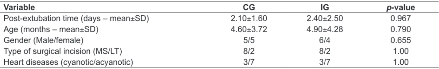
Table 1 - Demographic characteristics of the study patients
Variable CG IG p-value
Post-extubation time (days – mean±SD) 2.10±1.60 2.40±2.50 0.967 Age (months – mean±SD) 4.60±3.72 4.90±4.28 0.790
Gender (Male/female) 5/5 6/4 0.655
Type of surgical incision (MS/LT) 8/2 8/2 1.00
Heart diseases (cyanotic/acyanotic) 3/7 3/7 1.00
Then nasotracheal suctioning was performed (for approxi-mately 30 seconds)(16). This procedure lasted for ive minutes,
including the preparation of the materials, the beginning and the end of the maneuver, and the positioning of the infant on bed; afterwards, 15 additional minutes of rest were considered (visual observation by the examiner). Thus, the session had an overall duration of 30 minutes (manual chest vibrocompression, nasotracheal suctioning, and rest). The intervention was carried out only once, always by the same physiotherapist (MSA), who also conducted all the
described evaluations, always respecting the sequence of the procedures. The study considered the data of an only session for each infant.
Both groups were irst evaluated in terms of
cardiorespira-tory parameters (hr, rr and SpO2) and subsequently in terms
of signs of respiratory distress and pain, before and after intervention or rest (Tpre and Tpost respectively).
To evaluate cardiorespiratory parameters, hr and SpO2
were analyzed by checking the monitor available at the
cardiology ICU (Dixtal Monitor Dx2021®), recording
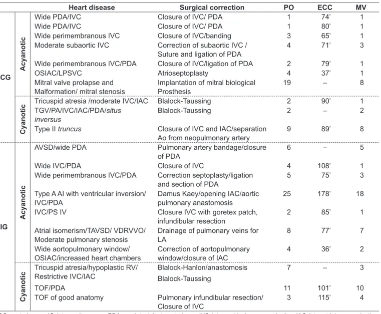
Table 2 - Description of cyanotic and acyanotic heart diseases according to groups, in terms of type of heart disease, surgical correction, postoperative time in days, extracorporeal circulation time in minutes and mechanical ventilation time in days
Heart disease Surgical correction PO ECC MV
CG
Acyanotic
Wide PDA/IVC Closure of IVC/ PDA 1 74’ 1
Wide PDA/IVC Closure of IVC/ PDA 1 80’ 1
Wide perimembranous IVC Closure of IVC/banding 3 65’ 1 Moderate subaortic IVC Correction of subaortic IVC /
Suture and ligation of PDA
4 71’ 3
Wide perimembranous IVC/PDA Closure of IVC/ligation of PDA 2 79’ 1
OSIAC/LPSVC Atrioseptoplasty 4 37’ 1
Mitral valve prolapse and Malformation/ mitral stenosis
Implantation of mitral biological Prosthesis
19 – 8
Cyanotic
Tricuspid atresia /moderate IVC/IAC Blalock-Taussing 2 90’ 1 TGV/PA/IVC/IAC/PDA/situs
inversus
Blalock-Taussing 2 – 2
Type II truncus Closure of IVC and IAC/separation Ao from neopulmonary artery
9 89’ 8
IG
Acyanotic
AVSD/wide PDA Pulmonary artery bandage/closure of PDA
6 – 5
Wide IVC/PDA Closure of IVC 4 108’ 1
Wide perimembranous IVC/PDA Correction septoplasty/ligation and section of PDA
5 75’ 3
Type A AI with ventricular inversion/ IVC/PDA
Damus Kaey/opening IAC/aortic pulmonary anastomosis
25 178’ 18
IVC/PS IV Closure IVC with goretex patch, infundibular resection
2 85’ 1
Atrial isomerism/TAVSD/ VDRVVO/ Moderate pulmonary stenosis
Drainage of pulmonary veins for LA
8 77’ 7
Wide aortopulmonary window/ OSIAC/increased heart chambers
Correction of aortopulmonary window/closure of IAC
4 36’ 2
Cyanotic
Tricuspid atresia/hypoplastic RV/ Restrictive IVC/IAC
Blalock-Hanlon/anastomosis
Blalock-Taussing
7 – 3
TOF/PDA 11 101’ 10
TOF of good anatomy Pulmonary infundibular resection/ Closure of IVC
3 115’ 4
CG: control group; IG: intervention group; PDA: persistent ductus arteriosus; IVC: interventricular communication; IAC: inter-atrial communication; OSIAC: ostiumsecundum inter-atrial communication; LPSV: left persistent superior vena cava; TGV: transposition of the great vessels; PA: pulmonary
atresia; AVSD: atrioventricular septal defect; TAVSD: total atrioventricular septal defect; Ao: aorta artery; AI: aortic insuficiency; PS IV: grade IV
the prevailing value during one minute. Rr was counted for one minute by observing infant’s chest and abdominal movements, in order to conirm the beginning and the end of each respiratory cycle.
Subsequently, signs of respiratory distress were investi-gated by the Bulletin of Silvermann-Andersen (BSA), used in other studies with infants(17-21) and described for use beyond
the neonatal period in the Série de Atualização de Reciclagem em Pneumologia of the Sociedade Paulista de Pneumologia e Tisiologia (SPPT), in 2011(22). The BSA assesses the
follow-ing items: expiratory gruntfollow-ing, nostril larfollow-ing, intercostal retraction, sternal retraction, and paradoxical breathing. Its score ranges from zero (no respiratory distress) to ten (maximum respiratory distress), with the score from one to ive being considered moderate distress, and, from six to ten, severe distress(23).
The Neonatal Infant Pain Scale (NIPS) was used to evalu-ate pain. This scale considers the following parameters: facial expression (zero or one point), cry (zero, one or two points), breathing patterns (zero or one point), position of legs (zero or one point), position of arms (zero or one point) and state of arousal (zero or one point). Pain is present when the score is higher than or equal to four(24).The NIPS was used
in infants based on the study by Pereira et al(25) and on the
document entitled Atenção à saúde do recém-nascido: Guia para proissionais de saúde(26). None of these references validate the
application of this scale beyond the neonatal period. Despite this limitation, its use is justiied by the lack of instruments of this nature in the aforementioned age group.
Data were analyzed using the Statistical Package for the Social Sciences (SPSS) software, version 20.0. Results were presented by descriptive and frequency statistics and expressed as means and standard deviation. The behavior
of the variables in the groups (IG and CG) and at the two time points of data collection, Tpre and Tpost, was assessed by analysis of variance (ANOVA) for repeated measures, with three main effects: time, group and the interaction of both, with signiicance set at p≤0.05.
Results
Twenty infants participated in the study, assigning ten in each group. Subjects’ age ranged from zero to 12 months,
with mean of 4.67±3.61 months in the CG (0.23 to 9.00)
and of 5.04±4.10 in the IG (0.50 to 11.00) (p=0.790). Table 1 shows age, sex distribution, extubation time, type of cor-rected heart disease and surgical incision.
Table 3 describes descriptive data for hr, rr and SpO2 in the IG and in the CG at the two study time points. There was no signiicant difference in SpO2 (p=0.77), hr (p=0.146),
rr (p=0.166), NIPS (p=0.4.88) and BSA (p=0.512) between
Tpre and Tpost, in any of the groups.
Considering the interaction between group and time, it was observed that the group in which the infant was as-signed at the two time points signiicantly interfered with the variation in SpO2 (p=0.016), with CG showing a decrease from 91.5±4.38% (at Tpre) to 90.5±6.22% (at Tpost); in the
IG, there was an increase from 89.0±8.73 to 91.2±8.26%,
at the Tpre and Tpost, respectively. On the other hand, with regard to the remaining parameters (hr, rr, NIPS and BSA),
group allocation did not have a signiicant effect (p=0.867;
0.585; 0.851; 0.170, respectively) (Table 3).
Considering the main effect on time, only rr showed a signiicant difference (p=0.001). In both groups, there was a decrease in this variable from Tpre to Tpost (from 56.2±51.0 to
51.0±11.65 in the IG and from 48.6±11.50 to 44.7±10.58
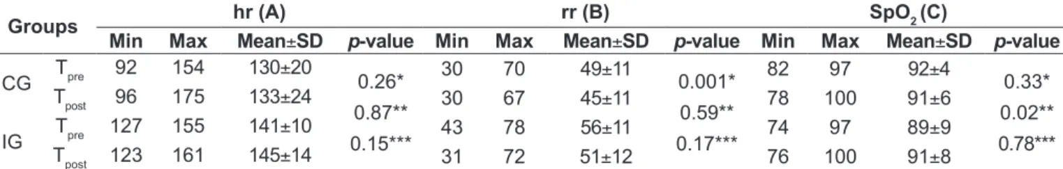
Table 3 - Descriptive data of the cardiorespiratory parameters hr, rr and SpO2, in IG and CG, before and after one of the
procedu-res (intervention or procedu-rest), and the procedu-result of the comparison of study parameters at Tpre and Tpost, according to the ANOVA test for
repeated measures
Groups hr (A) rr (B) SpO2 (C)
Min Max Mean±SD p-value Min Max Mean±SD p-value Min Max Mean±SD p-value
CG Tpre 92 154 130±20 0.26* 0.87** 0.15***
30 70 49±11
0.001*
0.59** 0.17***
82 97 92±4
0.33*
0.02** 0.78*** Tpost 96 175 133±24 30 67 45±11 78 100 91±6
IG Tpre 127 155 141±10 43 78 56±11 74 97 89±9 Tpost 123 161 145±14 31 72 51±12 76 100 91±8
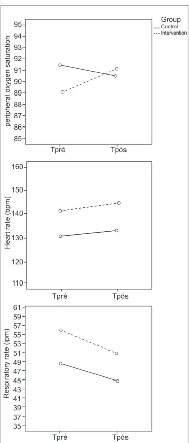
in the CG). However, this behavior was not observed in the other variables (Table 3). Figure 1 graphically represents the change in means for rr, hr and SpO2 before and after the procedures (rest or intervention), according to each group (CG and IG).
As for the main effect on the group, there was no signii-cant difference in any of the study variables: rr (p=0.166),
hr (p=0.146), SpO2 (p=0.777), NIPS (p=0.488) and BSA
(p=0.512) (Table 3).
In the IG, NIPS scores ranged from zero to seven (0.90±2.18); in the CG, this variation was also from zero to
seven points (1.70±2.11). In the IG, one infant had a score
compatible with pain at the beginning of the procedure and kept this pattern at the end of assessments. This also hap-pened with one infant in the CG. In addition, eight infants in this group did not have a score corresponding to pain, four of which did not change their score before and after the procedure. One infant who initially had a score compatible with the presence of pain had a score corresponding to the absence of the symptom at the end of the procedure.
As for BSA score for respiratory distress, three infants showed improvement in signs (from moderate to absence) and none of them showed moderate to severe distress after intervention in the IG. In the CG, four infants did not show changes in any of the time points and three showed improve-ment, with two cases from severe to moderate and one from moderate to absence.
Discussion
Despite the frequent recommendation of respiratory physiotherapy in the postoperative period of cardiac surgery and the wide range of physiotherapeutic techniques used to revert pulmonary dysfunction or negative pathophysiological changes(12,27) in this period, there is no consensus about the
best method to use(28).
Manual vibrocompression aims for a better lung dela-tion, to displace middle airways secretions to proximal airways and thus to the trachea, facilitating the process of
mucus elimination(29-31). This mechanism of action is an
important aspect to be considered when one aims to assess the eficacy of manual techniques in intensive care(32), like
in the present study.
In this study, manual vibrocompression was complement-ed with nasotracheal suctioning, because infants with heart disease have ineficient physiological mechanisms to remove secretions, and nasotracheal suctioning is recommended as Figure 1 - Behavior of mean values for respiratory rate (rr), heart
rate (hr) and peripheral oxygen saturation (SpO2) of infants with heart disease at the different time points (Tpre and Tpost), in each of the two study groups
Tpré
peripheral oxygen saturation
95 94
93 92
91 90
89 88
87 86
85
Heart rate (bpm)
110 120 130 140 150 160
Respiratory rate (ipm)
61 59 57 55 53 51 49 47 45 43 41 39 37 35
Tpós
Tpré Tpós
Tpré Tpós
Group
Control Intervention
an airway clearance method in these situations(16). The
con-cern to investigate the effect of combined physiotherapeutic techniques (manual vibrocompression and nasotracheal suc-tioning) had the purpose of portraying the routine care of physiotherapists in ICUs for the management of infants with heart disease, since compromised respiratory function and mechanics is a frequent inding in this clinical situation(11,33).
In view of the foregoing, it was observed that the results shown in this study did not have a negative impact on
cardio-respiratory parameters. There was an increase in SpO2 after
therapy, which did not happen with the group that did not receive the intervention (CG). Although this 2% increase (from 89% to 91%) has arguable clinical relevance, because it is a little variation that is within the margin of error of pulse oximetry, it become evident that physiotherapeutic procedures are not harmful for this type of patient, a topic that is routinely discussed in the postoperative manage-ment of patients with heart disease. This is because the physiotherapeutic procedures analyzed here did not cause clinical harms, such as hemodynamic instability, tachy-cardia or bradytachy-cardia, tachypnea and desaturation. On the contrary, there was an increase in oxygenation and reduction in respiratory rate, which deserves further investigations. The increase in sample size could reveal greater differences between the groups.
A study that evaluated the effects of manual vibration in infants with acute viral bronchiolitis, conducted by Pupin
et al(34), combined this maneuver with postural drainage and
compared them with the expiratory low increase technique (EFIT). In the sample of 27 infants who underwent manual vibration (intervention group), no beneit was observed in terms of oxygenation. None of the techniques used lead to an improvement in the cardiorespiratory parameters hr, rr
and SpO2. The authors reported that the effect of the two
therapies, when analyzed over time (ten minutes after the end of the procedure), seems to contribute to decrease only rr. Following this same line of studies evaluating the effects of manual vibration on cardiorespiratory parameters, Lanza
et al(35) evaluated the behavior of 13 preterm newborns after
performing the maneuver, for approximately 17 minutes. No detrimental effects were identiied after this therapy in terms of pain signs (according to the Neonatal Facial Coding
System scale) and of the parameters rr, hr and SpO2, which
remained within the normal range. The authors descriptively observed an increase in hr immediately after the maneuver,
as well as a decrease in rr and an increase in SpO2, which
remained after 30 minutes.
With regard to the effects of nasotracheal suctioning, Falcão and Silva(36) found a decrease in SpO
2 in 13 newborns
who underwent this procedure alone, compared to the group that received the same procedure, but combined with con-tainment maneuvers. Hr also had a greater variation after the use of nasotracheal suctioning alone.
It is important to emphasize that confronting the indings from the present research with studies involving different populations is justiied by the lack of clinical trials with children with heart diseases.
One of the few investigations on the subject was developed by Reines et al(37), who were not able to prove the beneits
of respiratory physiotherapy in patients who underwent surgical correction of congenital heart diseases. This study included 44 children, aged from three months to nine years and randomized into two groups. One of them underwent maneuvers of postural drainage, vibration, decubitus posi-tions, nasotracheal suctioning of airways, deep breathing, and requested or stimulated cough. In the other group, patients received only nasotracheal suctioning, deep breath-ing, and requested or stimulated cough, without physio-therapeutic maneuvers. According to the authors, respira-tory physiotherapy should be recommended to patients in whom its beneits have been proven, especially those with large amount of sputum production, and not as a routine for the prophylaxis of atelectasis in the postoperative period, because the group that received physiotherapeutic maneuvers developed atelectasis with a signiicantly higher frequency than the group that did not.
A more recent study involving infants with heart disease reported a series of 14 cases of babies diagnosed with acya-notic congenital heart diseases. They were randomized into three groups: control; placebo (only gentle manual contact in the chest); and intervention group, whose participants received two supports of the method of thoracic-abdominal rebalance (TAR) during ten minutes. Cardiorespiratory pa-rameters, respiratory distress and pain were assessed by the same instruments used in the present study. Results showed that the four infants in the intervention group had an increase in hr and SpO2 and a decrease in rr. None of the infants ex-pressed pain after the physiotherapeutic intervention with
TAR(38). Although this study analyzes a physiotherapeutic
technique different from that used in the present investiga-tion, both studies exhibit positive results with respiratory physiotherapy performed in this type of patient.
presence of respiratory distress, leads to changes in infant’s behavior. Therefore, its quantiication contributes to more comprehensive assessments. When it comes to heart diseases, pain is the main manifestation reported by patients who underwent cardiac surgery and has a multifactorial nature(39).
The factors that inluence in pain may be: surgical incision, intraoperative tissue retraction and dissection, multiple in-travenous cannulations, chest tubes, and invasive procedures that these patients undergo during their therapeutic regi-men(40).Considering that pediatric patients do not verbalize
their painful feeling and that it may be caused or potentiated by different procedures and manipulations, assessing it after the use of physiotherapeutic techniques is of great clinical relevance. In this sense, it was observed that manual vibro-compression and nasotracheal suctioning, applied in infants with heart disease, were not triggering factors for pain. The divergence between the indings from the present study and those from other publications may result from different clinical situations, patients’ age, and severity of the disease. Additionally, in this investigation, there was a concern about how to apply vibrocompression. Considering that the type of surgical incision may itself have an inluence on pain, the manual technique was carefully performed so that it was not applied with an aggressive manual pressure on infant’s chest. Regardless of the type of incision, both in lateral incision and in median sternotomy (which corresponded to 80% of the cases), we took the same level of care.
Another important issue refers to the scales used in this paper. Our small study sample included newborns and infants, applying scales for pain and respiratory distress validated for neonates (NIPS and BSA), since the literature does not provide speciic compatible tools that take exactly into account the second age group (infants). However, other researchers who evaluated infants used similar instruments in their investigations(17-21,25,26). These scales are simple, based
on the evaluation of clinical signs routinely observed in the management of the pediatric patient and, thus, we decided for its use in the entire sample, standardizing the assessment instrument. However, future studies should be conducted to
develop scales designed for this type of assessment in infants, contributing for the adequacy of methodology and of the results obtained in this line of research.
In view of the foregoing, the association of manual vibro-compression and nasotracheal suctioning was favorable in the postoperative of pediatric cardiac surgery, both due to
the increase in SpO2 and the decrease in rr and due to the
absence of detrimental effects in pain and respiratory dis-tress. Studies point out the effectiveness of physiotherapy in these cases, emphasizing its use as a routine procedure for the reduction in pulmonary complications(13,41,42). However,
there is little scientiic evidence about the most appropriate techniques and their effects in this context and on this age group. The existing studies also show small samples, vari-ability in therapeutic protocols and different clinical
situ-ations, which makes comparisons dificult. Its consensus(31)
that the choice for the pediatric therapy for airway clearance should have as basic principles anatomic and physiological differences, the existing pathological processes, and the speciicities of each age group(43).
It is important to point out some limitations of the study. Data collection was a dificult procedure, due to the complexity of the cases and the rigorous physiotherapeu-tic protocol. This prevented the sample from being larger. The classiication of patients’ clinical severity into mild, moderate or severe would also have contributed even more to the quality of the investigation. Another factor to be discussed refers to the use of neonatal scales (NIPS and BSA) in older children. The application of instruments speciic for infants may improve the accuracy of the results. Finally, the scarcity of investigations on the subject made it impossible to perform more detailed analyses and comparisons of the results obtained in this study with those obtained by other methods and conducts.
It can be considered that the physiotherapeutic techniques of manual vibrocompression and nasotracheal suctioning, applied to infants in the postoperative period of cardiac
surgery, did not alter SpO2 and rr in the treated group and
did not trigger pain signs and respiratory distress.
References
1. Silva ZM, Perez A, Pinzon AD, Ricachinewsky CP, Rech DR, Lukrafka JL et al. Factors associated with failure in ventilatory weaning of children undergone pediatric cardiac surgery. Rev Bras Cir Cardiovasc 2008;23:501-6. 2. Souza P, Scatolin BE, Ferreira LM, Croti UA. The nursing team relationship
with the child and the family in immediate postoperative period of congenital heart defects. Arq Cienc Saude 2008;15:163-9.
3. Vieira TC, Trigo M, Alonso RR, Ribeiro RH, Cardoso MR, Cardoso AC et al. Assessment of food intake in infants between 0 and 24 months with congenital heart disease. Arq Bras Cardiol 2007;89:219-24.
4. João PR, Faria Junior F. Immediate post-operative care following cardic surgery. J Pediatr (Rio J) 2003;79 (Suppl 2):S213-22.
Changes in respiratory mechanics among infants undergoing heart surgery. Anesth Analg 2004;98:49-55.
6. Sociedade Brasileira de Pediatria [homepage on the Internet]. Portal da Sociedade Brasileira de Pediatria [cited 2012 Dec 12]. Available from: http:// www.sbp.com.br/
7. Cavenaghi S, Moura SC, Silva TH, Venturinelli TD, Marino LH, Lamari NM. Importance of pre- and postoperative physiotherapy in pediatric cardic surgery. Rev Bras Cir Cardiovasc 2009;24:397-400.
8. Moerman D, Clément de Cléty S. La kinésithérapie respiratoire chez l’enfant après chirurgie cardiaque congénitale. Réanimation 2010;19:179-84. 9. Feltrim MI, Parreira VF. Fisioterapia respiratória. Proceedings of the 1ª
Conferência de Consenso em Fisioterapia Respiratória; 1994 Dec 2-3; Lyon, França. p. 8-47.
10. Zimmerman AT, Ibsen LM. Advances in postoperative care of pediatric cardiac patients. Curr Opin Anaesthesiol 2004;17:241-6.
11. Felcar JM, Guitti JC, Marson AC, Cardoso JR. Preoperative physiotherapy in prevention of pulmonary complications in pediatric cardic surgery. Rev Bras Cir Cardiovasc 2008;23:383-8.
12. Ribeiro IF, de Melo AP, Davidson J. Chest physical therapy in newborn infants with patent ductus arteriosus and pulmonary complications. Rev Paul Pediatr 2008;26:77-83.
13. Arcêncio L, Souza MD, Bortolin BS, Fernandes AC, Rodrigues AJ, Evora PR. Pre-and postoperative care in cardiothoracic surgery: a physiotherapeutic approach. Rev Bras Cir Cardiovasc 2008;23:400-10.
14. Armitage P, Berry G, Matthews JNS. The planning of statistical investigations. In: Armitage P, Berry G, editors. Statistical methods in medical research. 2nd ed. Oxford: Blackwell; 1987. p. 179-85.
15. Postiaux G. Fisioterapia respiratória pediátrica: o tratamento guiado por ausculta pulmonar. 2nd ed. Porto Alegre: Artmed; 2004.
16. Autoria não referida. AARC clinical practice guideline. Nasotracheal suctioning - 2004 revision & update. Respir Care 2004;49:1080-4.
17. Ajambuja AZ, Parazzi PL, Ries LG, Schivinski CI. Immediate effects of thoraco-abdominal rebalance in children with gastroesophageal relux disease – case series report. ConScientiae Saude 2012;11:607-17.
18. Barbié L, Caillat-Miousse JL, Vion V. La détresse respiratoire du nourrisson atteint de bronchiolite: aspiration ou désobstruction rhino-pharyngée? Rev Kinesither 2009;9:49-54.
19. Lanza FC, Cadrobbi C, Gazzotti MR, Faria R, Luque A, Solé D. Respiratory physiotherapy for nurslings with bronchiolitis: should we do it or not? Mundo Saude 2008;32:183-8.
20. Gómez-y-López RE, Hernández-Sierra JF, Torres-Ruvalcaba BA, Martínez-Puente E, Martínez-Garcia MC. Uso de dexametasona y salbutamol nebulizados en bronquiolitis aguda: estudio clínico comparativo. Gac Med Mex 2007;143:189-92.
21. Osnaya-Romero N, de Jesus Medina-Hernández T, Flores-Hernández SS, León-Rojas G. Clinical symptoms observed in children envenomated by scorpion stings, at the children’s hospital from the State of Morelos, Mexico. Toxicon 2001;39:781-5.
22. Liberali J, Lanza FC. Atuação da isioterapia respiratória no paciente pediátrico hospitalizado: novas perspectivas. In: Nápolis LM, Chiavegato LD, Nascimento O, editors. Fisioterapia Respiratória. Série Atualização e Reciclagem em Pneumologia - SPPT. São Paulo: Atheneu; 2011. p. 59-70.
23. Almeida MF, Kopelman BI. Rotinas médicas: disciplina de pediatria neonatal da Escola Paulista de Medicina. São Paulo: Atheneu; 1994.
24. Lawrence J, Alcock D, McGrath P, Kay J, MacMurray SB, Dulberg C. The development of a tool to assess neonatal pain. Neonatal Netw 1993;12:59-66. 25. Pereira e Silva Y, Gomez RS, Máximo TA, Simões e Silva AC. Pain evaluation
in Neonatology. Rev Bras Anestesiol 2007;57:565-74.
26. Brasil - Ministério da Saúde. Secretaria de atenção à saúde - Departamento de ações programáticas e estratégicas. Atenção à saúde do recém-nascido: guia para os proissionais de saúde – intervenções comuns, icterícia e infecções. Brasília: Ministério da Saúde; 2011.
27. Wallis C, Prasad A. Who needs chest physiotherapy? Moving from anecdote to evidence. Arch Dis Child 1999;80:393-7.
28. Renault JA, Costa-Val R, Rossetti MB. Respiratory physiotherapy in the pulmonary dysfunction after cardic surgery. Rev Bras Cir Cardiovasc 2008;23:562-9.
29. McIlwaine M. Physiotherapy and airway clearance techniques and devices. Paediatr Respir Rev 2006;7 (Suppl 1):S220-2.
30. Van der Schans CP. Airway clearance: assessment of techniques. Paediatr Respir Rev 2002;3:110-4.
31. Johnston C, Zanetti NM, Comaru T, Ribeiro SN, Andrade LB, Santos SL. I Brazilian guidelines for respiratory physiotherapy in pediatric and neonatal intensive care units. Rev Bras Ter Intensiva 2012;24:119-29.
32. Shannon H, Stiger R, Gregson RK, Stocks J, Main E. Effect of chest wall vibration timing on peak expiratory flow and inspiratory pressure in a mechanically ventilated lung model. Physiotherapy 2010;96:344-9. 33. Oliveira PM, Held PA, Grande RA, Ribeiro MA, Bobbio TG, Schivinski CI.
Proile of children undergoing congenital heart surgery and analysis of their respiratory complications. Rev Paul Pediatr 2012;30:116-21.
34. Pupin MK, Riccetto AG, Ribeiro JD, Baracat EC. Comparison of the effect that two different respiratory physical therapy techniques have on cardiorespiratory parameters in infants with acute viral bronchiolitis. J Bras Pneumol 2009;35:860-7.
35. Lanza FC, Kim AH, Silva JL, Vasconcelos A, Tsopanoglou SP. Does chest vibration during respiratory physiotherapy in neonates cause pain? Rev Paul Pediatr2010;28:10-4.
36. Falcão FR, Silva MA. Contenção durante a aspiração traqueal em recém-nascidos. Rev Cienc Med Biol 2008;7:123-31.
37. Reines HD, Sade RM , Bradford BF, Marshall J. Chest physiotherapy fails to prevent postoperative atelectasis in children after cardiac surgery. Ann Surg 1982;195:451-5.
38. Coelho R, Assumpção MS, Gonçalves RM, Mondo JM, Schivinski CI. Cardiac infants undergoing support to the method of Thoracic-Abdominal-Rebalance (TAR). Ter Man 2012;10:154-60.
39. Baumgarten MC, Garcia GK, Frantzeski MH, Giacomazzi CM, Lagni VB, Dias AS et al. Pain and pulmonary function in patients submitted to heart surgery via sternotomy. Rev Bras Cir Cardiovasc 2009;24:497-505. 40. Mueller XM, Tinguely F, Tevaearai HT, Revelly JP, Chioléro R, von Segesser
LK. Pain location, distribution, and intensity after cardiac surgery. Chest 2000;118:391-6.
41. Silva ME, Feuser MR, Silva MP, Uhlig S, Parazzi PL, Rosa GJ et al. Pediatric cardiac surgery: what to expect from physiotherapeutic intervention? Rev Bras Cir Cardiovasc 2011;26:264-72.
42. Padovani C, Cavenaghi OM. Alveolar recruitment in patients in the immediate postoperative period of cardic surgery. Rev Bras Cir Cardiovasc 2011;26:116-21. 43. Schechter MS. Airway clearance applications in infants and children. Respir