w w w . r b o . o r g . b r
Original article
Reconstruction of the anterior cruciate ligament in skeletally
immature patients: an individualized approach
夽
,
夽夽
Osmar Valadão Lopes Júnior
∗, Paulo Renato Saggin, Gilberto Matos do Nascimento,
André Kuhn, José Saggin, André Manoel Inácio
Instituto de Ortopedia e Traumatologia de Passo Fundo, Passo Fundo, RS, Brazil
a r t i c l e
i n f o
Article history:
Received 26 August 2012 Accepted 15 August 2013 Available online 2 May 2014
Keywords:
Reconstruction
Anterior cruciate ligament Orthopedic procedures
a b s t r a c t
Objective:to evaluate a series of skeletally immature patients who underwent three surgical techniques for anterior cruciate ligament (ACL) reconstruction according to each patient’s growth potential.
Methods:a series of 23 skeletally immature patients who underwent ACL reconstruction surgery at ages ranging from 7 to 15 years was evaluated prospectively. The surgical tech-nique was individualized according to the Tanner sexual maturity score. The surgical techniques used were transphyseal reconstruction, partial transphyseal reconstruction and extraphyseal reconstruction. Four patients underwent the extraphyseal technique, seven the partial transphyseal technique and twelve the full transphyseal technique, on the ACL. The postoperative evaluation was based on the Lysholm score, clinical analysis on the knee and the presence of angular deformity or dysmetria of the lower limb.
Results:the mean Lysholm score was 96.34 (±2.53). None of the patients presented dif-ferences in length and/or clinical or radiographic misalignment abnormality of the lower limbs.
Conclusion:ACL reconstruction using flexor tendon grafts in skeletally immature patients provided satisfactory functional results. Use of individualized surgical techniques according to growth potential did not give rise to physeal lesions capable of causing length discrepan-cies or misalignments of the lower limbs, even in patients with high growth potential.
© 2014 Sociedade Brasileira de Ortopedia e Traumatologia. Published by Elsevier Editora Ltda. All rights reserved.
Reconstruc¸ão do ligamento cruzado anterior em pacientes
esqueleticamente imaturos: uma abordagem individualizada
Palavras-chave:
Reconstruc¸ão
Ligamento cruzado anterior Procedimentos ortopédicos
r e s u m o
Objetivo:avaliar uma série de pacientes esqueleticamente imaturos submetidos a três téc-nicas cirúrgicas de reconstruc¸ão do ligamento cruzado anterior (LCA) de acordo com o potencial de crescimento de cada paciente.
夽
Please cite this article as: Lopes Júnior OV, Saggin PR, Matos do Nascimento G, Kuhn A, Saggin J, Inácio AM. Reconstruc¸ão do ligamento cruzado anterior em pacientes esqueleticamente imaturos: uma abordagem individualizada. Rev Bras Ortop. 2014;49:252–259.
夽夽
Work performed at the Knee and Foot Surgery Service, Instituto de Ortopedia e Traumatologia de Passo Fundo, RS, Brazil. ∗ Corresponding author.
E-mail: ovlopesjr@yahoo.com.br, scjp@iotrs.com.br (O.V. Lopes Júnior).
Métodos: foram avaliados prospectivamente 23 pacientes com idade de sete a 15 anos esqueleticamente imaturos submetidos à cirurgia de reconstruc¸ão do LCA. A técnica cirúr-gica foi individualizada de acordo com o escore de maturac¸ão sexual de Tanner. As técnicas cirúrgicas usadas foram a reconstruc¸ão transfisária (TF), a transfisária parcial (TFP) e a extrafisária (EF). Quatro pacientes foram submetidos à EF, sete à TFP e 12 à TF. Avaliac¸ão pós-operatória foi baseada no escore de Lysholm, na análise clínica do joelho e na presenc¸a de deformidade angular ou dismetria do membro inferior.
Resultados:a média do escore de Lysholm foi de 96,34 (±2,53). Nenhum paciente apresentou diferenc¸a de comprimento e/ou alterac¸ão clínica ou radiográfica de mau alinhamento dos membros inferiores.
Conclusão: a reconstruc¸ão do LCA com o uso de enxerto de tendões flexores em pacientes esqueleticamente imaturos proporcionou resultados funcionais satisfatórios. O uso de téc-nicas cirúrgicas individualizadas de acordo com o potencial de crescimento não ocasionou lesão fisária capaz de determinar discrepância de comprimento ou mau alinhamento dos membros inferiores, mesmo em pacientes com alto potencial de crescimento.
© 2014 Sociedade Brasileira de Ortopedia e Traumatologia. Publicado por Elsevier Editora Ltda. Todos os direitos reservados.
Introduction
Anterior cruciate ligament (ACL) injuries in skeletally imma-ture patients are still considered to be infrequent. Although some studies prior to the 1980s stated that ACL injuries in patients with open growth plates were rare findings, some authors of recent studies have reported greater incidence of complete ACL tears in skeletally immature patients, account-ing for 0.4% to 3.4% of the lesions.1–4 This increase in the incidence of ACL injuries observed in skeletally immature patients probably results from greater clinical suspicion, improved diagnostic methods and increased participation and demands on children in sports presenting risks of such injuries.
The ideal treatment for ACL tears in skeletally immature patients still remains controversial. Some series that have considered that conservative treatment should be used until the growth plates close have reported unsatisfactory results because the instability continues and secondary lesions develop on the menisci and joint cartilage.5–8Other authors have also highlighted that this group of patients presents low adherence to the care that is inherent to conservative treat-ment, particularly with regard to changes in physical activity, the need to use a brace or the frequency of attendance of muscle strengthening programs.9–11
Today, surgical treatment for anterior instability resulting from ACL injury is a promising reality based on evolution of the diagnostic methods and development of new operative techniques.12–18However, the time and the type of ACL recon-struction surgery to be performed on skeletally immature patients with a high potential for growth remains controver-sial matters. The presence of growth phases in the distal femur and the proximal tibia is a challenge for ACL reconstruction techniques in immature patients.
The growth cartilage of the distal femur and proximal tibia accounts for most of the lower-limb growth, and for the injuries. In such cases, injuries caused by constructing bone tunnels in ACL reconstruction surgery may result in prema-ture closure of the phase and in consequent unequal lengths and/or angular deformity around the knee.11
The present study had the aim of clinically evaluating a series of skeletally immature patients at different phases of growth who underwent three different ACL reconstruction techniques using autologous grafts from the flexor tendons. The techniques were chosen according to each patient’s growth potential.
Materials and methods
Twenty-three skeletally immature patients who underwent ACL reconstruction surgery to treat complete tears of the ligament were evaluated prospectively. The patients were operated between March 2005 and August 2010. Among the patients included, 19 were male and four were female. The inclusion criterion was the presence of extensively open growth phases at the time of the surgery. The exclusion criteria were a history of previous surgery on the knee involved, failure of the patient to return for clinical evaluations; and refusal to participate, expressed through the patient’s legal representa-tive. This study had previously been approved by the Research Ethics Committee of the University of Passo Fundo.
The patients’ mean age at the time of the surgery was 12.3 years (range: 7–15). Fourteen individuals had been injured on the right knee and nine on the left knee. The mean length of time between the ACL injury and the surgery was 4.8±2.9 months. Twenty individuals (86.9%) suffered ACL injuries by twisting their knee during sports activities: 15 in soccer, three in volleyball, one in handball and one in tennis. Two individ-uals (8.6%) suffered injuries through falls from bicycles and one (4.3%; the youngest patient) tore the ACL in a fall down the stairs.
ACL injury
Tanner ≤2
Epiphyseal tunnel in tibia
Transphyseal tunnel in tibia
Transphyseal tunnels femur and tibia
over-the-top no femur
Tanner=3
over-the-top no femur
Tanner 4 or 5
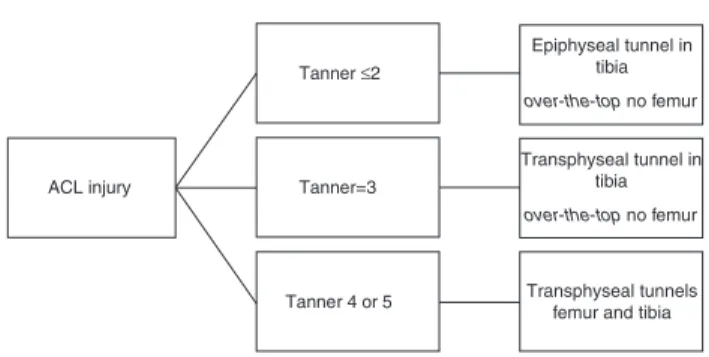
Fig. 1 – Protocol for choosing the surgical technique, according to age and Tanner’s sexual maturity score.
The surgical technique was individualized according to chronological age and Tanner’s sexual maturity score19(Fig. 1). In all cases, it was decided to use a quadruple graft from the gracilis and semitendinosus tendons, fixed in posts both in the femur and in the tibia. The surgical techniques used were transphyseal (TP), partial transphyseal (PTP) and extraphyseal (EP) reconstruction. Four patients underwent EP, seven PTP and 12 TP. The mean age was 9.25 years for the patients in the EP group, 12 for those in PTP and 13.5 for those in TP. The demographic data on the sample are laid out in Table 1.
Associated injuries were diagnosed and treated. Twelve patients presented meniscal injuries: eight in the medial meniscus and four in the lateral meniscus. In three cases (two in the lateral meniscus and one in the medial menis-cus); conservative treatment was instituted, given the stability of the injury.20 Partial meniscectomy was performed in
seven patients: five in the medial meniscus and two in the lateral meniscus. Two patients underwent suturing of the medial meniscus. Injury to the medial collateral ligament was diagnosed in two patients: conservative treatment was insti-tuted in one and surgical repair in the other.
The patients returned for postoperative clinical review after three, six and twelve months, and then annually until the end of their growth. At the end of the follow-up, all the patients underwent a functional assessment using the Lysholm score.21,22 Angular changes in the sagittal and/or coronal plane were evaluated clinically using the contralateral limb as the control. The lengths of the lower limbs were eval-uated by means of the distance between the anterosuperior iliac crest and the medial malleolus. It was considered that a difference of less than 10 mm in the lengths of the lower limbs would not represent a significant clinical discrepancy.
Surgical techniques
All the patients were positioned in dorsal decubitus under peridural anesthesia or general anesthesia in association with ipsilateral femoral nerve block. A pneumatic tourniquet was used on the proximal third of the thigh. After antisepsis and placement of sterile fields, the lower limb was placed at 45◦ of hip flexion and 90◦ of knee flexion. Arthroscopy
was performed on the knee by means of anterolateral and anteromedial portals. After confirmation of the diagnosis of tearing of the ACL, the associated injuries to the menisci and cartilage were duly diagnosed and treated. By means of an
Table 1 – Demographic data.
Techniquea Patient Age (years) Sex Side Tanner Length of time with
injury (months)b
EP 1 9 M R 1 3
2 7 M R 1 3
3 11 M L 2 4
4 10 M L 2 3
PTP 5 12 M R 3 3
6 12 M R 3 4
7 12 M R 3 3
8 12 M L 3 2
9 12 M R 3 2
10 12 M L 3 6
11 12 F L 3 4
TP 12 14 M R 4 5
13 14 M R 4 3
14 13 F R 4 12
15 13 M L 4 4
16 15 F R 4 4
17 13 M R 4 2
18 13 M L 4 7
19 14 M R 4 12
20 13 M L 4 6
21 14 M R 4 10
22 14 F L 4 5
23 13 M R 4 5
a EP, extraphyseal reconstruction of the ACL; PTP, partial transphyseal reconstruction of the ACL; TP, total transphyseal reconstruction of the
ACL.
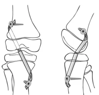
Fig. 2 – Illustration demonstrating the transphyseal (TP) technique for ACL reconstruction.
incision of approximately 2 cm in the anteromedial aspect of the proximal tibia, the gracilis and semitendinosus ten-dons were separated out and removed. Krackow sutures were performed23 using Ethibond 2-0 thread at the extremity of each tendon graft. The tendon grafts were folded over to form a quadruple graft of at least 10 cm in length. The graft diame-ter was measured proximally and distally. The remains of the ACL were identified and debrided. From this point onwards, the techniques varied according to the group of patients, as follows.
Transphyseal reconstruction technique (TP)
The centers of the femoral and tibial insertions of the ACL were identified. To construct the femoral tunnel, a guidewire was introduced through the anteromedial portal at the cen-ter of the femoral insertion of the ACL. With the knee flexed at 120◦, the femoral tunnel was constructed with a length of
approximately 20–30 mm and the same diameter as the prox-imal portion of the graft. The tibial tunnel was constructed by means of a guidewire that was introduced using a tib-ial guide at an angle of 55◦, centered on the tibial insertion
of the ACL. The tibial tunnel also presented the same diam-eter as the graft, measured previously. The graft was then passed through the bone tunnels and was fixed in a post in the femur, by means of a lateral accessory incision; and in the tibia, using a 4.5 mm cortical screw and washer. The tib-ial fixation was performed with the knee positioned at 30◦of
flexion (Fig. 2).
Partial transphyseal reconstruction technique (PTP)
In this technique, no femoral tunnel was constructed. The tibial tunnel was constructed in the same way as described in the TP technique. In relation to the femur, the graft was passed around posteriorly to the lateral condyle (over-the-top
Fig. 3 – Illustration demonstrating the partial transphyseal (PTP) technique for ACL reconstruction.
position), and was fixed on the lateral face of the femur in the same way as described previously (Figs. 3 and 4).
Extraphyseal reconstruction technique (EP)
In this technique too, no femoral tunnel was constructed. The tibial tunnel was constructed by means of a guidewire that was introduced above the proximal phase of the tibia, with the aid of fluoroscopy, and was directed to the center of the tibial insertion of the ACL (Fig. 5). The diameter of the tibial tunnel was the same as that of the graft. The graft was passed around posteriorly to the lateral condyle (over-the-top position) and was fixed using the same technique as described previously (Figs. 6 and 7).
Results
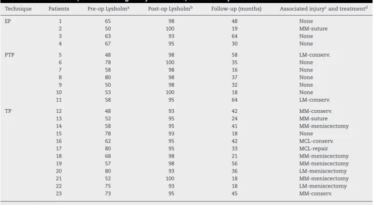
After a mean follow-up of 35.4±15.3 months, the mean score from the Lysholm functional evaluation was 96.3±2.5 (Table 2). All the patients presented a hard stop in the Lach-man test and only two (8.6%), who were both in the TP group, presented a +/4 result in the pivot test (patients 14 and 20). However, neither of these patients presented any complaint of clinical instability of the knee. Nineteen patients (82.6%) returned to their pre-injury physical activity levels and four (17.4%) reported that they had not returned to sports practice at the same level as before the injury, even though the knee was considered stable according to the clinical criteria and functional score. None of the patients presented dysmetria of the lower limbs greater than 10 mm. No clinical or radio-graphic sign of misalignment in the coronal and/or sagittal planes was observed.
Fig. 4 – Anteroposterior (A) and lateral (B) X-rays on a 12-year-old patient with Tanner 3 who underwent ACL reconstruction using the PTP technique. Note the verticalized and centralized tibial tunnel.
implant, which led to complete remission of the symptoms. Furthermore, one of the patients who underwent meniscal suturing presented recurrence of the lesion in the posterior cornu of the medial meniscus two years after the operation, and was then treated with partial meniscectomy of the medial meniscus (patient 13).
Discussion
Even with increasing numbers of cases, ACL injuries in skeletally immature patients are still considered to be uncommon.20This low incidence has meant that there is no consensus regarding its management, particularly in dealing with patients with a high potential for growth.
It is important to emphasize that although choosing con-servative treatment eliminates the iatrogenic possibilities of surgery, the results obtained are generally unsatisfactory.2,5,23 Kannus and Jarvinen23evaluated 33 patients with ACL tears that were treated conservatively and most of these patients presented unsatisfactory results because of the recurrent instability. One third of the patients already showed degen-erative alterations on radiological examinations at the end of the follow-up.
Surgical treatment of ACL injuries in patients with the open phase requires special attention because of the possibility of iatrogenic injury to the growth plate during construction of the bone tunnels. It is important to emphasize that each patient’s potential for growth, which is assessed from the physiologi-cal skeletal maturity, may have a standard deviation of around one year for each chronological age.24Skeletal maturity should be assessed before the surgery, and the risk of damage to the growth cartilage should be gauged. It is known that approx-imately 65% of the growth of the lower limb occurs around the knee,25and perforation of the physis may cause localized
epiphysiodesis and leads to growth discrepancy (complete epi-physiodesis) or angular deformity (partial epiepi-physiodesis). In a study using magnetic resonance imaging, Sasaki et al.26 showed that the growth physes around the knee did not show any sign of closure in patients under the age of 11 years. Only 34% of the physes of the knee were closed at the age of 13 years, and the physes were only completely closed over the age of 16 years. In this same study, the authors also emphasized that closure of the physis took place from the central portion toward the peripheral portion.
The ideal time for surgical intervention, and also the choice of technique to minimize the damage to the growth cartilage, is an important factor in the planning. Some authors have
Fig. 6 – Illustration demonstrating the extraphyseal technique (EP).
recommended that the reconstruction should be done closer to the time of skeletal maturity, in order to diminish the risk of damage to the growth cartilage.10,19,27Woods et al.27observed that there were 20% more meniscal injuries in patients whose surgery was postponed for more than six months, in com-parison with a group that underwent earlier reconstruction
with restrictions on activities. Millet et al.10showed that there was an association between medial meniscal injury and the length of time from the injury to the reconstruction. McIn-tosh et al.28did not observe any development of new meniscal injuries after ACL reconstruction, which justified their deci-sion to perform early surgical intervention. In the present study, 12 patients presented meniscal injury at the time of the surgery, which may have been related to greater chronicity among our cases.
Use of a graft composed only of soft tissues, without any presence of bone material, is another factor that contributes toward diminishing the risk of disturbances during the growth phase.17,19,29McIntosh et al.28obtained satisfactory results by using grafts from autologous flexor tendons, with only one case of discrepancy of length in the lower limbs.
In a systematic review, Vavken et al.30evaluated 47 stud-ies relating to top conservative and surgical treatment of ACL injuries in skeletally immature patients. The conservative treatment was correlated with unsatisfactory clinical results and with high incidence of secondary meniscal and carti-lage lesions. A few studies have correlated surgical treatment with growth disorders and have shown strong evidence that surgical treatment is correlated with good functional results and joint stability. In those authors’ evaluation, no specific surgical technique was shown to be superior. Among the stud-ies evaluated, only nine included patients who underwent techniques that preserved the growth cartilage. Thirty-one studies reported results from patients after ACL reconstruc-tion with at least one transphyseal tunnel. In this group of
Table 2 – Clinical results, evaluated using the Lysholm score, associated injuries and treatment.
Technique Patients Pre-op Lysholma Post-op Lysholmb Follow-up (months) Associated injurycand treatmentd
EP 1 65 98 48 None
2 50 100 19 MM-suture
3 63 93 64 None
4 67 95 30 None
PTP 5 48 98 58 LM-conserv.
6 78 100 35 None
7 58 98 16 None
8 80 98 37 None
9 50 98 32 None
10 53 100 18 None
11 58 95 64 LM-conserv.
TP 12 48 93 42 MM-conserv.
13 52 95 24 MM-suture
14 58 95 41 MM-meniscectomy
15 78 93 18 None
16 62 95 42 MCL-conserv.
17 80 95 33 MCL-repair
18 68 98 21 MM-meniscectomy
19 57 98 56 MM-meniscectomy
20 80 93 36 LM-meniscectomy
21 52 100 18 MM-meniscectomy
22 75 93 18 LM-meniscectomy
23 73 95 45 MM-conserv.
a Preoperative Lysholm score.
b Postoperative Lysholm score.
c Associated injuries: MM, medial meniscal injury; LM, lateral meniscal injury; MCL, medial collateral ligament injury.
Fig. 7 – X-ray (A) and MRI (B) of a nine-year-old patient with Tanner 1 who underwent EP reconstruction one year earlier.
approximately 500 patients of mean age 13.6±0.9 years, in whom at least one of the tunnels was constructed during the growth phase, only three patients presented angular deformi-ties and two showed growth disorders. Thus, the authors of the systematic review concluded that there was no significant difference in the results between patients with one and two transphyseal tunnels, and they considered the transphyseal technique to be safe.
Despite the evidence presented in the study by Vavken et al.,30 it has to be taken into consideration that the great majority of the published studies included presented evi-dence level IV and that, moreover, the patients’ mean age was approximately 13 years. There was only one study with evidence level II31 in this review. Few authors have solely evaluated patients with a high potential for growth (Tan-ner I and II).30 Liddle et al.31 and Streich et al.32 reported good clinical results using transphyseal techniques in patients with a high potential for growth, but the small number of patients in each series and the type of study design were limiting factors. In a recent retrospective study on 16 skele-tally immature patients with high potential for growth (Tanner 1 and 2), Hui et al.33 evaluated the results from arthro-scopic anatomical reconstruction using soft-tissue grafts. In this series, the authors did not find any alignment and growth disorders over a mean follow-up period of 25 months. All of the patients returned to their pre-injury activity levels.
In a recent systematic review, Moksnes et al.34emphasized the low quality of the studies available, according to the Cole-man methodology score. Out of the 31 studies included in this review, only two were prospective and no randomized clinical trials on this subject were found. In these authors’ opinion, greater attention needs to be given to study design and sam-ple size. There is a need for prospective observational studies to assess late and rare events.
In the study presented here, we considered that there was no strong scientific evidence that transphyseal ACL recon-struction techniques were completely safe in patients with high potential for growth. Thus, the surgical techniques were individualized according to each patient’s potential for growth. Individuals with high potential for growth should still
be operated on, but using techniques that present lower risk of damage to the growth cartilage.
Like most other studies involving treatment of ACL injuries in skeletally immature patients, this study presented some limitations: it was a case series with a relatively short follow-up and also involved a small number of patients. However, this is the first study in the Brazilian literature to describe different technical options among patients with different potentials for growth. We chose not to conduct a case-control study because of the current evidence that con-traindicates conservative treatment among these patients and the lack of strong evidence to show that transphyseal recon-struction would be safe for patients with a high potential for growth.
Conclusion
ACL reconstruction using grafts from flexor tendons in skele-tally immature patients produced satisfactory functional results. Use of techniques individualized according to the potential for growth, use of grafts composed only of soft tis-sue and fixation away from the physis are important factors to be born in mind in surgical management of ACL injuries in patients with a high potential for growth.
Conflicts of interest
The authors declare no conflicts of interest.
r e f e r e n c e s
1. DeLee JC, Curtis R. Anterior cruciate ligament insufficiency in children. Clin Orthop Relat Res. 1983;(172):112–8.
2. McCarroll JR, Rettig AC, Shelbourne KD. Anterior cruciate ligament injuries in the young athlete with open physes. Am J Sports Med. 1988;16(1):44–7.
3. Johnston DR, Ganley TJ, Flynn JM, Gregg JR. Anterior cruciate ligament injuries in skeletally immature patients.
4. Andrish JT. Anterior cruciate ligament injuries in the skeletally immature patient. Am J Orthop (Belle Mead, NJ). 2001;30(2):103–10. Review. Erratum in: Am J Orthop 2001;30(6):503.
5. Camanho GL, Olivi R, Camanho LF, Torres MR, Ribeiro Filho JE. Anterior cruciate ligament lesion of the knee in patients with immature skeleton. Acta Ortop Bras. 1999;7(4):152–8. 6. McCarroll JR, Shelbourne KD, Porter DA, Rettig AC, Murray S.
Patellar tendon graft reconstruction for midsubstance anterior cruciate ligament rupture in junior high school athletes. An algorithm for management. Am J Sports Med. 1994;22(4):478–84.
7. Mizuta H, Kubota K, Shiraishi M, Otsuka Y, Nagamoto N, Takagi K. The conservative treatment of complete tears of the anterior cruciate ligament in skeletally immature patients. J Bone Joint Surg Br. 1995;77(6):890–4.
8. Aichroth PM, Patel DV, Zorrilla P. The natural history and treatment of rupture of the anterior cruciate ligament in children and adolescents. A prospective review. J Bone Joint Surg Br. 2002;84(1):38–41.
9. Graf BK, Lange RH, Fujisaki CK, Landry GL, Saluja RK. Anterior cruciate ligament tears in skeletally immature patients: meniscal pathology at presentation and after attempted conservative treatment. Arthroscopy. 1992;8(2):229–33. 10. Millett PJ, Willis AA, Warren RF. Associated injuries in
pediatric and adolescent anterior cruciate ligament tears: does a delay in treatment increase the risk of meniscal tear? Arthroscopy. 2002;18(9):955–9.
11. Hawkins RJ, Misamore GW, Merritt TR. Followup of the acute nonoperated isolated anterior cruciate ligament tear. Am J Sports Med. 1986;14(3):205–10.
12. Kocher MS, Saxon HS, Hovis WD, Hawkins RJ. Management and complications of anterior cruciate ligament injuries in skeletally immature patients: survey of the Herodicus Society and The ACL Study Group. J Pediatr Orthop. 2002;22(4):452–7. 13. Angel KR, Hall DJ. Anterior cruciate ligament injury in
children and adolescents. Arthroscopy. 1989;5(3):197–200. 14. Dorizas JA, Stanitski CL. Anterior cruciate ligament injury in
the skeletally immature. Orthop Clin North Am. 2003;34(3):355–63.
15. Fehnel DJ, Johnson R. Anterior cruciate injuries in the skeletally immature athlete: a review of treatment outcomes. Sports Med. 2000;29(1):51–63.
16. Bisson LJ, Wickiewicz T, Levinson M, Warren R. ACL reconstruction in children with open physes. Orthopedics. 1998;21(6):659–63.
17. Lo IK, Kirkley A, Fowler PJ, Miniaci A. The outcome of operatively treated anterior cruciate ligament disruptions in the skeletally immature child. Arthroscopy. 1997;13(5): 627–34.
18. Edwards TB, Greene CC, Baratta RV, Zieske A, Willis RB. The effect of placing a tensioned graft across open growth plates. A gross and histologic analysis. J Bone Joint Surg Am. 2001;83(5):725–34.
19. Tanner JM. The development of the reproductive system. In: Growth at adolescence. 2nd ed. Oxford: Blackwell Scientific; 1962. p. 28–39.
20. Kellenberger R, von Laer L. Nonosseous lesions of the anterior cruciate ligaments in childhood and adolescence. Prog Pediatr Surg. 1990;25:123–31.
21. Lysholm J, Gillquist J. Evaluation of knee ligament surgery results with special emphasis on use of a scoring scale. Am J Sports Med. 1982;10(3):150–4.
22. Peccin MS, Coconelli R, Cohen M. Questionário específico para sintomas do joelho Lysholm Knee Score Scale – Traduc¸ão e validac¸ão para a língua portuguesa. Acta Ortop Bras. 2006;14(5):268–72.
23. Kannus P, Järvinen M. Knee ligament injuries in adolescents. Eight year follow-up of conservative management. J Bone Joint Surg Br. 1988;70(5):772–6.
24. Barber FA. Anterior cruciate ligament reconstruction in the skeletally immature high-performance athlete: what to do and when to do it? Arthroscopy. 2000;16(4):391–2.
25. Aronowitz ER, Ganley TJ, Goode JR, Gregg JR, Meyer JS. Anterior cruciate ligament reconstruction in adolescents with open physes. Am J Sports Med. 2000;28(2):
168–75.
26. Sasaki T, Ishibashi Y, Okamura Y, Toh S, Sasaki T. MRI evaluation of growth plate closure rate and pattern in the normal knee joint. J Knee Surg. 2002;15(2):
72–6.
27. Woods GW, O’Connor DP. Delayed anterior cruciate ligament reconstruction in adolescents with open physes. Am J Sports Med. 2004;32(1):201–10.
28. McIntosh AL, Dahm DL, Stuart MJ. Anterior cruciate ligament reconstruction in the skeletally immature patient.
Arthroscopy. 2006;22(12):1325–30.
29. Andrews M, Noyes FR, Barber-Westin SD. Anterior cruciate ligament allograft reconstruction in the skeletally immature athlete. Am J Sports Med. 1994;22(1):48–54.
30. Vavken P, Murray MM. Treating anterior cruciate ligament tears in skeletally immature patients. Arthroscopy. 2011;27(5):704–16.
31. Liddle AD, Imbuldeniya AM, Hunt DM. Transphyseal reconstruction of the anterior cruciate ligament in prepubescent children. J Bone Joint Surg Br. 2008;90(10):1317–22.
32. Streich NA, Barié A, Gotterbarm T, Keil M, Schmitt H. Transphyseal reconstruction of the anterior cruciate ligament in prepubescent athletes. Knee Surg Sports Traumatol Arthrosc. 2010;18(11):1481–6.
33. Hui C, Roe J, Ferguson D, Waller A, Salmon L, Pinczewski L. Outcome of anatomic transphyseal anterior cruciate ligament reconstruction in Tanner stage 1 and 2 patients with open physes. Am J Sports Med. 2012;40(5):1093–8.


