Effect of nanotechnology in self-etch bonding systems on the
shear bond strength of stainless steel orthodontic brackets
Shaza M. Hammad1, Noha El-Wassefy2, Ahmed Maher3, Shafik M. Fawakerji4
Objective: To evaluate the effect of silica dioxide (SiO2) nanofillers in different bonding systems on shear bond strength (SBS) and mode of failure of orthodontic brackets at two experimental times. Methods: Ninety-six intact premolars were divided into four groups: A) Con-ventional acid-etch and primer Transbond XT; B) Transbond Plus self-etch primer; and two self-etch bonding systems reinforced with silica dioxide nanofiller at different concentrations: C) Futurabond DC at 1%; D) Optibond All-in-One at 7%. Each group was allocated into two subgroups (n = 12) according to experimental time (12 and 24 hours). SBS test was performed using a universal testing machine. ARI scores were determined under a stereomicroscope. Scanning electron microscopy (SEM) and transmission electron microscopy (TEM) were used to determine the size and distribution of nanofillers. One-way ANOVA was used to compare SBS followed by the post-hoc Tukey test. The chi-square test was used to evaluate ARI scores. Results: Mean SBS of Futurabond DC and Optibond All-in-One were significantly lower than conventional system, and there were no significant differences between means SBS obtained with all self-etch bonding systems used in the study. Lower ARI scores were found for Futurabond DC and Optibond All-in-One. There was no significant difference of SBS and ARI obtained at either time points for all bonding systems. Relative homogeneous distribution of the fillers was observed with the bonding systems. Conclusion: Two nanofilled systems revealed the lowest bond strengths, but still clinically acceptable and less adhesive was left on enamel. It is advisable not to load the brackets immediately to the maximum.
Keywords: Nanotechnology. Self-etch. Shear bond strength. Scanning electron microscopy. Transmission electron microscopy.
1 Associate Professor of Orthodontics, Mansoura University School of Dentistry,
Mansoura, Egypt.
2 Assistant Professor of Dental Biomaterials, Mansoura University, School of
Dentistry, Mansoura, Egypt.
3 Assistant Professor of Orthodontics, Mansoura University, School of Dentistry,
Mansoura, Egypt.
4 Graduate student in Orthodontics department, Mansoura University, School of
Dentistry, Mansoura, Mansoura, Egypt.
Submitted: April 12, 2016 - Revised and accepted: September 12, 2016
DOI: http://dx.doi.org/10.1590/2177-6709.22.1.047-056.oar
How to cite this article: Hammad SM, El-Wassefy N, Maher A, Fawakerji SM. Effect of nanotechnology in self-etch bonding systems on the shear bond strength of stainless steel orthodontic brackets. Dental Press J Orthod. 2017 Jan-Feb;22(1):47-56. DOI: http://dx.doi.org/10.1590/2177-6709.22.1.047-056.oar
» The authors report no commercial, proprietary or financial interest in the products or companies described in this article.
Contact address: Shaza M. Hammad, Department of Orthodontics, School of Dentistry, Mansoura University, El Gomhoria Street, Mansoura, Egypt 35516 E-mail: [email protected]
Objetivo: avaliar o efeito das nanopartículas de dióxido de silício (SiO2), presentes em diferentes sistemas adesivos, na resistência ao cisalha-mento da colagem (RAC) e no modo de fratura de braquetes ortodônticos avaliados em dois mocisalha-mentos. Métodos: noventa e seis pré-mo-lares intactos foram divididos em quatro grupos: A) condicionador ácido convencional e primer Transbond XT; B) primer autocondicionador Transbond Plus; e dois sistemas adesivos autocondicionantes reforçados com nanopartículas de dióxido de silício em diferentes concentrações, C) DC Futurabond a 1%; D) Optibond All-In-One a 7%. Cada grupo foi dividido em dois subgrupos (n = 12), de acordo com o tempo para realização do teste (12 e 24 horas). O teste da RAC foi realizado em uma máquina universal de ensaios. Os resultados do índice de adesivo remanescente foram determinados com um estereomicroscópio. Para determinar o tamanho e a distribuição das nanopartículas, utilizou-se microscopia eletrônica de varredura (MEV) e microscopia eletrônica de transmissão (MET). O ANOVA a um critério foi usado para compa-rar a RAC, seguido pelo teste post-hoc de Tukey. O teste qui-quadrado foi usado para avaliar os índices de adesivo remanescente. Resultados: a RAC média do Futurabond DC e do Optibond All-In-One foi menor do que a do sistema convencional, de forma estatisticamente signi-ficativa; e não houve diferença estatisticamente significativa entre os níveis médios de RAC obtidos nos sistemas adesivos autocondicionantes avaliados nesse estudo. Os menores índices de adesivo remanescente foram observados com o Futurabond DC e o Optibond All-In-One. Não houve, entre os sistemas adesivos, diferença significativa na RAC e nos índices de adesivo remanescente obtidos nos dois tempos de aplicação. Foi observada uma distribuição relativamente homogênea das partículas nos sistemas adesivos. Conclusão: os dois sistemas com nanopartículas demonstraram menor RAC, mas ainda aceitável e com o menor índice de adesivo remanescente no esmalte. É, assim, aconselhá-vel não submeter os braquetes à carga máxima logo após a colagem.
INTRODUCTION
Bonding to enamel has always been a challenge in dental materials; Buonocore pioneered the use of acid-etch technique in 19551 and Newman was the irst to
recommend this technique in orthodontics.2 Despite
the reliability and acceptability of phosphoric acid-etch-ing technique, it is necessary to decrease the number of eroded enamel rods and reduce the chair time without afecting the bond strength.3
Self-etching primer (SEP) combining the acid-etch-ing and primer have been introduced in orthodontics. Beside being a more simple and time-efective tech-nique — by decreasing the bonding steps and dispensing the need for etching and priming —, it also avoids the undesirable efects of acid-etching and prevents salivary contamination bonding failure.4,5 However, traditional
multi-procedures etch and prime systems have shown higher shear bond strength (SBS).3,6
The development of dental material sciences has intro-duced nanotechnology in bonding systems.7
Nanotech-nology or molecular nanotechNanotech-nology is the production of functional structures and materials in the length scale of approximately 0.1-100 nanometers (1 nm = 10-9 m) by
vari-ous physical or chemical methods.8 Many theoretical
pred-ications based on the potential application of nanotechnol-ogy in dentistry have been made in the last 20 years, with varying levels of optimism.9
Recently, composite resins containing nanoillers were introduced to reduce shrinkage during polym-erization.10 It was claimed that nanoparticles (NPs)
provide more dimensional stability and reduce surface roughness. On the other hand, silica dioxide (SiO2) nanoparticles have been introduced in bonding sys-tems with diferent shapes, sizes and levels. Futur-abond DC (Voco, Cuxhaven, Germany) and Optibond All-in-One (Kerr, Orange, CA, USA) are self-etch bonding systems reinforced with diferent levels (1% and 7% respectively) of spherical (SiO2) nanoparticles. The manufacturers claimed that such particles enhance the bond strength to dentin and enamel.7
In order to make a successful orthodontic treatment, it is important to assure adequate bond strength. Stress created by clinical procedure, normal mastication forces and the microleakage of the adhesive lead oten to bond failure next to orthodontic brackets placement.11,12 Prior
studies reported that in order to resist short- and long-term forces in the oral cavity, materials must be
sui-ciently strong.13-15 Other studies highlighted the
neces-sity of early measuring the bond strength many times within 24 hours.16,17
There have been not enough studies to explain the in-luence of nanotechnology in bonding systems on orth-odontic procedures. So, the aim of the present study was to highlight the efect of this technology in self-etch adhe-sives on the shear bond strength and the mode of failure of stainless steel orthodontic brackets, in comparison with other SEP and conventional acid-etch with primer system ater two times intervals (12 - 24 hours).
MATERIAL AND METHODS
The present sample consisted of 96 human premo-lars that were recently extracted for orthodontic pur-pose. The inclusion criteria for selection was: Sound (intact) teeth without dental caries, cavities, restoration, hypomineralized lesion, enamel hypoplasia, fracture or cracks. Following extraction, residues on the teeth were removed and washed away, then the teeth were stored in 0.9 percent NaCl solution at room temperature.
The root of each tooth was completely embedded in acrylic resin up to cementoenamel junction leaving the crown exposed. Each tooth was oriented so that the buccal surface was parallel to the applied force dur-ing SBS test. Blocks were randomly divided into four main groups according to the utilized adhesive sys-tem (n = 24). Each group was then subdivided randomly into equal two subgroups (n = 12), according to the time of test (12 or 24 hours). Acrylic blocks were color-coded to identify each test group.
Buccal surfaces were cleaned using a slurry of non-luoridated pumice using rubber cup for 10 seconds, followed by rinsing with water spray and drying for 30 seconds. Stainless steel premolar brackets, 0.022-inch slot with mean area of each base =11.55 mm2 (3M
Uni-tek, California, USA) were bonded as follows.
Bonding procedures
The bonding systems used in this study are shown in Table 1.
Table 1 - Bonding materials used in the study.
the enamel surface of each tooth and pressed. Excess hesive was removed around the bracket base and the ad-hesive was light-cured using Elipar S10 LED curing light (3M ESPE, St. Paul, USA) for 40 seconds (10 seconds from each side, i.e. mesial, distal, occlusal and gingival).
For group B, Transbond Plus was used. A thin layer of SEP was rubbed on the enamel for 15 seconds and evaporat-ed with gentle air, then, the bracket was bondevaporat-ed as in group A. For group C, Futurabond DC single dose was used. It is a self-etch bonding system that consists of two liquids. A drop of liquid A and a drop of liquid B were mixed, then the mixture was rubbed on the enamel surface for 20 seconds, air-dried and light-cured for 10 seconds, then the bracket was bonded as previously described.
For group D, Optibond All-in-One adhesive was used, which is an one-step self-etching bonding system. At irst, the bottle was shaken, then the liquid was ap-plied and rubbed on the enamel surface of each tooth for 20 seconds (this procedure was repeated twice), dried with gentle air and light-cured for 10 seconds, then the bracket was bonded.
Shear bond strength (SBS) testing
Ater bonding, the specimens were immersed in distilled water and stored for 12 or 24 hours at 37oC.
SBS testing was done using an universal testing machine (Instron 3345, England). The chisel edge mounted on the cross-head of the machine contacted between the bracket base and occlusal wings of the bracket as close to the base as possible, at a speed of 1 mm/min. The brack-et debonding force was recorded in Newton and then the bond strength was calculated in megapascal (MPa) considering the surface area of the bracket.
Adhesive remnant index (ARI)
Ater debonding, all the specimens were examined under stereomicroscope (SZ-PT, Olympus, Japan) at X10 magniication, in order to assess adhesive remnants on tooth surfaces using the ARI.18 The ARI scores
were: 0 = no adhesive let on the tooth; 1 = less than half of the adhesive let on the tooth, 2 = more than half of the adhesive let on the tooth, and 3 = all the adhesive let on the tooth.
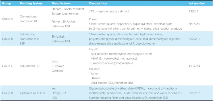
Group Bonding System Manufacturer Composition Lot number
Group A Conventional Transbond XT
Etchant - Ivoclar Vivadent,
Schaan, Liechtenstein 37% phosphoric acid as etchant. T30617
Primer - 3M Unitek California, USA
Primer:
Silane-treated quartz, bisphenol A, diglycidyl ether, dimethacrylate,
bis(2-hydroxyethyl ether), dichlorodimethyl silane, silica reaction products
N513350
Group B
Self-etching Transbond Plus
SEP
3M Unitek California, USA
Silane-treated quartz, glass reacted with hydrolyzed silane,
polyethylene glycol, dimethacrylate, citric acid, dimethacrylate oligomer,
silane-treated silica and bisphenol A, diglycidyl ether
B579201
Group C Futurabond DC
Voco
Cuxhaven Germany
Liquid 1:
Acid-modiied methacrylate (methacrylate ester) HEMA (2-hydroxyethyl methacrylate)
Camphorquinone (photoinitiator)
1505034 Liquid 2:
Water
Ethanol
Silica dioxide (SiO2) nanoiiler (1%)
Group D Optibond All-in-One Kerr
Orange, CA USA
Glycerol phosphate dimethacrylate (GPDM), mono- and di–functional
methacrylate, monomers, HEMA, ethanol, acetone and water as solvents, luoride-releasing illers and silica dioxide (SiO2) nanoillers (7%)
Scanning electron microscopy (SEM) examination
Ater ARI test, one specimen from each subgroup was randomly selected for scanning electron microscop-ic (SEM) evaluation to assess the distribution of bond-ing materials on enamel surface. The root and lbond-ingual part of the crown was dissected using Isomet low speed (Buehler, Lake Bluf, IL, USA) under abundant irriga-tion. The specimens were cleaned in distilled water with
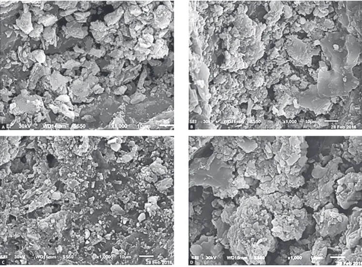
ultrasonic agitation for 30 minutes and gently air-dried, mounted on aluminum stubs and sputter coated with gold prior to SEM examination (JEOL, JSM-6510LV, Japan) operated at an accelerating voltage of 30 kV. The examination of all groups was done at X1000 mag-niication and at X5000 magmag-niication to determine the distribution of nanoillers more accurately. Represen-tative images of diferent specimens were digitally cap-tured and are presented in Figures 1 and 2.
A
C
B
D
Figure 1 - Representative SEM photomicrographs of: conventional bonding system after 12 hours (A) and 24 hours (B); Transbond Plus system after 12 hours (C)
Figure 2 - Representative SEM photomicrographs of the two nanofilled bonding systems: Futurabond DC after 12 hours (A) and 24 hours (B); Optibond All-in-One after 12 hours (C) and 24 hours (D).
A
C
B
D
Transmission electron microscopy (TEM) examination
A drop from each utilized bonding system was load-ed on carbon coatload-ed copper grid (200-mesh) and exam-ined by TEM (JOEL, JEM-2100, Japan) to determine the size and distribution of the illers within each bond-ing agent (Fig 3).
Statistical analysis
RESULTS
Shear bond strength (SBS)
The obtained SBS for the diferent four groups at 12 and 24 hours are given in Table 2. There were no statis-tically signiicant diferences within each group at either time points (p > 0.05). Although the mean SBS observed
with all diferent bonding systems at 12 hours were not statistically signiicant, the mean SBS obtained with the conventional system was statistically signiicant higher than that observed with Optibond All-in-One bonding system at 24 hours (p < 0.05).
Since there was no statistical signiicance of time points on the bond strength, the two subgroups (12 and 24 hours) were combined in Table 3. The combination showed that the SBS obtained with the two nanoilled systems used in this study were statistically signiicant lower than conventional system (p < 0.05), but there was no statistically signiicant diference between them. There was no statistically signiicant diference between the mean SBS obtained with Transbond Plus SEP and that obtained with other bonding systems.
A
C
B
D
Figure 3 - Representative TEM photomicrographs of the four used bonding systems; Transbond XT primer of conventional system (A), Transbond Plus (B), Futurabond
Adhesive remnant index (ARI) scores
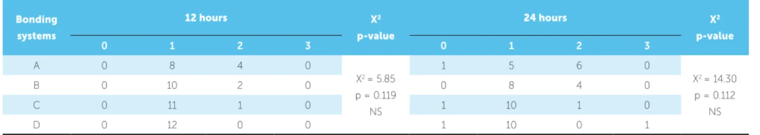
Table 4 shows that there was no statistically dif-ference between the observed ARI scores on the four groups at 12 and 24 hours (p > 0.05). In addition,
there was no statistically difference for each group at the two time points. The two nanofilled self-etch bonding systems showed a tendency towards the score 1, however score 2 was noted in conventional system and Transbond Plus more than nanofilled systems at the two time points. These results indi-cated that there was less adhesive left on the enamel surface when the nanofilled bonding systems were used in this study.
SEM examination
The selected scanning electron photomicrographs were in accordance with the ARI stereomicroscope results. SE photomicrographs of total-etch system showed nearly even particles size that illed the porosi-ties of etched enamel, and homogenous illers distribu-tion of bonding agent (Fig 1). Moreover, the scanning electron photomicrograph of Transbond Plus system showed a rather more condensed larger size particles with homogeneously distributed illers on enamel that illed most of its porosities (Fig 1). Scanning electron microscope examination of the two nanoilled bond-ing systems revealed that the particles illed enamel
po-Time point Bonding system Mean Standard deviation Maximum Minimum p-value
12 hours
A 8.86 2.69 13.24 4.37
p= 0.199
B 8.33 2.31 12.76 5.68
C 7.19 2.52 11.17 3.93
D 7.03 2.13 11.29 3.81
24 hours
A 10.06a 3.02 14.53 5.49
p= 0.021*
B 9.08 3.11 12.88 4.54
C 7.27 2.40 10.67 4.36
D 7.15b 1.61 9.43 4.78
Table 2 - Shear bond strength values in megapascals of the bonding systems at 12 and 24 hours
(A = 37% acid-etch and primer; B = Transbond plus; C = Futurabond DC; D = Optibond All-in-One). Groups with different superscript letters are statistically signifi-cantly different according to post-hoc Tukey HSD test, p = 0.043; *Significance was considered at p < 0.05.
Table 3 - Shear bond strength values in megapascals of the bonding systems used in the study.
(A = 37% acid-etch and primer; B = Transbond plus; C = Futurabond DC; D = Optibond All-in-One). Groups with superscript letters present statistically significant difference according to post-hoc Tukey HSD test (between A and C, p= 0.013; between A and D, p= 0.007).*Significance was considered at p < 0.05.
Bonding system N Mean Standard deviation Maximum Minimum P-value
A 24 9.46ab 2.86 14.53 4.37
p= .002*
B 24 8.71 2.71 12.88 4.54
C 24 7.23a 2.41 11.17 3.93
D 24 7.08b 1.85 11.29 3.81
Table 4 - Distribution frequency of ARI scores for the bonding systems at debonding after 12h and 24h.
NS, non-significant (A = 37% acid-etch and primer; B = Transbond plus; C = Futurabond DC; D = Optibond All-in-One). Bonding
systems
12 hours X2
p-value
24 hours X2
p-value
0 1 2 3 0 1 2 3
A 0 8 4 0
X2 = 5.85
p = 0.119 NS
1 5 6 0
X2 = 14.30
p = 0.112 NS
B 0 10 2 0 0 8 4 0
C 0 11 1 0 1 10 1 0
rosities to an extent. The enamel porosities appeared smaller and shallower. These bonding agent systems seemed less homogeneous and had a tendency to ag-gregate in crowds and clusters. Photomicrograph of Optibond All-in-One showed larger size globular structures with higher concentration than DC and have more trends to accrue (Fig 2). SE microphoto-graph within each group showed that the bonding materials distribution was more homogeneous and showed more constancy when the debonding proce-dures were done ater 24 hours in comparison with 12 hours (Fig 1 and 2).
TEM examination
Figure 3 shows transmission electron Microscope (TEM) results for the bonding systems used in the study. TEM photomicrograph of Transbond XT primer in conventional system and Transbond Plus showed larger size of separated and distributed fillers (0.1 µm and more) and lesser filler concentration in comparison with nanofilled systems.
In nanofilled bonding systems (Futurabond DC and Optibond All-in-One), spherical nano-sized fillers had an average size of 20 nm. Futurabond DC system had less concentration of nanofiller, which had more homogeneous distribution within the bonding agent, while Optibond All-in-One system had higher concentration of nanofillers that accu-mulate in dense clusters.
DISCUSSION
The bond strength of orthodontic brackets has to be appropriate to support masticatory and orth-odontic forces. Although acid-etching technique is a common useful technique for direct bonding of orthodontic brackets to enamel, the developed SEP adhesive systems can reduce chair time, eliminate the risk of contamination and provide intact enamel surface at the end of treatment. By the time, modern technologies utilizing new materials and substances are constantly developing to improve the quality of the bracket bonding to tooth structures, such as nanotechnology.19
Nanoparticles (NPs) of various composition rep-resent the most widespread use of nanotechnology in dentistry.7 Incorporated nanoillers in adhesives
com-positions increased resistance against fracture and
wear, provided higher dimensional stability and pro-duced higher SBS than conventional adhesives.20
Oth-er studies reported comparable21,22 or lower but still
acceptable shear bond strength,23,24 when nanoilled
adhesives were used for bonding orthodontic brackets. This study was conducted to evaluate the ef-fect of spherical silicon dioxide (SiO2) nanoparticles with average size of 20 nm added to two self-etch adhesive systems by the manufacturers with differ-ent concdiffer-entrations (Futurabond DC; 1% by weight and Optibond All-in-One; 7% by weight) on the shear bond strength and mode of failure of stainless steel orthodontic brackets bonded to human enamel. The manufacturers claim that nanoparticles enhance bond strength, since they act as cross-links that pro-mote the bond strength to enamel and dentin. In this regard, nano-sized fillers lead to entire permeation of the bond so they improve the bond strength.
Previous studies25,26 found that the shrinkage of
polymerization was affected by the filler content, and another study7 indicated that greater shrinkage was
produced by smaller nanofillers size, which leads to decreased bond strength of self-etch bonding systems.
Mean SBS values obtained in this study with the two nanofilled self-etch systems (Futur-abond DC, 7.23 ± 2.41 MPa; Optibond All-in-One, 7.08 ± 1.85 MPa) were insignificantly smaller than mean SBS value of Transbond Plus (8.71 ± 2.71), but they were significantly smaller than the conventional system (9.46 ± 2.86 MPa). This might be attributed to the higher polymerization shrinkage produced by nano-sized particles. Moreover, SEM and TEM ex-amination revealed that the nano-sized filled bonding systems had less homogeneous distribution than the others, so less number of enamel porosities was filled. It was revealed that less noticeable etching of enamel surface was acquired by self-etching primer system, and bonding resulted in smaller and fewer resin tags.27
The results observed in this study showed that Trans-bond Plus Trans-bonding system had comparable SBS with con-ventional system, this is similar to the indings of previ-ous studies.4,5 On the other hand, self-etching bonding
systems used in the study had a clinically acceptable mean SBS values since they were almost within the range (6-8 MPa) recommended by Reynolds.28 However, only
clini-cal testing can ensure cliniclini-cal usefulness.
Testing of SBS at 24 hours ater bonding proce-dure is generally preferred because it has been widely reported and allows comparison with other in vitro stud-ies. However, initial stable time is highly important for clinical orthodontic practice, in which the archwire is usually placed ater bracket bonding.29 Regarding our
results, there were no statistically signiicant difer-ences between SBS values obtained with each tested bonding systems at the two time points. Conventional system revealed values of 8.86 ± 2.69 and 10.06 ± 3.02 MPa; Transbond Plus, 8.33 ± 2.31 and 9.08 ± 3.11 MPa; Futurabond DC, 7.19 ± 2.52 and 7.27 ± 2.40 MPa; and Optibond All-in-One, 7.03 ± 2.13 and 7.15 ± 1.61 MPa at 12 and 24 hours, respectively. However, SEM eval-uation showed more homogeneity and constancy in bonding materials ater 24 hours. From a clinical point of view, it is therefore advisable not to load the brackets immediately to the maximum.
An ideal orthodontic adhesive should have ade-quate bond strength while maintaining unblemished enamel after debonding. The ARI is one of the most commonly used methods of assessing the quality of adhesion between the composite and tooth, as well as between the composite and bracket base.19 Regarding
the present study, ARI scores were not significantly different from each other when time and bonding system were considered as variables.
Incomplete resin polymerization below the metal base of bracket usually occurs because the curing light cannot reach the adhesive behind the bracket mesh; for light-cured adhesive, most of the failures occurred at the adhesive-bracket interface, which were similar to other indings.30
On the other hand, a score 2 of ARI was showed with conventional system and Transbond Plus in combination with a score 1. However, the two nanoilled self-etch sys-tems had a high tendency towards a score 1. This could be clinically advantageous, because, when brackets fail at the enamel-adhesive interface, less adhesive remains, and tooth cleanup is likely to be easier and faster.19
CONCLUSION
» The two nano-filled bonding systems revealed clinically acceptable SBS and presented lower ARI scores than the other bonding systems tested.
1. Buonocore MG. A simple method of increasing the adhesion of acrylic illing materials to enamel surfaces. J Dent Res. 1955 Dec;34(6):849-53.
2. Newman G. Current status of bonding attachments. J Clin Orthod. 1973 July;7(7):425-34 passim.
3. Mandall NA, Millett DT, Mattick CR, Hickman J, Worthington HV,
Macfarlane TV. Orthodontic adhesives: a systematic review. J Orthod. 2002 Sept;29(3):205-10; discussion 195.
4. Buyukyilmaz T, Usumez S, Karaman AI. Efect of self-etching primers on bond strength--are they reliable? Angle Orthod. 2003 Feb;73(1):64-70. 5. Grubisa HS, Heo G, Raboud D, Glover KE, Major PW. An evaluation and
comparison of orthodontic bracket bond strengths achieved with self-etching primer. Am J Orthod Dentofacial Orthop. 2004 Aug;126(2):213-9; quiz 255.
6. Silverstone LM, Saxton CA, Dogon IL, Fejerskov O. Variation in the pattern of acid etching of human dental enamel examined by scanning electron microscopy. Caries Res. 1975;9(5):373-87.
7. Başaran G, Ozer T, Devecioğlu Kama J. Comparison of a recently developed nanoiller self-etching primer adhesive with other self-etching primers and conventional acid etching. Eur J Orthod. 2009 June;31(3):271-5.
8. Mitra SB, Wu D, Holmes BN. An application of nanotechnology in advanced dental materials. J Am Dent Assoc. 2003 Oct;134(10):1382-90.
9. Saunders SA. Current practicality of nanotechnology in dentistry. Part 1: Focus on nanocomposite restoratives and biomimetics. Clin Cosmet Investig Dent. 2009 Nov 30;1:47-61. Print 2009.
10. Argueta-Figueroa L, Scougall-Vilchis RJ, Morales-Luckie RA, Olea-Mejía OF. An evaluation of the antibacterial properties and shear bond strength of copper nanoparticles as a nanoiller in orthodontic adhesive. Aust Orthod J. 2015 May;31(1):42-8.
11. Katona TR. Stresses developed during clinical debonding of stainless steel orthodontic brackets. Angle Orthod. 1997;67(1):39-46.
12. Stanford SK, Wozniak WT, Fan PL. The need for standardization of test protocols. Semin Orthod. 1997 Sept;3(3):206-9.
13. Whitlock BO 3rd, Eick JD, Ackerman RJ Jr, Glaros AG, Chappell RP. Shear strength of ceramic brackets bonded to porcelain. Am J Orthod Dentofacial Orthop. 1994 Oct;106(4):358-64.
14. Mitchell CA, O’Hagan E, Walker JM. Probability of failure of orthodontic brackets bonded with diferent cementing agents. Dent Mater. 1995 Sept;11(5):317-22.
15. Trites B, Foley TF, Banting D. Bond strength comparison of 2 self-etching primers over a 3-month storage period. Am J Orthod Dentofacial Orthop. 2004 Dec;126(6):709-16.
16. Bin Abdullah M, Rock WP. The efect of etch time and debond interval upon the shear bond strength of metallic orthodontic brackets. Br J Orthod. 1996 May;23(2):121-4.
REFERENCES
17. Turk T, Elekdag-Turk S, Isci D. Efects of self-etching primer on shear bond strength of orthodontic brackets at diferent debond times. Angle Orthod. 2007 Jan;77(1):108-12.
18. Artun J, Bergland S. Clinical trials with crystal growth conditioning as an alternative to acid-etch enamel pretreatment. Am J Orthod. 1984 Apr;85(4):333-40.
19. Sharma S, Tandon P, Nagar A, Singh GP, Singh A, Chugh VK. A comparison of shear bond strength of orthodontic brackets bonded with four diferent orthodontic adhesives. J Orthod Sci. 2014 Apr;3(2):29-33.
20. Hegde MN, Hegde P, Bhandary S, Deepika K. An evalution of compressive strength of newer nanocomposite: An in vitro study. J Conserv Dent. 2011;14(1):36-9.
21. Ahn SJ, Lee SJ, Kook JK, Lim BS. Experimental antimicrobial orthodontic adhesives using nanoillers and silver nanoparticles. Dent Mater. 2009 Feb;25(2):206-13.
22. Silva CFLM, Correa MA, Correr Sobrinho L, Moro A, Moresca RC, Correr GM. Shear bond strength of nanoilled lowable resins used for indirect bracket bonding. Braz J Oral Sci. 2012;11(4):458-62.
23. Uysal T, Yagci A, Uysal B, Akdogan G. Are composites and nano-ionomers suitable for orthodontic bracket bonding? Eur J Orthod. 2010 Feb;32(1):78-82.
24. Machado CT, Borges BC, Araujo GJ, Santos AJ, Dametto FR, Pinheiro FH. Inluence of adhesion promoters and curing-light sources on the shear bond strength of orthodontic brackets. Indian J Dent Res. 2012 Nov-Dec;23(6):747-52.
25. James JW, Miller BH, English JD, Tadlock LP, Buschang PH. Efects of high-speed curing devices on shear bond strength and microleakage of orthodontic brackets. Am J Orthod Dentofacial Orthop. 2003 May;123(5):555-61.
26. Faltermeier A, Rosentritt M, Faltermeier R, Reicheneder C, Müssig D. Inluence of iller level on the bond strength of orthodontic adhesives. Angle Orthod. 2007 May;77(3):494-8.
27. Fjeld M, Øgaard B. Scanning electron microscopic evaluation of enamel surfaces exposed to 3 orthodontic bonding systems. Am J Orthod Dentofacial Orthop. 2006 Nov;130(5):575-81.
28. Reynolds I. A review of direct orthodontic bonding. Br J Orthod. 1975;2(3):171-8.
29. Yamamoto A, Yoshida T, Tsubota K, Takamizawa T, Kurokawa H, Miyazaki M. Orthodontic bracket bonding: enamel bond strength vs time. Am J Orthod Dentofacial Orthop. 2006 Oct;130(4):435.e1-6.