Fixed Lingual Mandibular Growth Modificator:
A new appliance for Class II correction
Osama Hasan Alali1
Introduction: This article demonstrates the description and use of a new appliance for Class II correction. Mate-rial and Methods: A case report of a 10-year 5 month-old girl who presented with a skeletally-based Class II divi-sion 1 maloccludivi-sion (ANB = 6.5°) on a slightly low-angle pattern, with ML-NSL angle of 30° and ML-NL angle of 22.5°. Overjet was increased (7 mm) and associated with a deep bite. Results: Overjet and overbite reduction was undertaken with the new appliance, Fixed Lingual Mandibular Growth Modificator (FLMGM). Conclusion:
FLMGM may be effective in stimulating the growth of the mandible and correcting skeletal Class II malocclusions. Clinicians can benefit from the unique clinical advantages that FLMGM provides, such as easy handling and full integration with bracketed appliance at any phase.
Keywords: Orthodontic appliance design. Functional orthodontic appliances. Angle Class II malocclusion.
How to cite this article: Alali OH. Fixed Lingual Mandibular Growth Mod-ificator: A new appliance for Class II correction. Dental Press J Orthod. 2013 July-Aug;18(4):70-81.
Submitted: September 06, 2010 - Revised and accepted: June 20, 2011 » The patient displayed in this article previously approved the use of her facial and intraoral photographs.
» The authors report no commercial, proprietary or financial interest in the prod-ucts or companies described in this article.
1 MDS in Orthodontics, Teaching Assistant, Graduate-PhD.
Contact address: Osama Hasan Alali P.O.box: 10256 – Aleppo, Syrian Arab Republic E-mail: [email protected]
Introdução: esse artigo apresenta, por meio de um relato de caso, a descrição e o uso de um novo aparelho (o Aparelho Lingual Fixo para Alteração do Crescimento Mandibular, ou Fixed Lingual Mandibular Growth Modificator, o FLMGM) para correção de má oclusão de Classe II. sendo a paciente do sexo feminino, de 10 anos e 5 meses de idade, com má oclusão esquelética de Classe II, divisão 1, (ANB = 6,5°), com ângulos levemente baixos: ângulo SN.GoGn de 30° e ângulo PP.GoGn de 22,5°. A paciente apresentava sobressaliência aumentada (7mm) associada a mordida profunda. Re-sultados: a sobressaliência e a sobremordida foram reduzidas com o novo aparelho descrito. Conclusão: o FLMGM pode ser efetivo na estimulação do crescimento da mandíbula e na correção da má oclusão esquelética de Classe II. O dentista clínico pode beneficiar-se das vantagens clínicas exclusivas do FLMGM, tais como a facilidade de manejo e a total integração com aparelhos com braquetes em qualquer fase do tratamento.
INTRODUCTION
Many diferent appliances are now available for cor-recting skeletal Class II malocclusions. Some are re-movable1 such as activator and double-plate; and others
are ixed2 such as Herbst appliance and MARA.
Double plate system, that was introduced for the irst time by Schwarz in the early 1940s,3 is a reliable
means for treating Angle Class II, Division 1 maloc-clusions4. Sander et al.5,6,7 have veriied the
efective-ness of this system in a number of studies, providing detailed information on the biomechanics and thus on the working principles. This efectiveness prompted the author to develop a ixed double-plate system, Fixed Lingual Mandibular Growth Modiicator (FLMGM), that would not require patient compliance; especially because several studies8-11 have proven that ixed
func-tional appliances are much more eicient in stimulating skeletal mandibular growth than removable ones.
From a clinical perspective, the FLMGM ofers the following advantages:
1) Permanent effect, independent of patient compli-ance, as it is fixed.
2) Esthetics, as it is small and lingually located. 3) Eliminates the need for two separate treatment
phases, as it is suitable for use in parallel with com-plete multibracket appliance in both arches. 4) Flexibility in treatment timing, as it can be used
any-time during the mixed and permanent dentition. 5) No interference with occlusal development. 6) Wide and comfortable range of mastication
move-ments, as the appliance consists of two separate parts with no permanent and physical intermaxil-lary connection.
7) Construction bite is unnecessary because of the easy and quick chairside reactivation and progressive ad-vancement of the mandible in small increments. 8) Easy to handle because its insertion, clipping, and
removal are very simple.
9) Economic and cost effective, because it does not involve ready-made components, and only one appliance is necessary for entire orthopedic phase.
APPLIANCE DESIGN
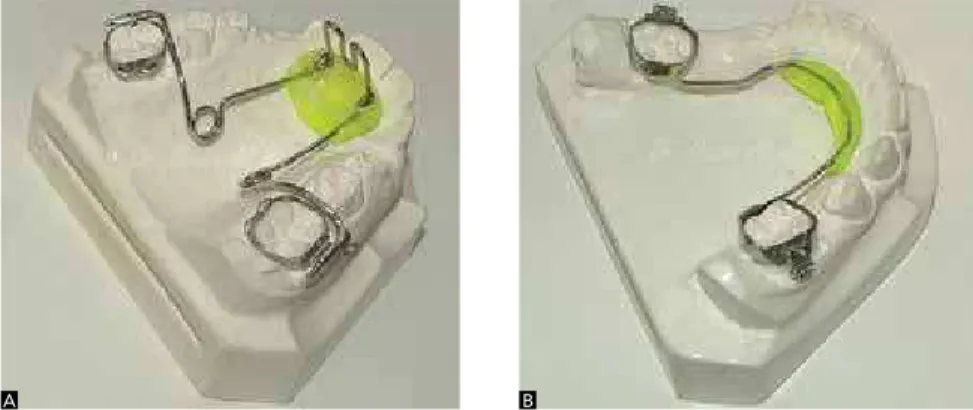
The FLMGM consists of two separate and ixed parts. The upper one is palatally positioned but bucal-ly clipped to traditional upper molars bands (Fig 1A), while the lower is lingually welded to traditional lower molars bands (Fig 1B).
Maxillary part
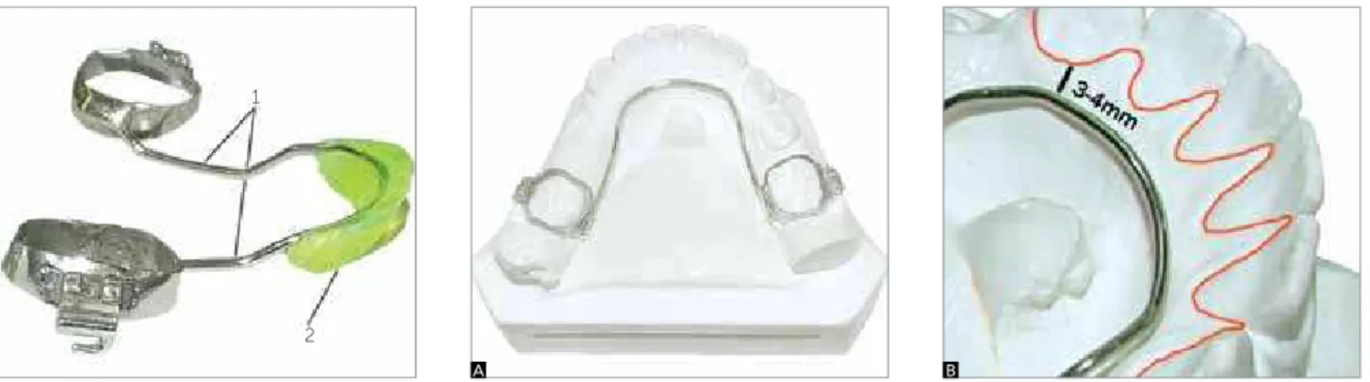
It has the following components (Fig 2):
1) Acrylic button: it is similar to Nance button, and designed to connect wire elements of the maxillary part, preventing them from embedding into the mucosa. This button is seated on the anterior area of the palate as anterior as possible with no contact with front teeth (1-2 mm away from the gingival margin). Its dimensions should be as small as possible to allow mucosal and periodontal hygiene.
2) Two retention wires: one in each side, speciically designed by the author to give excellent retention, facili-tate dealing with the appliance, and to enhance oral hy-giene condition. They are fabricated with round 1 mm thick stainless steel orthodontic wire. The wires are ante-riorly embedded into the acrylic button, run posteante-riorly without any contact with palatal mucosa, and each one contains an “U” loop with coil (giving some lexibility
Figure 1 - Fixed lingual mandibular growth modificator (FLMGM). No physical attachment between maxil-lary (A) and mandibular (B) parts.
Figure 2 - Components of maxillary part: acrylic button (1); retention wires (2); retention hooks (3) and advancement loops (4).
1 3
2
4
to help in easy insertion and removal) at the level of sec-ond upper premolar. At er the coils, the wires should run perpendicular to the midline towards the vestibule at the level of the mesial surface of upper i rst molar through the interdental space. Then, in the vestibule, the wires should run posteriorly to enter into the headgear tube.
3) Two retention hooks: one in each side, have a ball end to avoid irritation, and are directed anteriorly and welded to the retention wire before entering the headgear tube.
4) Advancement loops: wire projection embedded in the acrylic button and extended towards the man-dible. They consist of two consecutive long “U” loops, contain small protection coils where the wire exits the acrylic button, and are fabricated with round 1 mm thick hard stainless steel wire. The inclination of this advancing loops to the occlusal plane is about 70 °, mea-sured posteriorly.12
Mandibular part
It is made in a similar manner to a standard lingual arch with 1.0 mm stainless steel hard wire welded to the lingual aspect of i rst molars bands (Fig 3), and has the following features:
1) Its level in the anterior region must be 3-4 mm below the gingival margins of the incisors (Fig 4).
2) It includes an inclined guiding plane, made of acryl-ic resin, i xed on the anterior part of lingual arch, seated on the lingual alveolar mucosa below the level of incisors necks till the level of mouth l oor, and it is smooth to allow sliding against the advancement loops during man-dibular closing movement to reach its anterior position.
CLINICAL PROCEDURE
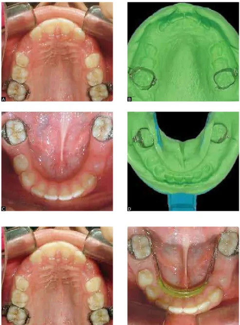
Separators are placed mesial and distal to the lower i rst molars. Subsequently,bands are selected and placed in position (Fig 5A and C). Optionally, construction bite can be taken in an edge-to-edge mandible position. Up-per and lower good alginate impressions are taken over the bands, with a good extension in the lingual sulcus. Bands are removed from the mouth and seated accurately in the impressions (Fig 5B and D). The impressions are sent to the laboratory for appliance fabrication. In the sequence, upper molar bands are cemented in position (Fig 6A). The inclined plane supported by lingual arch must be checked for stability, then the lower molar bands are cemented (Fig 6B). On the next day, maxillary part of corrector is attached to the upper molar bands (Fig 7A) by inserting the posterior ends of the retention wires into the headgear tubes. To ensure good stabilization of the maxillary part, an elastomeric ligature is used to tie the band hook and the retention hook together (Fig 7B).
Reactivation visits
Reactivation is generally required about every 2-3 months, and is carried out at the chairside, intra- or ex-traorally, by bending the advancement loops using orth-odontic pliers.
CASE REPORT
Diagnosis
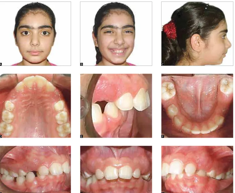
A 10-year 5-month-old girl presented with the chief complaints of the appearance of proclined upper inci-sors, retro-positioned chin, and dissatisfaction with her gingival smile. The family was concerned about the
Figure 4 - Level of the lingual arch 3-4 mm below the gingival margin.
Figure 3 - Components of mandibular part: lin-gual arch (1) and inclined guiding plane (2).
1
2
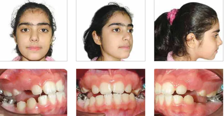
and slightly protrusive in appearance. There was some tipping of the lower right canine, irst premolar and irst permanent molar into the second primary molar extraction site. Apart from this slight constriction in the lower right buccal segment, the lower arch had a good form. In occlusion, the incisor relationship was Class II division I with an increased overjet of 7 mm and a traumatic deep bite of 5 mm (Fig 8E and H). The upper dental midline was coincident with the fa-cial midline, whilst the lower centerline was displaced 1 mm to the right. The molar relationship was an end-on (half unit) Class II on the let side and Class I on the right (Fig 8G and I).
lack of facial harmony and anxious for its improvement. The patient reported a history of bad oral habit, low-er-lip sucking, many years ago and an early loss of four lower primary molars.
On examination, she presented with a Class II di-vision 1 incisor relationship on a moderate Skeletal II bases. Clinical examination showed a convex-type fa-cial proile with a decreased anterior lower fafa-cial height and obviously obtuse nasolabial angle (Fig 8A to C). Pretreatment intraoral examination of her dentition (Fig 8D to I) revealed that the pattern of oral hygiene was good and that she was in the late mixed dentition. The upper and lower labial segments were well aligned
Figure 5 - Molar bands placed in situ (A and C), and repositioned in alginate impressions (B and D).
Figure 6 - The upper bands and the mandibular part cemented in place.
A B
Figure 7 - Maxillary part inserted into headgear tubes (A) and an elastomeric ligature placed be-tween the band hook and the retention hook to clip the maxillary part (B).
Figure 8 - Pretreatment facial and intraoral photographs (age 10 years and 5 months). Increased overjet and overbite.
A B
A B C
D E F
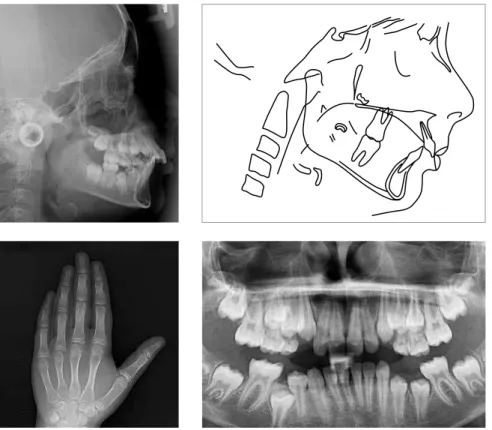
Figure 9 - Pretreatment radiographs and cepha-lometric tracing.
Cast analysis revealed arch-length discrepancies of +3 mm and -3 mm in the upper and lower arches, respectively. The lower right irst molar was mesially drited. The lateral cephalometric radiograph analysis (Fig 9 and Table 1) conirmed that, skeletally, the patient had a moderate Class II jaw relationship, ANB was 6.5°, and a reduced maxillomandibular plane angle, the ML-NL was 22.5°. Dentally, there was a degree of bimaxillary proclination and the interincisal angle was 120.5°. Sot tis-sue proile showed that the upper lip was slightly behind ‘E’ line, and the nasolabial angle was obviously obtuse (124°).
Assessment of hand-wrist radiograph (Fig 9) indi-cated considerable skeletal growth potential remaining. The patient was in the MP3cap stage.
Panoramic radiograph (Fig 9) revealed no obvious pathology present. The lower right second premolar was impacted. In addition, the third molars were developing.
Treatment objectives
The objectives of nonextraction treatment for the patient were identiied as follows: (1) Reduce the an-teroposterior skeletal disharmony; (2) Establish ideal overjet and overbite relationship; and (3) Achieve a functional occlusion with good interdigitation.
Treatment plan
Two-phase nonextraction approach was planned. For the irst phase (orthopedic), treatment goal was to stimulate mandibular growth, improving facial
appear-variables T0 T1 ∆(T1-T0)
SNA (degrees) 84 83 -1
SNB (degrees) 77.5 80 +2.5
ANB (degrees) 6.5 3 -3.5
NSAr (degrees) 128.5 126 -1.5
SNL.NL (degrees) 7.5 7 -0.5
SNL.ML (degrees) 30 31 +1
NL.ML (degrees) 22.5 24 +1.5
U1.SNL (degrees) 108 104.5 -3.5
U1.NL (degrees) 116 111.5 -4.5
L1.ML (degrees) 101.5 98.5 -3
Interincisal angle (degrees) 120.5 126 +5.5
Upper lip (mm) -1 -2 -1
Lower lip (mm) 1 1 0
Nasolabial angle (degrees) 124 127.5 +3.5
Table 1 - Cephalometric summary.
T0: Pre-treatment records (Fig 9),
ance and proile, and reduce the overjet and overbite to an acceptable level (with re-evaluation ater 8 months). During the second phase (orthodontic), the goal would be to maintain the improvement achieved in the ortho-pedic phase, and to obtain a functional occlusion with a Class I molar and canine relationships. Retainers would be placed immediately ater appliances were removed for retention purposes.
Treatment progress
At 10 years and 5 months, FLMGM was used to im-prove skeletal Class II by stimulating mandibular growth.
Figure 10 - Design of FLMGM: Posterior (A) and lateral (B) views.
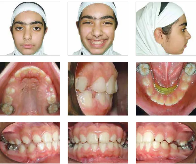
The patient was very motivated with the treatment. An additional motivating factor is the noticeable im-provement of facial appearance when FLMGM is it-ted. The patient was instructed to bite in an edge-to-edge position and to keep her lips in touch as much as possible (Fig 11). The patient was seen regularly at 6 weeks intervals, and at each visit, the occlusion was checked. The lip seal was maintained throughout treat-ment. At 11 years and 3 months, to assess the orthope-dic changes, only the maxillary part was removed, and a set of facial and intraoral photographs was taken (Fig 12); and radiographs were requested (Fig 13).
Figure 11 - Beginning of treatment, the FLMGM in place. Facial and intraoral photographs. The incisors were in an edge-to-edge bite and the lips touching.
Figure 12 - Facial and intraoral photographs immediately after removal of the maxillary part, promoting a normal incisor relationship, with buccal segments partially out of occlusion.
Results achieved
Through the course of FLMGM correction, about 8 months, the overjet was reduced from 7 mm to approxi-mately 2 mm, normal incisor and canine relationships were established, vertical eruption of lateral segments was enhanced, the lower midline coincided with the sot tissue midline, and a nice improvement in the facial esthetics and balance were achieved (Fig 12).
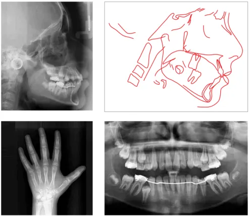
Data derived from cephalometric analysis (Fig 13 and Table 1) and superimpositions (Fig 14) reveal that there was continued vertical growth with valuable change in the anteroposterior relationship. Skeletally, ANB
Figure 13 - Radiographs and cephalometric trac-ing after orthopedic treatment.
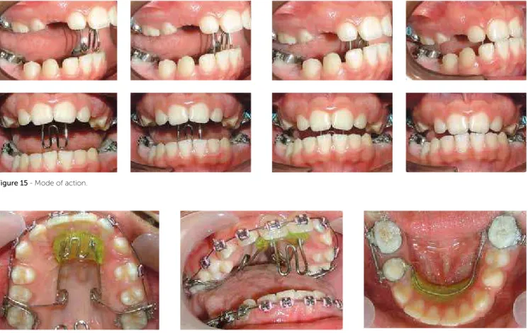
Figure 15 - Mode of action. DISCUSSION
In a growing patient, better aesthetic result would ideally be obtained by using orthopedic appliances to accelerate mandible development.13-17 The FLMGM has
efectively been used by the author in skeletal Class II division 1 patients, and a PhD clinical research is now in progress to identify its efectiveness. In the present case, the author attempted to stimulate mandible growth us-ing FLMGM that was well tolerated without compli-cations. Within 8 months of orthopedic treatment, the overjet was considerably enhanced, and the facial har-mony was good. The uprighting of incisors was favor-able, and this is believed to be resulting from the mecha-nism of breaking balance between the tongue and lips. While the vertical loops work as a shield relieving the tongue pressure on the incisors, only lingually-directed functional forces generated by the sealed lips afect the incisors and cause lingual crowns tipping.
Conceptually, FLMGM represents an exercise de-vice for the facial and masticatory muscles, in the same way as other functional appliances. It is proven that muscular training is important factor in the normal development of bone.18 Ater the appliance is placed,
the patient is asked to keep his mouth closed as long as possible. Once the patient closes his mouth, the inclined plane and the advancement loops will come in contact in the anterior area of the mouth. By con-tinuation of mouth closing, the inclined plane will be forced to slide against the loops, and eventually the mandible will take a predetermined forced ante-rior position (Fig 15). Functional force, generated by muscles that attempt to return the mandible posteri-orly to its original position,19 causes the upper acrylic
button and the lower acrylic inclined plane to apply pressure on the oral mucosa, and this in turn is sup-posed to cause proprioceptive response that reposi-tions the mandible forward in the same way as the Fränkel II Regulator20. In the first period, the patient
avoids collision between the loops and the inclined plane by forward-mandibular movement, and with time, the mandible functions without interference in the desired position and the patient will automati-cally, via neuromuscular reprogramming, close com-fortably into the protrusive position. The repetition of the new closure pattern results in orofacial muscu-lature reeducation and induces skeletal adaptation.19
Technically, the FLMGM represents a ixed ver-sion of the removable double-plate appliance because the two appliances follow an identical mechanism of action, based on incorporating an inclined plane in the mandible and guide bars in the maxilla. The most important two advantages of FLMGM over remov-able double-plate are: (1) FLMGM is active full-time, regardless of patient cooperation; (2) With FLMGM, the functional appliance phase is not separated from but completely integrated with the bracketed appliance phase (Fig 16). In clinical practices, although not many orthopaedic appliances are suitable for this integration,
Dynamax and MARA do approach it.11,21
When FLMGM alone in place during orthopaedic or retention phases, fully or partially bracketed appli-ances can be bonded. Another alternative is to integrate the FLMGM with an existing bracketed appliance. This coordination is a signiicant feature allowing maximum skeletal Class II correction without extending treatment time and delaying the progress of treatment by eliminat-ing a major drawback of many orthopedic appliances, where there is oten a need for an additional interim sta-bilizing phase, to avoid the relapse which may be seen if the orthopedic phase is abruptly discontinued. Leveling and aligning the dentition and erupting the buccal seg-ment to achieve good interdigitation using ixed appli-ances, while the FLMGM is maintaining mandibular advancement, is a crucial factor in stability of skeletal correction outcomes.
Extra advantages
• The vertical growth of the alveolus is enhanced during the FLMGM treatment phase, this may be as-cribed to two reasons: (1) the upper and lower teeth
are free because the FLMGM is supported only by the permanent molars; (2) When the mandible is in a pro-truded position, there is adequate interocclusal space available for spontaneous eruption.
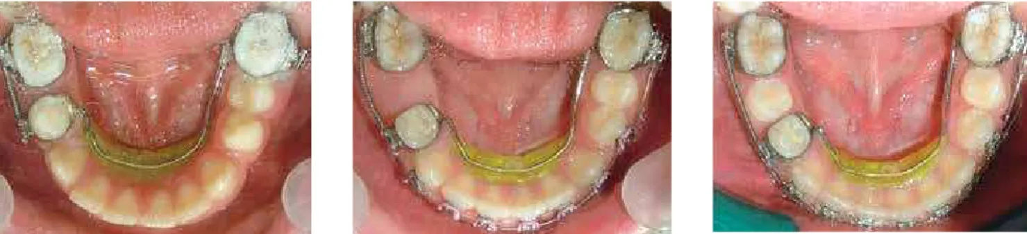
• The need for permanent extractions may be re
-duced during mixed dentition FLMGM treatment be-cause the lingual arch maintains the leeway space, and in some cases, it can be modiied to help in molar distaliza-tion (Fig 17).
• In case of still-active thumb-sucking habit, ad
-vancement loops efectively can block the thumb from making contact with the palate.
• In cases associated with lower lip sucking, a lip bumper may be buccally added to the mandibular part of corrector to modify the sot tissue as well.
Disadvantages
• The patient may complain of irritation from the vertically extended advancement loops.
• Swallowing, eating and speaking could be cum
-bersome, in contrast with buccally positioned appliance, yet the patient accepts it and gets used to it soon.
• Oral hygiene is somewhat diicult in the lower lingual anterior area, but has not been a problem.
CONCLUSIONS
The FLMGM appliance is economic, can be used starting from the mixed dentition and may be efective in stimulating the growth of the mandible and correct-ing skeletal Class II malocclusions associated with bi-maxillary dentoalveolar protrusion. Clinicians can ben-eit from the unique clinical advantages the FLMGM provides, such as easy handling and full integration with bracketed appliance at any phase.
1. Bishara S. Functional appliances: a review. Am J Orthod Dentofacial Orthop. 1989;95(3):250-8.
2. McSherry PF, Bradley H. Class II correction-reducing patient compliance: a review of the available techniques. J Orthod. 2000;27(3):219-25.
3. Altuna G, Schumacher HA. Schmuth and Muller Double Plates. J Clin
Orthod. 1985;19(6):422-5.
4. Lisson JA, Tränkmann J. Efects of Angle Class II, Division 1 treatment with Jumping-the-Bite appliances: a longitudinal study. J Orofac Orthop. 2002;63(1):14-25.
5. Sander FG. Indikation für die Anwendung der Vorschubdoppelplatte.
Prakt Kieferorthop. 1988;2:209-22.
6. Sander FG. Der Nachtefekt bei der Anwendung der
Vorschubdoppelplatte. Prakt Kieferorthop. 1989;3:97-106.
7. Sander FG, Lassak C. Die Beeinlussung des Wachstums
mit der Vorschubdoppelplatte im Vergleich zu anderen funktionskieferorthopädischen Geräten. Fortschr Kieferorthop. 1990;51:155-64.
8. Cozza P, Baccetti T, Franchi L, De Tofol L, McNamara JA Jr.
Mandibular changes produced by functional appliances in Class II malocclusion: a systematic review. Am J Orthod Dentofacial Orthop. 2006;129(5):599.e1-12; discussion e1-6.
9. McNamara JA Jr, Howe RP, Dischinger TG. A comparison of the Herbst
and Fränkel appliances in the treatment of Class II malocclusion. Am J Orthod Dentofacial Orthop. 1990;98(2):134-44.
10. Nelson C, Harkness M, Herbison P. Mandibular changes during functional appliance treatment. Am J Orthod Dentofacial Orthop. 1993;104(2):153-61.
REFERENCES
11. Pangrazio-Kulbersh V, Berger JL, Chermak DS, Kaczynski R, Simon ES, Haerian A. Treatment efects of the mandibular anterior repositioning appliance on patients with Class II malocclusion. Am J Orthod Dentofacial Orthop. 2003;123(3):286-95.
12. Kinzinger G, Ostheimer J, Förster F, Kwant PB, Reul H, Diedrich P. Development of a new ixed functional appliance for treatment of skeletal Class II malocclusion: irst report. J Orofac Orthop. 2002;63(5):384-99.
13. Fränkel R. Decrowding during eruption under the screening inluence of vestibular shields. Am J Orthod. 1974;65(4):372-406.
14. Graber TM, Rakosi T, Petrovic A. Dentofacial orthopedics with functional appliances. St. Louis: C.V. Mosby; 1997.
15. Bass NM. Orthopedic coordination of dento-facial development in skeletal class II malocclusion in conjunction with edgewise therapy. Part I. Am J Orthod. 1983;84(6):466-90.
16. Clark WJ. Twin block functional therapy: applications in dentofacial orthopaedics. London: Mosby-Wolf; 1995.
17. Pancherz H. The Herbst appliance: its biological efects and clinical use. Am J Orthod. 1985;87(1):1-20.
18. Fränkel R. Concerning recent articles on Fränkel appliance therapy. Am J Orthod. 1984;85(5):441-7.
19. Graber TM, Neuman B. Removable orthodontic appliances. Philadelphia: WB Saunders; 1984.
20. McNamara JA. On the Fränkel appliance. Part I. Biological basis and appliance design. J Clin Orthod. 1982;16:320-37.