Sensorimotor speech disorders in
Parkinson’s disease
Programming and execution deficits
Karin Zazo Ortiz1, Natalia Casagrande Brabo2, Thais Soares C. Minett3
ABSTRACT. Introduction: Dysfunction in the basal ganglia circuits is a determining factor in the physiopathology of the classic signs of Parkinson’s disease (PD) and hypokinetic dysarthria is commonly related to PD. Regarding speech disorders associated with PD, the latest four-level framework of speech complicates the traditional view of dysarthria as a motor execution disorder. Based on findings that dysfunctions in basal ganglia can cause speech disorders, and on the premise that the speech deficits seen in PD are not related to an execution motor disorder alone but also to a disorder at the motor programming level, the main objective of this study was to investigate the presence of sensorimotor disorders of programming (besides the execution disorders previously described) in PD patients. Methods: A cross-sectional study was conducted in a sample of 60 adults matched for gender, age and education: 30 adult patients diagnosed with idiopathic PD (PDG) and 30 healthy adults (CG). All types of articulation errors were reanalyzed to investigate the nature of these errors. Interjections, hesitations and repetitions of words or sentences (during discourse) were considered typical disfluencies; blocking, episodes of palilalia (words or syllables) were analyzed as atypical disfluencies. We analysed features including successive self-initiated trial, phoneme distortions, self-correction, repetition of sounds and syllables, prolonged movement transitions, additions or omissions of sounds and syllables, in order to identify programming and/or execution failures. Orofacial agility was also investigated. Results: The PDG had worse performance on all sensorimotor speech tasks. All PD patients had hypokinetic dysarthria. Conclusion: The clinical characteristics found suggest both execution and programming sensorimotor speech disorders in PD patients.
Key words: Parkinson’s disease, motor disorders, speech disorders, dysarthria.
DISTÚRBIOS SENSÓRIOS-MOTORES DA FALA NA DOENÇA DE PARKINSON
RESUMO. Introdução: Na doença de Parkinson (DP) a disfunção dos circuitos dos núcleos da base é um fator determinante na fisiopatologia dos sinais clássicos da DP e a disartria hipocinética é uma das manifestações da doença. No que se refere aos distúrbios da fala associados à DP, os modelos recentes de processamento de fala complicam a visão antiga da disartria como um déficit apenas de execução motora. Baseado nos achados de que as disfunções nos gânglios basais podem causar alterações de fala e que os distúrbios não estão apenas relacionados aos déficits de execução motora, mas também de programação motora, o objetivo deste estudo foi investigar a presença de distúrbios sensórios-motores da programação motora além dos de execução motora já descritos na fala de pacientes com DP. Métodos: O estudo é transversal e se baseou numa amostra composta por 60 adultos pareados por sexo, idade e escolaridade: 30 adultos diagnosticados com DP idiopática e 30 adultos sadios (grupo controle). Dados obtidos em um estudo prévio que analisou alterações de fluência em indivíduos com DP foram reanalisados acrescentando-se todos os tipos de manifestações/erros na fala, a fim de verificar falhas de programação e/ou execução motora. Os pacientes também realizaram avaliação da apraxia orofacial. Resultados: Todos os pacientes tinham disartria hipocinética. O grupo com DP obteve pior desempenho em todas as tarefas de fala. Conclusão: As características clínicas das manifestações/ erros de fala encontrados em pacientes com DP são sugestivas de déficits de execução e de programação motora.
Palavras-chave: doença de Parkinson, distúrbios motores, distúrbios da fala, disartria.
This study was conducted at the Department of Human Communication Sciences. Universidade Federal de São Paulo, SP, Brazil.
1Specialist, Master and PhD in Human Communication Disorders. Postdoctoral Fellow in Neuroscience. Associate Professor, Department of Human Communication Sciences, Universidade Federal de São Paulo, SP, Brazil. 2Speech Therapist, Master in Sciences, Department of Human Communication Sciences, Universidade Federal de São Paulo, SP, Brazil. 3Neurologist, PhD. Department of Radiology, University of Cambridge, Institute of Public Health, University of Cambridge.
Karin Zazo Ortiz. Departamento de Fonoaudiologia / UNIFESP – Rua Botucatu, 802 – 04023-900 São Paulo SP – Brazil. E-mail: [email protected] Disclosure: The authors report no conflicts of interest.
Received June 07, 2016. Accepted in final form August 04, 2016.
INTRODUCTION
P
arkinson’s disease (PD) is characterized by a degen-eration of neurons in the substantia nigra of the mesencephalon, leading to a fall in dopamine produc-tion. Dysfunction in the basal ganglia circuits is a deter-mining factor in the physiopathology of the classic signs, and hypokinetic dysarthria is commonly related to PD.1Regarding speech disorders associated with PD, the latest four-level framework of speech sensorimo-tor control2 proposed complicates the traditional view
of dysarthria as just a motor execution disorder. his model proposes diferent phases of the transformation of speech code involving the diferent neural structures. hese phases are identiied as linguistic-symbolic plan-ning, which is a nonmotor (or premotor) process, motor planning, motor programming and execution. According to the cited author,2 Linguistic Symbolic Planning is the
phase where linguistic rules of language are involved and this level of processing is nonmotor in nature so typical symptoms are aphasia signs. During the Motor planning phase a gradual transformation of symbolic units (pho-nemes) into a code that can be handled by the motor system takes place. Speech signs and symptoms result-ing from disorders in motor plannresult-ing can include slow, struggling speech with distortions and even apparent substitutions. Motor programming is a phase that deter-mines the spatiotemporal and force dimensions such as the amount of muscle tension needed, velocity, direction and range. A disorder at this level can result in impair-ment in these aspects and repeated initiation. Finally, during the execution phase, the hierarchy of plans and programs is inally transformed into non-learned auto-matic motor adjustments.2
he role of the structures such as the basal ganglia and the lateral cerebellum in both motor programming and execution suggests the possibility of dual symptom-atology in certain types of dysarthria, particularly in the parkinsonian (hypokinetic) type.
It is well known that the circuits in the basal ganglia play a fundamental role in the mechanisms of stuttering commonly present in these patients.3 It is important to
recognize that neurogenic stuttering is totally diferent from the other kinds of stuttering. Disluencies in PD patients may be analogous to limb motor symptoms such as diiculty with the initiation of motor move-ments and festination of gait observed in walking.4 In
this case, these failures could be more related to pro-gramming than execution deicits. In a previous study,5
a Speech Fluency Assessment Protocol6 was applied to
classify typology of disruptions into typical or atypical disluencies. he atypical disluencies such as repetitions
of syllables; repetition of sounds; prolongation; block-ing; pauses (over two seconds) and intrusions of sounds or segments and episodes of palilalia, characterized by the presence of repetitions of syllables (over four times) and words (over three times), with or without accelera-tion of speech rate were analysed. he authors found that PD subjects had a signiicantly higher number of speech disluencies overall compared to control subjects. In light of this, most of the characteristics described by the authors might be related to motor programming problems, especially considering the current view that the most prominent disluency type in PD is sound repetition, followed by initial syllable and word repeti-tions and some prolongarepeti-tions.7 In order words, some
of these characteristics could be analysed as program-ming deicits. Apraxia of speech is believed to result from a motor planning deicit. In a previous study on apraxia of speech in PD, the authors found that half of the PD patients presenting dysarthria also had apraxia of speech.8
Another approach is the use of Nonspeech Assess-ment for understanding the speech production mech-anism. Darley et al.,9 in their presentation of Motor
Speech Disorders, recommended several nonspeech observations and maneuvers during the assessment. here is continuing debate over the utility of nonspeech tasks for informing clinical diagnosis.10 According to
Bal-lard et al.,11 studies have reached diferent conclusions.
he authors stated that, nonspeech tasks can provide useful information about the functioning of the motor system. A study investigating the association between speech and orofacial apraxia found an association in 48% of cases studied.12 Although the classiication of
these speech disorders difered to that currently in use, the possibility of an association between these two con-ditions cannot be ruled out.
Based on indings that dysfunctions in basal ganglia can cause luency of speech deicits, and on the premise that the speech deicits seen in PD are not related to an execution motor disorder alone but also to a disorder at the motor programming level, the main objective of this study was to investigate the presence of sensorimotor disorders of programming (besides the execution disor-ders previously described) in PD patients.
METHODS
his study was approved by the Research Ethics Committee of the Universidade Federal de São Paulo
Casuistic. A cross-sectional study was conducted in a sample of 60 adults matched for gender, age and education: 30 adult patients diagnosed with idiopathic PD attended at the Sector for Motor Disorders of the Neurology Department of the Universidade Federal de São Paulo, and 30 healthy adults (control group) that were companions or family members of the patients assessed.
he general inclusion criteria for both groups were as follows: age ≥ 50 years; education ≥ 4 years; absence of personal or family history of developmental or psy-chogenic stuttering or language disorders; absence of history of stroke or previous traumatic brain injury; absence of alcoholism or use of illegal drugs; visual or hearing impairments which could afect performance on the tasks given; normal performance on the MMSE for educational level, according to the standards estab-lished for the Brazilian population,13 thus excluding
sub-jects with dementia from the sample and ensuring that impairments in cognitive aspects did not interfere with the speciic assessment.
he patients participating in the study were diag-nosed with PD, had not undergone neurosurgery, were at stages 2, 2.5 or 3 on the Hoehn & Yahr,14 and
in use of medication for PD. hus, subjects at initial or advanced stages of the disease were excluded from the sample because individuals at the initial stage may not have impaired speech while, in advanced cases, speech samples may be unintelligible or insuicient.
All patients were at the ‘on’ phase of the medication during the assessment.
Instruments. First, the patients were submitted to the Protocol for Dysarthria Assessment.15 Respiration,
phonation, articulation, resonance and prosody were evaluated in order to check for the presence of Hypoki-netic Dysarthria.
For the sensorimotor speech disorders assessment, the subjects told a story based on sequences of pictures composed of seven drawings and also described a typi-cal day to produce a suicient speech sample for subse-quent analysis.
he oral agility subtest of the Boston Diagnostic Aphasia Examination (BDAE) was used to evaluate speech and orofacial praxis.16 his test includes six tasks
of orofacial agility and seven involving speech agility. he orofacial agility task comprises oral commands such as tongue to alternate corners of the mouth, pro-trude and retract tongue, tongue alternately to upper and lower teeth, purse lips and release, open and close mouth, retract and release lips. he subject must
per-form the movements correctly in terms of programming and timing. On the speech agility task, the subject has to repeat words as fast as they can in a correct fashion. he score is given according to correct repetition and timing. Speech errors were analysed using the same criteria as presented below.
Data collection was carried out on an individual basis. he discourse produced was recorded using a dig-ital camera (SONY Cyber – shot 6.0 mega pixels) and later transcribed. he data were obtained from a sam-ple of a previous study5 in which luency disorders were
analysed. In that study, episodes of palilalia, number of hesitations; interjections; revisions; uninished words; repetition of words, segments and sentences, repeti-tions of syllables; repetition of sounds; prolongation; blocking; pauses and intrusions of sounds or segments and also speech rate, were analyzed as luency disorders.
In the present study, all types of articulation errors were reanalyzed to investigate the nature of these errors. In this new analysis, interjections, hesitations, repetitions of words or sentences (during discourse) were considered typical disluencies; blocking, episodes of palilalia (words or syllables) were analysed as atypical disluencies. We analysed features including successive self-initiated trial, phoneme distortions, self-correction, repetition of sounds and syllables, prolonged movement transitions, addition or omissions of sounds and sylla-bles, all of which can be related to programming disor-ders of sensorimotor control of speech. It is noteworthy that successive self-initiated trial, phoneme distortions, addition and omission can also been found in planning disorders. he features present on each test were scored with 1 point. Total score was calculated by summing all feature scores.
Statistical analysis. Categorical data were compared using the Chi-squared (c2) test (without Yates comparison)
with application of Fisher’s exact test when Cochran’s restrictions were present.
A probability (p) of less than 0.05 was considered statistically signiicant and all tests were two-tailed. Diferences among means were calculated for a ninety-ive percent conidence interval (95%CI). All statistical analyses were carried out using the software SPSS (Sta-tistical Package for the Social Science) version 11.5.1 for Windows.
RESULTS
speech assessment. Of this total, 10 were not included in the sample because they did not attend the sched-uled session. hus, a total of 30 patients followed the protocol, in addition to 30 controls. he data from these 60 subjects were considered in the subsequent analyses.
General characteristics. he age of subjects in the sample ranged from 50 to 75 years, with a mean age 62.3±7.0 years, and in terms of gender, 82% were men.
here were no statistically signiicant diferences between the Control group (CG) and the Parkinson’s disease group (PDG) for age (62.4±6.9 versus 62.2±7.1 years; t(58)=0.13; 95%CI= –3.4 to 3.9; p=0.898), educa-tion (8.7±4.2 versus 8.4±4.2 years; t(58)=0.21; 95%CI= –2.0 to 2.4; p=0.832), MMSE score (28.5±1.2 versus
28.4±1.4; t(58)=0.29; 95%CI= –0.6 to 0.8; p=0.770) or gender (83% men versus 80% men; c2(1)=0.11; p=0.739).
Clinical characteristics of PD patients. Disease duration ranged from 2 to 20 years (mean=9.9, SD=4.4), 20% of patients had a score of 2 on the Hoehn and Yahr scale, 37% scored 2.5 and the remainder scored 3. A total of 90% of the patients were in use of Levodopa, 37% Amantadine, 10% Selegiline, 60% Pramipexole and 13% Biperiden. Of the 30 patients in the sample, 24 (80%) were in use of combined medications whereas 6 (20%) used a single medication. Of the single users, ive used levodopa and one pramipexole.
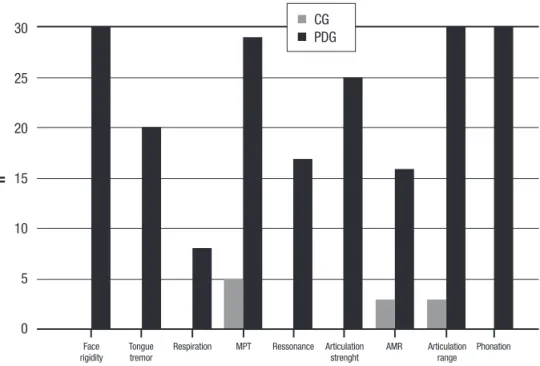
Dysarthria assessment results. he distribution of changes, according to study group: face rigidity, tremor
of tongue, increased respiration, decreased maximum phonation time (MPT), altered resonance, reduced articulation strength, slow alternate motion rate (AMR), reduced articulation amplitude and change in voice quality are shown in Figure 1.
he CG had signiicantly better performance than the PDG for all dysarthria features (1.1±0.7 versus 6.7±1.3; t(58)= –20.2; 95%CI= –6.0 to –4.9; p<0.001).
Assessment of non-verbal and verbal praxis. he CG had signiicantly better performance than the PDG for both non-verbal (7.6±1.8 versus 4.9±1.6; t(58)=5.88; 95%CI=1.76 to 3.57; p<0.001) and verbal (12.0±0.6 versus 11.0±1.1; t(58)=4.26; 95%CI=0.53 to 1.47; p<0.001) praxis.
For the purposes of intragroup comparison of two types of apraxia, we calculated the proportion of correct responses on the oral agility tests of each individual to standardize the results.
his comparison revealed that the proportion of cor-rect responses on the task assessing verbal praxis was signiicantly higher than on the tasks assessing non-verbal praxis in both groups.
• CG: 0.63±0.15 versus 0.86±0.04; t(29)= –8.47; 95%CI= –0.28 to –0.17; p<0.001
• PDG: 0.41±0.14 versus 0.37±0.14 years; t(29)= –15.09; 95%CI= –0.42 to –0.32; p<0.001
he total features found for spontaneous speech in the CG was signiicantly lower than in the PDG (4.8±2.6 versus 8.9±6.7; t(58)= –3.12; 95%CI= –6.7 to 1.4; p=0.003). he CG had signiicantly better performance
30
25
20
15
10
5
0
n
n CG
n PDG
Figure 1. Distribution of types of changes in dysarthrias according to study group.
than the PDG on the verbal agility task from the Boston test (12.0±0.6 versus 11.0±1.1; t(58)=4.26; 95%CI=0.53 to 1.47; p<0.001).
DISCUSSION
he most relevant inding of this study was that analysis of all features of speech clearly suggested impairments at the motor programming and execution level in the patients with Parkinson’s disease.
he idea of reanalyzing separately all types of errors had the principal goal of identifying the occurrence of programming disorders.
In relation to dysarthria, only AMR, MPT and reduced range of articulation were seen in some CG individuals (Figure 1). he inding of these alterations in a few individuals may be related to aging. In the PD group, alterations were observed in all motor bases and it was clearly possible to statistically diferentiate the two groups. All PD patients presented dysarthria.
In Table 1, it can be observed that diferent speech errors were more evident in the PD group. Speech errors were identiied in three speech samples: telling the story, describing a day, and the agility task of the Boston test.
An analysis of errors committed on the oral agil-ity task showed that eight of the 30 patients from the PDG had motor programming of speech deicits, not observed in the control group. During this task,
sylla-ble repetition was the only feature present in the PDG. Although the syllable repetition featured by the PDG patients can be present in both neurogenic stuttering and speech apraxia (nowadays regarded as a motor plan-ning disorder), making it hard to diferentiate between the conditions, some considerations should be taken into account. First, stuttering associated with acquired neurological disorders can mask the presence of other communication problems.7 Over the years, various
sub-groups of neurogenic stuttering have been proposed, such as diferentiations between dysarthric stutter-ing, apraxic stuttering and dysnomic stuttering.17 More
recently, further subdivisions have been suggested based on underlying lesion location18 and stuttering
associ-ated with extrapyramidal disease has been described.19
On this point, the most prominent disluency type is sound repetition and in this study we found more syl-lable repetition, features more related to programming disorders. However, accurately distinguishing between these syndromes remains challenging.
Some authors20 in a study review airmed that,
although the onset of stuttering in luent speaking adults has been discussed in the literature for over a century, it remains unclear whether acquired stutter-ing is a distinct disorder or an epiphenomenon of speech deicits such as apraxia of speech. Although the exact nature of repetition is unclear, in this case it would be
SPEECH FEATURES IN CG AND PDG
Table 1. Statistical data for groups studied according to speech characteristics.
CG PDG
Mean SD Min Max Mean SD Min Max
Typical Disfluencies 0.84 1.11 0 5 0.76 1.16 0 5
Atypical disfluencies 0 0 0 0 1 2.11 0 8
Self-correction 0.53 0.78 0 3 0.43 0.77 0 3
Self-initiated trials 0.17 0.46 0 2 0.1 0.31 0 1
Prolonged movement transitions 0 0 0 0 0.93 1.66 0 8
Repetition of syllables 0.03 0.18 0 1 0.2 0.48 0 2
Repetition of sounds 0 0 0 0 0.07 0.37 0 2
Phoneme distortions 0 0 0 0 0.23 0.57 0 2
Addition of sounds 0 0 0 0 0.1 0.31 0 1
Omissions of sounds 0 0 0 0 0 0 0 0
considered, according to the latest four-level framework of speech sensorimotor control, a programming disorder and not a planning disorder.
Besides, given that patients performed the test more slowly, other speech errors may not have been mani-fested on this task.
Previous studies that considered apraxia a program-ming disorder, state that the speech deicits occurring in PD are not related only to the muscle control level, causing dysarthria, but also to the speech programming level, with the condition of apraxia.8 In the cited study,
the apraxic patient comprised a subgroup of a group with dysarthria, leading authors to believe that per-haps speech apraxia does not exist in PD without being associated with dysarthria. he authors concluded that dysarthria is twice as frequent as apraxia in PD. In our study, we found that all patients presented dysarthria and some presented speech errors that suggested pro-gramming deicits.
Non-verbal and verbal apraxias have been previously described in other neurodegenerative diseases that occur with parkinsonian syndrome. Cases of individuals with corticobasal degeneration (CBD) and progressive supranuclear palsy (PSP) that presented impairments such as speech apraxia, non-luent aphasia or a combina-tion of both disorders, have been reported.21 he authors
stated these disorders are often not detected at disease onset but become evident at more advanced stages and can be associated with pathologic diagnoses of CBD and PSP. According to the authors, patients with these neu-rodegenerative disorders also exhibit initial changes of speech apraxia and non-luent aphasia and in general, the condition progresses rapidly compared to the clas-sic picture characteristic of PD. In a study involving 35 patients with CBD, three had speech apraxia.22
he quest for a better understanding of the process of programming has primarily sought a comprehensive formulation of the role of the diferent neural structures involved in the programming phase of motor process-ing. he motor areas involved in motor programming comprise the basal ganglia, lateral cerebellum, supple-mentary motor area, motor cortex, and the frontolimbic system.2 It is generally accepted that the basal ganglia,2,23
and the lateral cerebellum2,24 in particular, are involved
in programming, and these parts perform complemen-tary functions.2,25 he exact role of each, however, is
not yet fully understood.2 Parkinson’s disease causes
delayed initiation, slowed execution, abnormal sequen-tial complex movements and an inability to automati-cally execute learned motor plans.2,26 Dysarthria due to
Parkinson’s disease also indicates that the basal ganglia
may play a role in initiation, temporal synchroniza-tion, timing and automatized production of speech,2,27
as observed in all motor tasks analyzed in the current study.
During the repetition tasks, we observed that eight of the 30 patients from the PDG presented symptoms such as: syllable repetitions, besides episodes of acceler-ated speech while performing the task, whereas controls did not. Phoneme substitutions, distorted substitutions, omissions and additions were not found in this sample, probably because motor planning was preserved in these patients.
Comparison of performance of the two groups ana-lyzed revealed a statistically signiicant diference in total score obtained on the tasks for both non-verbal and verbal praxis, i.e. the PD patients had signiicantly worse performance on both tasks.
Intragroup comparison of the two types of apraxia revealed that the proportion of correct responses on the task assessing verbal praxis was signiicantly higher than on the tasks assessing non-verbal praxis in both groups. his result suggests that these tasks may be more sen-sitive for the early detection of cases that progress to programming disorders.
We noted that all individuals performed the move-ments with impaired velocity, although 15 subjects, besides slowness in performing the movements, also exhibited praxic deicits, i.e. in motor programming, evidenced by non-performance or partial performance of the movements.
he need to demonstrate the movements, known to facilitate motor programming, was frequent in the PDG whereas CG subjects did not require this aid. herefore, we concluded that the poorer performance seen in the PD group on the task assessing non-verbal praxis can be explained by deicits in programming and sequencing movements, i.e. non-verbal praxis. Other explanations include the diiculty in motor execution present in PD and the presence of both these deicits, as observed in 15 subjects from the PDG. hus, it is notable that the task proposed, although originally intended to assess apraxia, was also sensitive for assessing dysarthria-related motor aspects. his was the case because velocity is one of the elements of the assessment procedure and allowed co-occurrence of apraxia and dysarthria-related motor aspects that hamper the performance of move-ments to be identiied.
con-comitantly with the dysarthric condition in some cases. hus, all of the apraxic patients in this study were dysar-thric but not necessarily the other way around.
To conclude, the PDG had worse performance on all sensorimotor speech tasks. All PD patients had hypoki-netic dysarthria. he clinical characteristics found sug-gest both execution and programming sensorimotor speech disorders in PD patients.
Author contribution. Karin Zazo Ortiz supervised the data collection and drafted the paper. Natália Casagrande Brabo collected, analyzed and interpreted the data.
hais Soares C. Minett supervised the data collection and performed the statistical analyses of the data
Support. his research was supported by the CAPES – the Brazilian Federal Agency for the Support and Evalu-ation of Graduate EducEvalu-ation and by FAPESP (proc. 2012/23587-4).
Acknowledgements. We would like to thank Professor Henrique Ballalai Ferraz for the opportunity to conduct this study with patients attended at the Sector for Motor Disorders of the Department of Neurology of the Universidade Federal de São Paulo.
REFERENCES
1. Darley FL, Aronson A, Brown JR. Motor speech disorders. Philadelphia,
London, Toronto: Saunders; 1975.
2. Merwe AVD. A Theorical Frmework for the Characterization of
Patho-logical Speech Sensoriomotor Control. In: McNeil M.R. Clinical Manage-ment of SEnsoriomotor Speech Disorders. 2a ed, Thieme; 2009:3-18.
3. Alm P. Stuttering and the basal ganglia circuits: a critical review of
possible relations. J Commun Disord. 2004;37:325-369.
4. Duffy JR. Motor speech disorders: substrates, differential diagnosis and
management. 2nd ed. Louis, MO: Elsevier Mosby; 2005.
5. Brabo NC, Minett TSC, Ortiz KZ. Fluency in Parkinson’s disease: disease
duration, cognitive status and age. Arq Neuropsiquiatr. 2014;72(5): 349-355.
6. Andrade CRF. Fluência. In: Andrade CRF, Lopes DMB, Fernandes FDM,
Wertzner E (Eds). ABFW – teste de linguagem infantil nas áreas de fono-logia, vocabulário, fluência e pragmática. 2ed. Carapicuiba: Pró-Fono, H.F; 2004;51-81.
7. De Nil, LF, Rochon E, Jokel R. Adult Onset Neurogenic Suttering. In:
McNeil M.R. Clinical Management of Sensoriomotor Speech Disorders. 2a ed , Thieme; , 2009:235-248.
8. Howard LA, Binks MG, Moore AP, Player JR. The contribuition of apraxic
sppech to working memory déficits Parkinson’s Disease. Brain Lang. 2000;74(2):269-88.
9. Darley FL, Aronson A, Brown J. Motor Speech Disorders. Philadelphia,
W.B Saunders; 1975.
10. Weismer G. Philosophy of research in motor speech disorders. Clin Linguist Phon. 2006;20(5):315-49.
11. Ballard KJ, Solomon NP, Robin DA, Moon JB, Folkins JW. Nonspeech Assessment of the Speech – Production Mechanism. In: McNeil M.R. Clinical Management of Sensoriomotor Speech Disorders. 2a ed, Thieme; 2009:30-45.
12. Dronkers NFA. A new brain region for coordinating speech articulation. Nature. 1996;384:159-161.
13. Brucki SMD, Nitrini R, Caramelli P, Bertolucci PHF, Okamoto IH. Sugestões para o uso do Mini-Exame do Estado Mental no Brasil. Arq Neuropsiquiatr. 2003;61:777-781
14. Hoehn MM, Yahr MD. Parkinsonism: onset, progression and mortality. Neurology. 1967;17:427-442.
15. Ortiz KZ. Avaliaçao das Disatrias In: Ortiz KZ. Distúrbios neurológicos
adquiridos: fala e deglutição. 2a ed, Barueri, Ed. Manole; 2010. p.73-96.
16. Goodglass H, Kaplan EF. The Assessment of Aphasia and Related Disor-ders. Philadelphia: Lea and Febiger; 1983.
17. Rosenbek JC. Stuttering secondary to Nervous System Damage. In: RF Curlee, Perkins WH. Nature and Treatment of Stuttering: New Directions. San Diego, College Hill;1984:31-48.
18. Van Borsel J, Van Der Made S, Santens P. Thalamic stuttering: a distinct clinical entity? Brain Lang. 2003;85:185-89.
19. Helm-Estabrooks N, Yeo R, Geschwind N, Freedman M. Stuttering: disappearance and reappearance with acquired brain lesions. Neurology. 1986;36:1109-1112.
20. Lundgren K, Helm-Estabrooks N, Klein R. Stuttering Following Acquired Brain Damage: A Review of the Literature. J Neurolinguistics. 2010:23(5): 447-454.
21. Josephs KA, Duffy JR. Apraxia of speech and nonfluent aphasia: a new clinical marker for corticobasal degeneration and progressive supra-nuclear palsy. Curr Opin Neurol. 2008;21(6):688-92.
22. Kertesz A, Martinez-Lage P, Davidson W, Munoz DG. The corticobasal degeneration syndrome overlaps progressive aphasia and frontotemporal dementia. Neurology. 2000;55:1368-1375.
23. Johnson AM, Vernon PA, Almeida QJ, Grantier LL, Jog MS. The role of basal ganglia in movement: the effect of precues on discrete biderectional movements in Parkinson’s disease. Motor Control. 2003; 7(1):71-81.
24. Miall RC, Weir DJ, Stein JF. Visuo-motor tracking during reversible inac-tivation of the cerebellum. Exp Brain Res. 1987;65:455-464.
25. Dreher J, Grafman J. The roles of cerebellum and basal ganglia in timing and error prediction. Eur J Neurosci. 2002;16:1609-1620.
26. Marsden CD. The enigma of the basal ganglia and movement. Trends Neurosci.1980;3:284-287.