Dement Neuropsychol 2015 December;9(4):424-427
424
424 Creutzfeldt-Jacob disease imaging features Valente et al.
Case Report
Magnetic ressonance imaging in the
diagnosis of Creutzfeldt-Jakob disease
Report of two cases
Alan Peres Valente1, Paula da Cunha Pinho2, Leandro Tavares Lucato3
ABSTRACT. Creutzfeldt-Jacob disease (CJD) is a rare condition caused by a pathogenic prion protein that evolves with rapidly progressive dementia and death. The clinical presentation may sometimes be misleading. Magnetic Resonance Imaging (MRI) aids diagnosis with patterns that can guide or confirm clinical hypotheses. Two cases of rapidly progressive dementia with ataxia, myoclonus and restricted diffusion on MRI in cortical/basal ganglia are presented to draw attention to CJD.
Key words: Creutzfeldt-Jakob disease, prionic disease, prion, prionic disease, rapidly progressive dementia, MRI, diffusion, DWI, basal ganglia, cortex.
RESSONÂNCIA MAGNÉTICA NO DIAGNÓSTICO DA DOENÇA DE CREUTZFELDT-JAKOB: RELATO DE DOIS CASOS
RESUMO. Doença de Creutzfeldt-Jacob (CJD) é uma rara doença relacionada a uma proteína priônica patogênica que evolui com demência rapidamente progressiva e morte. Por vezes, a apresentação clínica é inespecífica e desafiadora. A ressonância magnética contribui para o diagnóstico com padrões de imagem que podem orientar ou confirmar as hipóteses diagnósticas baseadas na clínica. Serão apresentados dois casos de pacientes com a forma esporádica da doença.
Palavras-chave: Creutzfeldt-Jakob, doença priônica, demência rapidament progressiva, ressonância magnética, difusão, gânglios da base, córtex.
INTRODUCTION
P
rion diseases (formerly known as spongiform encephalopathies) were irst des cribed in the beginning of last century. In 1966, a “transmissible in a viruslike manner” hypothesis conferred this group of diseases with the unique characteristic of being both infectious and inherited, with long incuba tion periods.1,2 CreutzfeldtJakob disease
(CJD), kuru, variant CreutzfeldtJakob dis ease (vCJD), GerstmannSträusslerScheinker syndrome (GSS), and fatal familial insomnia (FFI) are the ive human prion diseases cur rently recognized.
Sporadic form CreutzfeldtJacob disease (sCJD), a condition with rapidly progres sive dementia and a fatal outcome, accounts for the majority of these very rare entities.
he iatrogenic (iCJD) form (related to dural implants / cornea transplants) and the familial form (fCJD) both have, in common with sCJD, the pathological human prion protein (PrPsc) related to the prion protein gene.1 Variant
form (vCJD) is caused by the transmission of the bovine spongiform encephalopathy agent to humans and is considered a distinct entity.
Progressive mental deterioration and myoclonus are the most important and typi cal symptoms for diagnosis.3 Cerebellar and
oculomotor symptoms are common, and sometimes signs of pyramidal/extrapyrami dal dysfunction can also be present. Electro encephalogram patterns (EEG), especially periodic sharp wave complexes, and detec tion of 1433 protein in cerebrospinal luid can be adjunctive in the diagnosis of sCJD,
This study was conducted at the Instituto de Radiologia do Hospital das Clínicas da Faculdade de Medicina da USP.
1,2,3Setor de Neurorradiologia Diagnóstica. Instituto de Radiologia do Hospital das Clínicas da Faculdade de Medicina da USP.
Leandro Tavares Lucato. Hospital das Clínicas da Faculdade de Medicina da USP / Instituto de Radiologia / Ressonância Magnética. – Av. Dr. Enéas de Carvalho Aguiar 255 – 05403010 São Paulo SP – Brasil.
Disclosure: The authors report no conflits of interest.
Received August 15, 2015. Accepted in final form October 30, 2015.
Dement Neuropsychol 2015 December;9(4):424-427
425
Valente et al. Creutzfeldt-Jacob disease imaging features
with good sensitivity and speciicity. Magnetic resonance imaging (MRI), particularly the technique using difu sionweighted images (DWI),also plays an important diagnostic role.3,4
In the present study, we present two sCJD cases with classic clinical presentations and imaging indings.
CASE 1
A 51yearold female patient with no previous medical history presented at the emergency department with gait disturbance and rapidly progressive cognitive decline, which developed over 4 months. he patient was disoriented with psychomotor slowing. She could not assume orthostasis or walk without assistance although the strength of her lower limbs was preserved. Myoclonus were evident and she had cerebellar symp toms (ataxia and dysdiadochokinesia). Sensitivity was low in the feet and ingertips.
MRI disclosed restricted difusion in the basal ganglia (caudate and putamen), medial portion of the posterior thalami, frontal lobe cortex, anterior portion of the tem poral lobes and insula in a symmetrical pattern. Posterior fossa structures were preserved. he majority of these areas had corresponding high signal on T2weighted and FLAIR images.
Laboratory investigation was extensive, and entirely negative. CSF exam disclosed no remarkable indings.
Based on MRI pattern and clinical scenario, a diagno sis of sCJD was reached.
CASE 2
A 61yearold male patient was admitted to our service with a 5month history of diiculty walking, writing and speaking, together with muscular spasms and limi tation in daily activities. EEG showed disorganized slow waves while 1433 protein was present in CSF samples.
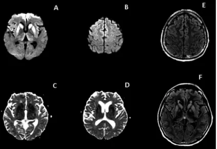
Figure 1. Case 1. Axial T2-weighted (A) and FLAIR (B) im-ages: hyperintensity in basal ganglia, with less evident hyperintensity in cortex. Axial post-contrast T1-weighted image (C) shows no enhancement. Axial DWI (D, E): hyper-intensity more evident than on T2/FLAIR images, involving the caudate nuclei bilaterally, putamen, medial portion of the thalami and both frontal and insular cortices. DWI-hy-perintensity in basal ganglia and thalami have correspond-ing restricted diffusion on the ADC map (F).
Dement Neuropsychol 2015 December;9(4):424-427
426 Creutzfeldt-Jacob disease imaging features Valente et al. No other relevant laboratory indings were present.
MRI showed symmetrical restricted difusion in the basal ganglia (caudate and putamen), medial and ante rior portion of the cortex of frontal lobes and insula. halami were also involved.
DISCUSSION
MRI plays an increasingly important role in the diag nosis of sCJD and, in combination with clinical, EEG and 1433 protein, is part of the WHO criteria for prob able and possible CJD.5 Deinitive diagnosis is reached
only by histopathological study. Sensitivity and speci icity for typical MRI indings lie in the 8392% and 8795% ranges, respectively.6,8
Although standardized protocol exists, T2weighted and FLAIR images are fundamental, together with difu sionweighted image (DWI) sequences, for greater sen sitivity and speciicity. hese sequences are now usually part of routine protocols.
Hyperintensity on T2weighted and FLAIR images involving cortex and basal ganglia (especially the head of the caudate nucleus and putamen) associated with progressive brain atrophy are typical, but sometimes the hyperintensity is very subtle or not detectable in early phases. Other structures may also exhibit signal abnor malities such as the globus pallidus, thalamus, white matter and cerebellar cortex.
DWI is a fast sequence and widely used in MRI proto cols. It has a greater sensitivity (range 80100%)4,6,7 than
T2/FLAIR images, especially for cortical involvement in early stages of the disease.4,6,8 DWI sequences may pre
cede EEG and laboratory tests, when dealing with early diagnosis of sCJD.
A low apparent difusion coeicient (ADC) may be associated with DWI hyperintensity.9 Some authors have
sought to correlate low ADC values, particularly in the thalamus, with spongiform changes and accumulation of the pathologic form of PrP, but there is some controversy on this matter.10,11 ADC values vary dynamically with
disease progression, and can be low even when normal signal intensities are found on other sequences such as FLAIR and T2weighted. his inding might be correlated to a rapid change in composition of diseased tissue. High ADC values are more commonly associated with atrophy and gliosis.
Regarding regions of involvement, cortex is the most prevalently afected in the literature.4 Some distinct pre
sentations have been deined based on clinical neurologi
cal indings and pattern of MRI involvement, such as subtypes with mainly cerebellar (OppenheimerBrownell variant) and occipital/visual cortex (Heidenhain variant) changes. Cortical hyperintensity is described in the lit erature as the ribboning sign, and can occur in a sym metrical or asymmetrical fashion.12
Basal ganglia (caudate and putamen) are the second most prevalent region of signal disturbance on MRI, although some authors claim this is the most prevalent. Association with cortical changes are suggestive of CJD. he globus pallidus is seldom involved, being more typi cally afected in late disease. Abnormal periaqueductal gray matter and posterior deep white matter changes may be present in vCJD.13
halamic involvement, initially described as a char acteristic feature of vCJD, can also be found in sCJD, and histopathological indings of thalamic lesions in post mortem specimens is very common.9 Nonspeciic
thalamic hyperintensity appears to be more frequent in sCJD (present in around 13% of cases).4 On the other
hand, pulvinar hyperintensity, especially when more evi dent than in other structures, appears to be more speciic for vCJD (sometimes characterizing the “hockey stick sign”), and is part of WHO criteria for this variant. he pulvinar sign can also be found in sCJD and is therefore not exclusive to vCJD.9,1214
Molecular classiication based on polymorphism at codon 129 of the prion protein gene has been proposed by some authors,15 claiming good correlation with clinical
presentation and MRI patterns.16
Atypical presentations have been described, difering to classical cortical and basal ganglia DWI restriction.
To sum up, MRI is vital for the diagnosis of sCJD, helping to diferentiate it from several other conditions and dementias. Images typically show abnormal signal on DWI sequences in the cortex, putamen and head of the caudate nucleus; but unusual patterns can also occur. Sensitivity and speciicity for typical MRI indings lie in the 8392% and 8795% ranges, respectively.6,8 he pri
mary objective of this article was to explore the classical MRI indings of sCJD.
In conclusion, characteristic MRI indings, especially using difusion weighted images, play an important role in the diagnosis of sCJD.
Dement Neuropsychol 2015 December;9(4):424-427
427
Valente et al. Creutzfeldt-Jacob disease imaging features
REFERENCES
1. Puoti G, Bizzi A, Forloni G, Safar JG, Tagliavini F, Gambetti P. Sporadic human prion diseases: molecular insights and diagnosis. Lancet Neurol. 2012l;11:618-628.
2. Masters CL, Harris JO, Gajdusek DC, Gibbs CJ Jr, Bernoulli C, Asher DM. Creutzfeldt-Jakob disease: patterns of worldwide occurrence and the significance of familial and sporadic clustering. Ann Neurol. 1979; 5:177-188.
3. Haywood AM. Transmissible Spongiform Encephalopathies. N Engl J Med 1997;337:1821-1828.
4. Kallenberg K, Schulz-Schaeffer WJ, Jastrow U. Creutzfeldt-Jakob Disease: Comparative Analysis of MR Imaging Sequences. AJNR Am J Neuroradiol 2006;27:1459-62.
5. Zerr I, Kallenberg K, Summers DM, et al. CDC’s Diagnostic Criteria for Creutzfeldt-Jakob Disease (CJD), 2010. http://www.cdc.gov/ncidod/ dvrd/cjd/resources/CDCs_Diagnostic_Criteria_for_CJD-2010.pdf 6. Shiga Y, Miyazawa K, Sato S, et al. Diffusion-weighted MRI abnormalities
as an early diagnostic marker for Creutzfeldt-Jakob disease. Neurology 2004;63:443-449.
7. Demaerel P, Sciot R, Robberecht W, et al. Accuracy of diffusion-weighted MR imaging in the diagnosis of sporadic Creutzfeldt-Jakob disease. J Neurol 2003;250:222-225
8. Young GS, Geschwind MD, Fischbein. Diffusion-weighted and fluid-attenuated inversion recovery imaging in Creutzfeldt-Jakob disease: high sensitivity and specificity for diagnosis. AJNR Am J Neuroradiol 2005;26:1551-1562.
9. Tschampa HJ, Mürtz P, Flacke S, Paus S, Schild HH, Urbach H. Thalamic
involvement in sporadic Creutzfeldt-Jakob disease: a diffusion-weighted MR imaging study. AJNR Am J Neuroradiol. 2003;24:908-915. 10. Mittal S, Farmer P, Kalina P, Kingsley PB, Halperin J. Correlation of
diffusion-weighted magnetic resonance imaging with neuropathology in Creutzfeldt-Jakob disease. Arch Neurol 2002;59:128-134.
11. Haïk S, Dormont D, Faucheux BA, Marsault C, Hauw JJ. Prion protein deposits match magnetic resonance imaging signal abnormalities in Creutzfeldt-Jakob disease. Ann Neurol 2002;51:797-799.
12. Renard D, Castelnovo G, Campello C, et al. Review Article Thalamic Lesions: A Radiological Review. Behav Neurol 2014;2014:154631. 13. Kallenberg K, Schulz-Schaeffer WJ, Jastrow U, Creutzfeldt-Jakob
disease: comparative analysis of MR imaging sequences. AJNR Am J Neuroradiol 2006;27:1459-1462.
14. Collie DA, Summers DM, Sellar RJ, et al. Diagnosing variant Creutzfeldt-Jakob disease with the pulvinar sign: MR imaging findings in 86 neuropathologically confirmed cases. AJNR Am J Neuroradiol 2003;24:1560-1569.
15. Parchi P, Giese A, Capellari S. Classification of sporadic Creutzfeldt-Jakob disease based on molecular and phenotypic analysis of 300 subjects. Ann Neurol 1999;46:224-233.
16. Meissner B, Kallenberg K, Sanchez-Juan P. MRI lesion profiles in sporadic Creutzfeldt-Jakob disease. Neurology. 2009;72:1994-2001. 17. Samman I, Schulz-Schaeffer WJ, Wöhrle JC, Sommer A, Kretzschmar