Letters to the Editor
Radiol Bras. 2016 Nov/Dez;49(6):406–413
407
http://dx.doi.org/10.1590/0100-3984.2015.0115
Rodolfo Mendes Queiroz1, Paula Puty e Costa1, Nara Yamada Fabril de Oliveira1, Juliana Alves Paron1, Eduardo Miguel Febronio1
1. Documenta – Hospital São Francisco, Ribeirão Preto, SP, Brazil. Mailing address: Dr. Rodolfo Mendes Queiroz. Documenta – Centro Avançado de Diagnóstico por Imagem. Rua Bernardino de Campos, 980, Centro. Ribeirão Preto, SP, Brazil, 14015-130. E-mail: [email protected]. structures, capable of detecting small UDs and identifying
neo-plasms(1,2,4–6,8). In T-2 weighted MRI sequences, UDs show
hyperintense signals, although they can be hypointense if they have thick content(1,2,4,6). Solid tumor components present as
vegetative lesions with intermediate signals on T1- and T2-weighted sequences, potentially restricting the diffusion, and show significant enhancement after intravenous administration of con-trast(1,2).
REFERENCES
1. Chou CP, Levenson RB, Elsayes KM, et al. Imaging of female urethral diverticulum: an update. Radiographics. 2008;28:1917–30.
2. Chaudhari VV, Patel MK, Douek M, et al. MR imaging and US of female urethral and periurethral disease. Radiographics. 2010;30:1857–74. 3. Tines SC, Bigongiari LR, Weigel JW. Carcinoma in diverticulum of the
female urethra. AJR Am J Roentgenol. 1982;138:582–5.
4. Khati NJ, Javitt MC, Schwartz AM, et al. MR imaging diagnosis of a urethral diverticulum. Radiographics. 1998;18:517–22.
5. Hosseinzadeh K, Furlan A, Torabi M. Pre- and postoperative evaluation of urethral diverticulum. AJR Am J Roentgenol. 2008;190:165–72. 6. Dwarkasing RS, Dinkelaar W, Hop WCJ, et al. MRI evaluation of
ure-thral diverticula and differential diagnosis in symptomatic women. AJR Am J Roentgenol. 2011;197:676–82.
7. Ahmed K, Dasgupta R, Vats A, et al. Urethral diverticular carcinoma: an overview of current trends in diagnosis and management. Int Urol Nephrol. 2010;42:331–41.
8. Grimsby GM, Wolter CE. Signet ring adenocarcinoma of a urethral di-verticulum. J Surg Case Rep. 2011;2011:2.
Chronic kernicterus: magnetic resonance imaging findings
Dear Editor,
A 3-year-old male child who had developed bilirubin encepha-lopathy in the neonatal period, due to Rh incompatibility, presented with delayed neuromotor/psychomotor development and involun-tary movements. The prenatal and perinatal periods had been free of complications. Serology for cytomegalovirus, toxoplasmosis, and HIV were negative, as was the VDRL test. The results of a com-plete blood count, serum ceruloplasmin, electrolytes, and thyroid function were all within the limits of normality. Magnetic reso-nance imaging (MRI) of the brain showed bilateral, symmetrical hyperintense signals on FLAIR and T2-weighted sequences, af-fecting the globus pallidus and subthalamic nuclei, with no mass effect, with no diffusion restriction or evidence of gadolinium enhancement (Figure 1). Those imaging findings, together with the clinical and biochemical history, confirmed the suspected di-agnosis of chronic kernicterus.
Recent studies conducted in Brazil have highlighted the im-portance of MRI studies to improving the diagnosis of central ner-vous system disorders(1–5). Kernicterus, also known as bilirubin
encephalopathy, is a rare complication of hyperbilirubinemia in childhood, occurring when serum bilirubin levels in the neonate are in excess of 20 mg/dL at term or even lower values in
prema-ture infants, which result in bilirubin deposition in the globus pallidus, subthalamic nuclei, hippocampus, putamen, thalamus, and cranial nerves, primarily the third, fourth, and sixth cranial nerves(6). Symptoms include drowsiness, hypotonia, opisthotonus,
rigidity, and seizures. The factors involved in its pathogenesis are hyperbilirubinemia, reduced serum bilirubin binding capacity, changes in the permeability of blood-brain barrier, and neurotox-icity. Although the main causes of kernicterus are ABO and Rh mismatches, it can also be caused by sepsis and other types of hemolytic anemia such as glucose-6-phosphate dehydrogenase deficiency(7). The clinical symptoms and signs can regress
com-pletely if properly treated with phototherapy and blood transfu-sions(6); without treatment, permanent damage can occur,
gen-erating encephalopathy with symptoms related to the basal nu-clei, including involuntary movements, asymmetric spasticity, ri-gidity, ataxia, and hearing loss(8).
The MRI findings in kernicterus are characterized by a hyperintense signal on T1-weighted sequences in the globus pallidus, progressing chronically to a shift from a hyperintense signal on T1-weighted sequences to a bilateral, symmetrical hyperintense signal on T2-weighted and FLAIR sequences in the globus pallidus and subthalamic nuclei(7,9–11), corresponding to
the areas of preferential deposition of unconjugated bilirubin, characterizing chronic kernicterus, as in the case presented here.
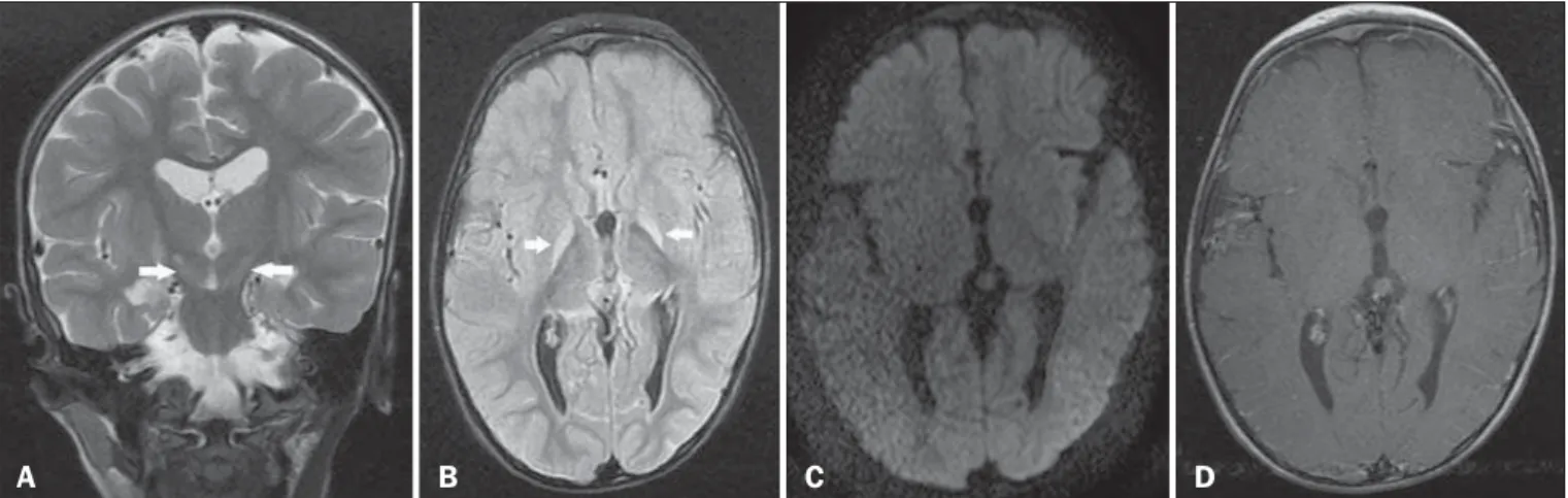
Figure 1. A: Coronal T2-weighted MRI sequence showing a bilateral, symmetrical hyperintense signal in the subthalamic nuclei (arrows), without a mass effect. B:
Axial FLAIR MRI sequence showing a bilateral, symmetrical hyperintense signal in the globus pallidus (arrows). C: Axial diffusion-weighted MRI sequence showing no diffusion restriction. D: Axial T1-weighted MRI sequence showing no evidence of gadolinium enhancement.
Letters to the Editor
Radiol Bras. 2016 Nov/Dez;49(6):406–413
408
http://dx.doi.org/10.1590/0100-3984.2015.0190 There is a broad spectrum of diagnoses of bilateral lesions in
the basal ganglia in the pediatric population. The main causes cited are hypoxic-ischemic encephalopathy; hypoglycemia; en-cephalitis; inborn errors of metabolism; water and electrolyte turbances; carbon monoxide poisoning; and demyelinating dis-orders. The correlation with clinical and laboratory data is funda-mental for making the definitive diagnosis(7,12,13).
In conclusion, the possibility of acute or chronic kernicterus should be considered when clinical symptoms, biochemical data, and MRI findings are suggestive of the disease, the chronic pre-sentation and permanent, irreversible profile being promoted by bilirubin neurotoxicity.
REFERENCES
1. Alfenas R, Niemeyer B, Bahia PRV, et al. Parry-Romberg syndrome: findings in advanced magnetic resonance imaging sequences – case report. Radiol Bras. 2014;47:186–8.
2. Bimbato EM, Carvalho AG, Reis F. Toxic and metabolic encephalopa-thies: iconographic essay. Radiol Bras. 2015;48:121–5.
3. Castro FD, Reis F, Guerra JGG. Intraventricular mass lesions at mag-netic resonance imaging: iconographic essay – part 1. Radiol Bras. 2014;47:176–81.
4. Ono SE, Carvalho Neto A, Gasparetto EL, et al. X-linked adrenoleukodys-trophy: correlation between Loes score and diffusion tensor imaging parameters. Radiol Bras. 2014;47:342–9.
5. Barbosa JHO, Santos AC, Salmon CEG. Susceptibility weighted imag-ing: differentiating between calcification and hemosiderin. Radiol Bras. 2015;48:93–100.
Bruno Niemeyer de Freitas Ribeiro1, Gabriela de Almeida Lima1, Nina Ventura1, Emerson Leandro Gasparetto1, Edson Marchiori2
1. Instituto Estadual do Cérebro Paulo Niemeyer, Rio de Janeiro, RJ, Brazil. 2. Universidade Federal do Rio de Janeiro (UFRJ), Rio de Janeiro, RJ, Brazil. Mailing address: Dr. Bruno Niemeyer de Freitas Ribeiro. Instituto Estadual do Cérebro Paulo Niemeyer – Departamento de Radiologia. Rua do Rezende, 156, Centro. Rio de Janeiro, RJ, Brazil, 20231-092. E-mail: [email protected]. 6. Turkel SB, Miller CA, Guttenberg ME, et al. A clinical pathologic
re-appraisal of kernicterus. Pediatrics. 1982;69:267–72.
7. Parashari UC, Singh R, Yadav R, et al. Changes in the globus pallidus in chronic kernicterus. J Pediatr Neurosci. 2009;4:117–9.
8. Perlstein MA. The late clinical syndrome of posticteric encephalopathy. Pediatr Clin North Am. 1960;7:665–87.
9. Martich-Kriss V, Kollias SS, Ball WS Jr. MR findings in kernicterus. AJNR Am J Neuroradiol. 1995;16(4 Suppl):819–21.
10. Coskun A, Yikilmaz A, Kumandas S, et al. Hyperintense globus pallidus on T1-weighted MR imaging in acute kernicterus: is it common or rare? Eur Radiol. 2005;15:1263–7.
11. Govaert P, Lequin M, Swarte R, et al. Changes in globus pallidus with (pre)term kernicterus. Pediatrics. 2003;112(6 Pt 1):1256–63. 12. Hegde AN, Mohan S, Lath N, et al. Differential diagnosis for bilateral
abnormalities of the basal ganglia and thalamus. Radiographics. 2011; 31:5–30.
13. Khanna PC, Iver RS, Chaturvedi A, et al. Imaging bithalamic pathol-ogy in the pediatric brain: demystifying a diagnostic conundrum. AJR Am J Roentgenol. 2011;197:1449–59.
Renal lymphangiectasia: know it in order to diagnose it
Dear Editor,
Here, we report the case of a 9-year-old girl with hyperpar-athyroidism. Ultrasound showed renal cysts and increased echo-genicity of the parenchyma in both kidneys. The diagnostic hy-pothesis was hyperparathyroidism secondary to chronic/polycystic kidney disease. The patient presented with gradually worsening kidney function and hypertension, and new imaging scans were requested. The ultrasound showed anechoic, multiloculated im-ages in the pyelocaliceal region of both kidneys, and perirenal, subcapsular cysts. A computed tomography (CT) scan was ac-quired, although no contrast agent was used, which precluded an accurate characterization. Nevertheless, the CT scan revealed changes similar to those observed on ultrasound. We also per-formed magnetic resonance imaging (MRI), which showed pyelocaliceal, perirenal cysts, with altered intensity of the signal of the renal parenchyma and loss of corticomedullary differentiation (Figure 1A), confirming, in conjunction with the clinical and bio-chemical data, the diagnosis of renal lymphangiectasia (RL).
RL is a rare benign disease that occurs because of miscom-munication between the renal lymphatic drainage system and the retroperitoneal lymphatic system(1). As a result, there is
accumu-lation of lymph in the renal lymph ducts, making them ectatic and forming simple or multiloculated, typically asymmetric and bilateral, collections in the pyelocaliceal, perinephric, or parenchy-mal regions, although, in some cases, only a part of one kidney is affected (Figure 1—B,C). There is no predilection for a given gender or age group. As of 2005, only 40 cases had been de-scribed(1,2).
In most cases, RL is an incidental finding, with or without signs and symptoms of pain, increased abdominal volume, he-maturia, ascites, edema of the lower limbs, hypertension, erythro-cytosis with renal vein thrombosis, and, rarely, chyluria(3). Such
manifestations can be explained by the distention of the renal
fascia and compression of the renal parenchyma by cysts, fistuli-zation to the pelvic cavity, and changes in the renin-angiotensin system(2–4). In rare cases, chronic kidney disease has been
re-ported(5). To our knowledge, there have been no specific reports
of clinical evolution to hyperparathyroidism, although a relation-ship with chronic kidney disease can be assumed.
A CT scan can reveal expansive perirenal formations with fluid attenuation, bounded by the renal fascia, that conform to
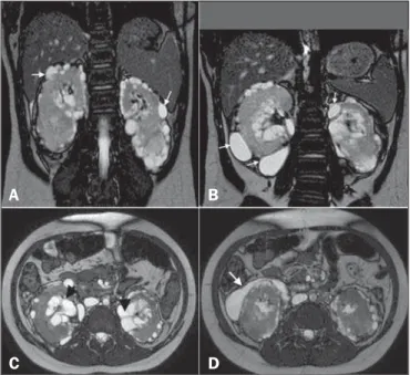
Figure 1. A: Coronal T2-weighted MRI sequence showing a loss of corticomedullary differentiation in both kidneys and multiple cystic lesions, with thin walls, located in the cortex (arrows). B: Cystic formations in the subcapsular cortex (arrows). C:
Axial T2-weighted MRI sequence showing cysts located in the renal sinuses (ar-rowheads) and perinephric spaces, simulating pelvic dilatation. D: The same images simulating cystic collections in the subcapsular cortex (arrow).