AR
TIGO ORIGINAL / ORIGINAL AR
TICLE
NEW “INTRODUCER” PEG-GASTROPEXY
WITH T FASTENERS:
a pilot study
Fernanda Prata
MARTINS
1, Maris Celia Batista de
SOUSA
2and Angelo Paulo
FERRARI
3ASBTRACT – Context - Enteral feeding is indicated for patients unable to maintain appropriate oral intake, and percutaneous endoscopic gastrostomy (PEG) is the most adequate long-term enteral access. Peristomal infections are the most common complications of PEG, occurring in up to 8% of patients, despite the use of prophylactic antibiotics. The ‘‘introducer’’ PEG-gastropexy technique avoids PEG tube passage through the oral cavity, preventing microorganisms’ dislodgment to the peristomal site. Objectives - To compare the incidence of peristomal wound infection at 7-day post-procedure after conventional “pull” technique versus a new “introducer” PEG-gastropexy kit. Secondary outcomes included success rates, procedure time, and other complications. Methods - Eighteen patients referred for PEG placement between June and December 2010 were randomly assigned to “pull” PEG with antibiotics or
“introducer” PEG-gastropexy technique without antibiotics. Results - Overall success rate for both methods was 100%, although
mean procedure duration was higher in the “introducer” PEG-gastropexy group (12.6 versus 6.4 minutes, P = 0.0166). Infection
scores were slightly higher in patients who underwent “pull” PEG with antibiotics compared with “introducer” PEG-gastropexy without antibiotics (1.33 ± 0.83 versus0.75 ± 0.67, P = 0.29). Conclusion - Although procedure duration was longer in the “introducer” PEG-gastropexy, infection scores were marginally higher in the “pull” PEG technique.
HEADINGS – Enteral nutrition. Gastrostomy, methods. Gastrostomy, adverse effects.
INTRODUCTION
Enteral feeding is indicated for patients with an intact gastrointestinal tract who are unable to maintain appropriate oral caloric intake mostly secondary to neurological impairment, malignancy, hypercatabolic
status and extensive burn injury(3).
The percutaneous endoscopic gastrostomy (PEG) technique has irstly been described in 1980 by Gauderer
et al.(5) and since then became the most standard
procedure for providing long-term enteral nutrition. The ‘‘pull’’ placement technique is the most commonly practiced worldwide.
PEG-site infection is the most common procedure-related complication. Prophylactic intravenous antibiotic is recommended 30 minutes before the procedure, an approach that signiicantly reduces
such complication(2).
A recent meta-analysis that included more than 1000 patients revealed that, even with antibiotic prophylaxis, the incidence of peristomal infection can
be as high as 8%(8).
The key point of the ‘‘introducer’’ PEG technique is to avoid the PEG tube passage through the oropharynx preventing microorganisms’ dislodgment to the peristomal site. However, its introduction 22 years ago was associated with several complications related to
The authors declare that they have no conflict of interest. Hospital Israelita Albert Einstein, São Paulo, SP, Brasil.
Correspondence: Dr. Fernanda Prata Martins – Rua Ruggero Fasano, s/n – Piso Intermediário 4 – Morumbi – 05653-120 - São Paulo, SP, Brasil. E-mail: [email protected]
delation or rupture of the balloon anchoring system in the stomach that could result in gastric contents
leakage into the peritoneum(12).
Recently, an improved introducer PEG technique with endoscopic gastropexy was shown, in a prospective randomized trial, to be both safe and easy to perform, resulting in fewer infectious complications compared
with the conventional pull PEG(10).
The aim of this pilot study was to compare the incidence of peristomal wound infection at 7-day post-conventional “pull” technique versus a new “introducer” PEG gastropexy kit (Kimberly-Clark* MIC-KEY* G Introducer Kit). Secondary outcomes included success rates, procedure times, and other complications.
METHODS
This pilot study protocol was approved by our Institutional Ethics Committee. Written informed consent was obtained from all patients before the procedure.
severe coagulation disorders, peritonitis, ascitis, peritoneal carcinomatosis or inability to achieve transillumination. Patients in the “pull” technique group received prophylactic IV antibiotics, unless they were undergoing continuous treatment with antibiotics. No prophylactic antibiotics were administered for patients in the “introducer” group.
All procedures were performed by two endoscopists, with patients in the supine position under monitored assisted care anesthesia with propofol. Procedure’s lengths were recorded.
The initial procedure phase was similar for both techniques. After performing an upper endoscopy, the stomach was insuflated and a safe location for PEG tube placement was determined by abdominal wall transillumination and inger indentation seen during endoscopy in the gastric wall. After the skin was scrubbed in a sterile fashion, local anesthesia was applied.
In the “pull” PEG group, conventional technique was used. A 1-cm incision was made at the identiied site and gastric access was achieved with a larger-bore needle with a catheter followed by wire passage through the access catheter into the stomach. The wire was then grasped with a snare and withdrawn through the patient’s mouth along with the endoscope. The wire was knotted to a 24Fr “pull” type tube and tracked from
the abdominal access until the abdominal wall(5).
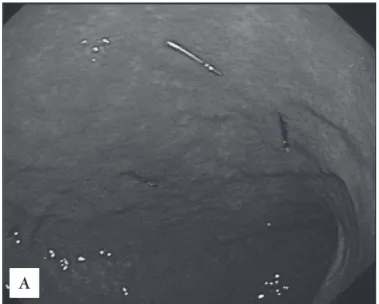
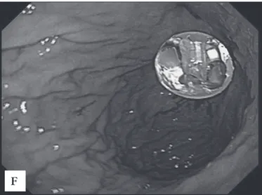
In the “introducer” PEG group, after site identiication, a 3-point gastropexy in a triangle shape was performed, using T-fasterners (Figure 1A), to ensure gastric wall ixation to anterior abdominal wall. An introducer needle was used to puncture the gastric wall (Figure 1B) and advance a J-guidewire into the gastric lumen. The introducer needle was then removed (keeping the J-guidewire in place), a small skin incision was performed and the serial passage dilator advanced over the guidewire (Figure 1C). After dilation and stoma length measure (Figure 1D), the dilator and J-guidewire were removed, leaving the peel-away sheath in the stomach for gastrostomy low-proile tube placement (16 or 20 Fr)
(Figure 1E and F)(3).
FIGURE 1A. Endoscopic view showing the triangle shape performed using T-fasterners delivered in the gastric anterior wall
A
FIGURE 1D. Stoma length measure
D
FIGURE 1C. Stoma dilation performed by advancing the serial dilator over the guidewire
C
FIGURE 1B. The introducer needle used to puncture the abdominal and gastric wall
The results were evaluated by using descriptive analysis including calculations of means, standard deviation and ranges of all continuous variables, as well as frequency and
percentage of categorical variables. An unpaired Student t
test was performed for continuous parameters and categorical
data was examined with the Chi-square test. A P value
equal or less than 0.05 was considered evidence of statistical signiicance.
RESULTS
A total of 18 patients were enrolled in the study: 10 in the “pull” group and 8 in the “introducer” group. PEG was successfully performed in all patients. The age of patients ranged from 60 to 97 years old and the main indication for PEG was neurological impairment. Patient’s demographic characteristics are summarized in Table 1.
“Pull” PEG group
“Introducer”
PEG group P
n 10 8
Mean age (range) 77.6 (64 – 97) 80.4 (60 – 91) NS* Gender (male:female) 6:4 7:1 NS** PEG indication
Neurological impairment 10 (100%) 8 (100%) NS** Current antibiotic use 4 2 NS ** Prophylactic antibiotic 6 0 N/A
Previous NG tube 8 6 NS**
Technical success 10 (100%) 8 (100%) N/A Procedure time (min) 6.40 12.6 0.0166*
Range 4 to 12 5 to 22
Peristomal infection score
Mean (SD) 1.33 (0.83) 0.75 (0.67) 0.29*
TABLE 1. Patient’s demographics characteristics
PEG = percutaneous endoscopic gastrostomy; NG tube = nasogastric tube; NS = non-signiicant; N/A = non-applicable. * Student t test; **Chi-square
FIGURE 1E. The sheath is peeled-away for gastrostomy low-proile tube placement
E
FIGURE 1F. Final endoscopic view with the low-proile tube placed
F
Enteral feeding was started within 6 hours after the procedure for both groups.
The peristomal site was evaluated 7 days after PEG placement and scored for erythema (0 = none, 1 = <5 mm, 2 = 6 to 10 mm, 3 = 11 to 15 mm, 4 = >15 mm), induration (0 = none, 1 = <10 mm, 2 = 11 to 20 mm, 3 = >20 mm), and exudate (0 = none, 1 = serous, 2 = serosanguinous, 3 = sanguinous, 4 = purulent). Infection was considered present if the combined score was 8 or higher, or in the presence of
suppurating exudate(9).
Statistical analysis
This study was intended as a pilot study, because due to costs containments only a few gastrostomy kits were available to perform the study, and therefore neither sample nor power calculations were performed.
The mean time for the “pull” PEG procedure (6.40 min,
range 4 to 12 min) was statistically signiicant (P = 0.0166)
shorter compared to that for the “introducer” PEG group (12.6 min, range 5 to 22 min).
The mean peristomal infection score was slightly higher in patients who underwent PEG by the “pull” technique then
in the “introducer” group (1.33 ± 0.83 versus0.75 ± 0.67,
P = 0.29). However, there was no statistically signiicant
difference between the two groups in terms of infection score. A combined score of 8 or higher indicating peristomal infection was not encountered in any patient at day 7-post procedure. All peristomal reactions were successfully treated with local wound care.
No other complications were observed in these patients.
DISCUSSION
oral intake. PEG-site infection is the most common procedure related complication, described initially in as high as 30%
to 41% of patients(1, 11). After prophylactic antibiotics were
routinely recommended, the infection rate decreased to 8%
in the most recent meta-analysis(8).
The pull method, described by Gauderer et al.(5), has the
disadvantage of carrying oral bacterial to the stoma site, increasing the risk of infectious complications. Based on this theory a valid approach to reduce the risk of infection would be to avoid the passage of the gastrostomy tube by the oral cavity.
Stomal infectious complications have been demonstrated to be signiicantly less frequent in the “introducer” PEG-placement
technique compared to the “pull” placement(6, 7, 10, 13, 14).
Maetani et al.(10) compared the two techniques (all
patients received prophylactic antibiotics) in a prospective randomized study, in which the introducer PEG resulted in signiicantly fewer infectious complications (0% vs 31%;
P<0.0001). Shastri et al.(13) compared the incidence of wound
infection after an “introducer” PEG gastropexy technique and demonstrated that it could be safely performed without prophylactic antibiotics.
In the present pilot study, although there was no patient considered to have stomal infection (score 8 or higher), the mean peristomal infection score was slightly higher in patients who underwent PEG by the “pull” technique than in the “introducer” group, similar to published results.
Another “introducer” PEG advantage is to reduce the risk for tumor implantation in the gastrostomy site in patients in whom the tube has to be passed through a neoplasic lesion
(larinx, esophagus)(4). We did not have any patient with neck
or esophageal tumor in our series.
The “introducer” PEG-placement technique was initially associated to complications related to balloon delation or rupture, breaking the stomach anchoring system possibly resulting in gastric contents leakage into the peritoneum. This problem was minimized by anchoring the stomach wall to the abdominal wall using T-fasteners.
The success rates for both methods were 100%, although mean procedure duration was higher in the “introducer” PEG-gastropexy group.
The results of the present study demonstrate that PEG-gastropexy can be placed safely, without any prophylactic antibiotics. Although such delay could represent a disadvantage, in the “introducer” technique the patient can be discharged with a ‘button’ instead of a PEG tube, what could also represent some improvement in quality of life.
In conclusion, the infection scores were marginally higher in patients who were undergoing “pull” PEG with antibiotics compared with “introducer” PEG-gastropexy without antibiotics. However, this is a pilot study and the conclusions are limited by the sample size. There is a need for larger multicenter randomized controlled trial to substantiate these results.
Martins FP, Sousa MCB, Ferrari AP. Gastrostomia endoscópica percutânea pela técnica de introdução com gastropexia com T-tags: um estudo piloto. Arq Gastroenterol. 2011;48(4):231-5.
RESUMO – Contexto - A nutrição enteral está indicada para pacientes incapazes de manter aporte voluntário adequado e a gastrostomia endoscópica percutânea (GEP) é a via preferencial para acesso enteral de longa duração. As infecções periostomais são as principais complicações da GEP, ocorrendo em até 8% dos pacientes, a despeito do uso de antibiótico proilático. A GEP pela técnica de introdução com gastropexia evita a passagem da sonda de gastrostomia pela cavidade oral, prevenindo contra o deslocamento de microorganismos ali presentes até o sítio da ostomia. Objetivo -
Comparar a incidência de infecção periostomal no 7º dia após GEP por técnica de tração versusGEP pela técnica de introdução com gastropexia.
Objetivos secundários incluíram: taxa de sucesso, tempo de procedimento e outras complicações. Métodos - Dezoito pacientes encaminhados ao
setor de endoscopia do Hospital Albert Einstein, São Paulo, SP, para realização de GEP entre junho e dezembro de 2010, foram randomizados para realização de gastrostomia pela técnica de tração com antibioticoterapia proilática ou pela técnica de introdução com gastropexia sem antibiótico
proilaxia. Resultados - A taxa de sucesso para ambos os métodos foi de 100%, apesar do tempo do procedimento ter sido mais longo no grupo da
técnica de introdução (12,6 versus 6,4 min, P = 0,0166). Os índices de infecção foram discretamente superiores no grupo de GEP por tração, com antibioticoterapia proilática, em comparação ao grupo GEP por introdução com gastropexia (1,33 ± 0,83 versus0,75 ± 0,67, P = 0,29). Conclusão
- Apesar da duração do procedimento ter sido mais longa no grupo GEP por introdução com gastropexia, a taxa de infecção foi discretamente mais elevada no grupo GEP por tração.
REFERENCES
1. Akkersdijk WL, van Bergeijk JD, van Egmond T, Mulder CJ, van Berge Henegouwen GP, van der Werken C, van Erpecum KJ. Percutaneous endoscopic gastrostomy (PEG): comparison of push and pull methods and evaluation of antibiotic prophylaxis. Endoscopy. 1995;27:313-6.
2. ASGE Standards of Practice Committee, Banerjee S, Shen B, Baron TH, Nelson DB, Anderson MA, Cash BD, Dominitz JA, Gan SI, Harrison ME, Ikenberry SO, Jagannath SB, Lichtenstein D, Fanelli RD, Lee K, van Guilder T, Stewart LE. Antibiotic prophylaxis for GI endoscopy. Gastrointest Endosc. 2008;67:791-8. 3. ASGE Technology Committee, Kwon RS, Banerjee S, Desilets D, Diehl DL,
Farraye FA, Kaul V, Mamula P, Pedrosa MC, Rodriguez SA, Varadarajulu S, Song LM, Tierney WM. Enteral nutrition access devices. Gastrointest Endosc. 2010;72:236-48.
4. Cruz I, Mamel JJ, Brady PG, Cass-Garcia M. Incidence of abdominal wall metastasis complicating PEG tube placement in untreated head and neck cancer. Gastrointest Endosc. 2005;62:708-11; quiz 752, 753.
5. Gauderer MW, Ponsky JL, Izant RJ Jr. Gastrostomy without laparotomy: a percutaneous endoscopic technique. J Pediatr Surg. 1980;15:872-5.
6. Hiki N, Maetani I, Suzuki Y, Washizawa N, Fukuda T, Yamaguchi T. Reduced risk of peristomal infection of direct percutaneous endoscopic gastrostomy in cancer patients: comparison with the pull percutaneous endoscopic gastrostomy procedure. J Am Coll Surg. 2008;207:737-44.
7. Horiuchi A, Nakayama Y, Tanaka N, Fujii H, Kajiyama M. Prospective randomized trial comparing the direct method using a 24 Fr bumper-button-type device with the pull method for percutaneous endoscopic gastrostomy. Endoscopy. 2008;40:722-6.
8. Jafri NS, Mahid SS, Minor KS, Idstein SR, Hornung CA, Galandiuk S. Meta-analysis: antibiotic prophylaxis to prevent peristomal infection following percutaneous endoscopic gastrostomy. Aliment Pharmacol Ther. 2007;25:647-56.
9. Jain NK, Larson DE, Schroeder KW, Burton DD, Cannon KP, Thompson RL, DiMagno EP. Antibiotic prophylaxis for percutaneous endoscopic gastrostomy. A prospective, randomized, double-blind clinical trial. Ann Intern Med. 1987;107:824-8. 10. Maetani I, Tada T, Ukita T, Inoue H, Sakai Y, Yoshikawa M. PEG with introducer
or pull method: a prospective randomized comparison. Gastrointest Endosc. 2003;57:837-41.
11. McClave SA, Chang WK. Complications of enteral access. Gastrointest Endosc. 2003;58:739-51.
12. Russell TR, Brotman M, Norris F. Percutaneous gastrostomy. A new simpliied and cost-effective technique. Am J Surg. 1984;148:132-7.
13. Shastri YM, Hoepffner N, Tessmer A, Ackermann H, Schroeder O, Stein J. New introducer PEG gastropexy does not require prophylactic antibiotics: multicenter prospective randomized double-blind placebo-controlled study. Gastrointest Endosc. 2008;67:620-8.
14. Tucker AT, Gourin CG, Ghegan MD, Porubsky ES, Martindale RG, Terris DJ. ‘Push’ versus ‘pull’ percutaneous endoscopic gastrostomy tube placement in patients with advanced head and neck cancer. Laryngoscope. 2003;113:1898-902.