Arquivos Brasileiros de Cardiologia - Volume 87, Nº 2, August 2006
Case Report
Case Report
Mailing Address: Edmundo Clarindo Oliveira • Rua Teodomiro Cruz, 65/102 - 30240-530 – Belo Horizonte, MG - Brazil
E-mail: [email protected] Received on 10/04/05 • Accepted on 12/12/05
Percutaneous Closure of Atrial Septal Defect Using
Transhepatic Puncture
Edmundo Clarindo Oliveira, Helder Machado Pauperio, Bráulio Muzzi Ribeiro Oliveira, Rogério Augusto Pinto da Silva, Fabrício Maia Torres Alves, Gustavo Lobato Adjuto
Hospital Luxemburgo de Belo Horizonte - Belo Horizonte, MG - Brazil
absent, and that it continued as an azygos towards the superior vena cava (SVC). We then punctured the right internal jugular vein and positioned the guide wire into the left inferior pulmonary vein. We then performed all the classic steps to close the ASD, without succeeding in obtaining a good position to release the prosthesis. In view of this, the procedure was interrupted, and re-planned. After obtaining the authorization of the patient and her parents, the procedure was carried out two weeks later by transhepatic puncture of the hepatic vein following the steps below: 1) General anesthesia with endotracheal tube. 2) Transesophageal echocardiogram. 3) Abdominal ultrasound. 4) IV cephalothin 2.0 grams. 5) Preparation of the region of the right hypochondrium and transhepatic puncture in the upper part of the inferior third of the liver at the level of the anterior axillary line using a Chiba 0.018 needle (Cook Inc.) (fi gure 2). The needle was positioned in parallel to the fl oor of the hemodynamics room and directed towards the spine. It was kept 2 cm away from it. The stylet was removed and a 5 ml-syringe with nonionic contrast medium was connected. The needle was slowly withdrawn and we aspirated until blood began to fl ow. At this point, we manually injected a small amount of contrast medium, and confi rmed the position in the hepatic vein (fi gure3). A 0.035 guide wire was then introduced and positioned in the right atrium. We then performed the dilation with a 7F introducer, then introduced the 7F hemaquet, administered 5,000 U/kg of IV heparin, measured the pressures with a multipurpose catheter, positioned the catheter in the left superior Percutaneous atrial septal defect (ASD) closure is done
by inferior vena cava (IVC) routinely. Occasionally this access is not possible due to obstruction or congenital absence of IVC. This case report shows the percutaneous implantation of an Amplatzer device to close large ASD in a patient with congenital absence of IVC. The procedure was done by a transhepatic punction of the hepatic vein with a Chiba needle (Cook Inc.) with no complications, taking the same amount of time, as the usual access. The transhepatic punction is a good option to central venous access in patients without other alternatives. The hepatic veins are large which make it possible to use large sheaths safely, even in neonates with low weight. This technique should be remembered, when it is impossible to use the usual venous access.
C
ASER
EPORTAn eighteen-year-old female patient was referred for percutaneous closure of atrial septal defect (ASD). On physical examination she had signs of right ventricle enlargement, fi xed splitting of the second heart sound, ejection systolic murmur in the upper left sternal edge (LSE) and mesodiastolic murmur in the lower LSE.
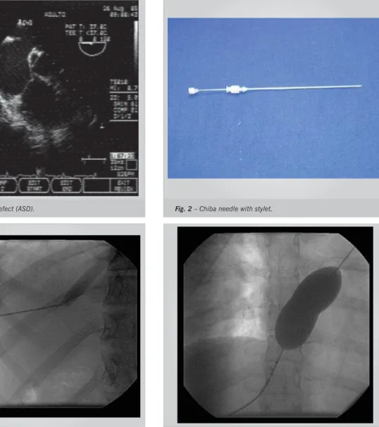
Supplementary tests evidenced sinus rhythm and second degree right branch block on electrocardiogram, slight increase of heart area on thorax x-ray, and OS-ASD with 18 mm of diameter on transesophageal echocardiogram (TE-ECHO) (fi gure 1).
We planned to perform the procedure according to the traditional method. After puncture of the femoral vein, we identifi ed that the suprahepatic segment of VCI was
K
EY WORDSASD occlusion, hepatic vein, transhepatic punction. The percutaneous closure of ostium secundum (OS) atrial septal defect (ASD) is a well-established procedure and is today considered the treatment of choice due to its good results and low morbidity and mortality. The procedure is routinely performed through the inferior vena
Arquivos Brasileiros de Cardiologia - Volume 87, Nº 2, August 2006 pulmonary vein (SLPV), and introduced a rigid guide wire
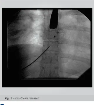
which was maintained in this position. We measured the distended size of the ASD using a number 34 balloon (AGA Medical) (fi gure 4), introduced directly in the skin over the guide wire. Later the balloon was replaced by a number-12 long sheath with the introducer advanced until the SLPV, the introducer was withdrawn, and the sheath was maintained at the entrance of the hepatic vein into the right atrium (RA). After verifying the return of blood, the sheath was introduced up to the left atrium (LA). We then continued the procedure following routine steps: release of the fi rst disc in the LA, release of the second disc in the RA, verifi cation of the position with TE-ECHO in several positions, and release of the prosthesis (fi gure 5). The distended diameter of ASD was 27.5 mm, and we implanted a number 30 Amplatzer prosthesis. The procedure was uneventful, with 10 minutes spent in the puncture of the hepatic vein and 55 minutes for the whole procedure. The sheath was removed and 50% of
Fig. 3 – Confi rmation of hepatic vein puncture. Fig. 4 – Balloon positioned for measuring ASD distended diameter.
Fig. 1 – Atrial septal defect (ASD). Fig. 2 – Chiba needle with stylet.
the heparin dose was neutralized with protamine. The right hypochondrium was manually compressed for 10 minutes. 6) The patient was sent to the Intermediate Care Unit (ICU) and kept in right lateral decubitus for two hours.
The patient complained of pain in the shoulder and in the right hypochondrium and was administered IV morphine at 2 mg every 6 hours in the fi rst 24 hours and 50 mg of diclofenac sodium three times a day for 48 hours. An abdominal ultrasound was performed six hours after the procedure and showed no alterations. The patient progressed well and was discharged from the ICU in the following day, and from hospital after 48 hours. At present, in the fi fth week of follow-up, she continues asymptomatic, and takes 200 mg de ASA. The patient was advised to carry out prophylaxis for endocarditis, to take ASA for six months and to have clinical check-ups one, six and twelve months after the procedure.
Arquivos Brasileiros de Cardiologia - Volume 87, Nº 2, August 2006
D
ISCUSSIONThe percutaneous closure of the OS-ASD is a safe and efficacious procedure, with low morbidity and mortality rates, and is therefore the treatment of choice for this condition. Many prostheses have been used, and the Amplatzer prosthesis is the most widely used worldwide due to its ease of handling, the high rate of success and the possibility of removal after it has been released. The procedure has been performed routinely through femoral vein puncture, using the normal ICV connection with the left atrium. Occasionally this classic route cannot be used due to ICV obstruction or congenital absence of its connection with the RA, which requires other alternatives. The superior vena cava can be used, through its tributaries1, but this alternative is not always
successful, especially when the ASDs are large.
Hepatic veins are large, and are a good alternative in these cases, as well as for interventions in low-weight prematures, where the procedure would not be possible through the routes commonly used, given the small caliber of vessels. Sheaths between 7 and 10F may be used for children2, including prematures.
The puncture can be guided by abdominal ultrasound or by the insertion of a catheter into a hepatic vein through the superior cava vein. However, these measures are not indispensable. In the case presented, the puncture was performed using the fi rst technique as described below, without using the ultrasound, although it was available.
Many puncture techniques have been described: 1) Insert the needle in the upper part of the inferior third of the liver or midway between the diaphragm and the lower edge of the liver, guiding it by means of fl uoroscopy or ultrasound, at the level of the anterior axillary line. Advance the insertion of the needle parallel to the fl oor of the room until approximately 2 cm away from the spine. Remove the needle’s stylet, pulling it slowly, while
Fig. 5 – Prosthesis released.
a small amount of contrast medium is injected. After you are certain that the hepatic vein has been reached, introduce the guide wire, dilator and sheath and follow the usual steps to perform the catheterization and the intervention2,3. 2) Puncture at the level of the median
axillary line below the ribs, slightly directing the needle upwards and backwards towards the spine4,5. Employ
extra care to avoid puncturing the gallbladder. Then proceed with the steps previously described. 3) Similar to the above, but with the puncture made at the level of the anterior axillary line6.
Most authors recommend that the catheter be slowly removed with small injections, and once it is out of the hepatic vein, coils or Gelfoam should be introduced to prevent bleeding2,6. Others simply maintain the patient
in right lateral decubitus for two to four hours as we did7. Occasionally the transhepatic puncture has to be
performed at the level of the left hypochondrium due to the position of the liver7 in this region or predominantly
in this region.
Patients usually complain of abdominal and shoulder pain in the fi rst 24 hours following the procedure, and it is necessary to medicate them.
If the usual venous access routes are not available, the transhepatic puncture may be performed for prolonged venous access, as is the case for parenteral nutrition or chemotherapy8, repeated myocardial biopsies9 and
pacemaker implantation10. Transhepatic puncture may
also be used as the fi rst option to perform diagnostic or therapeutic heart catheterization in prematures, as it allows the use of larger sheaths. Complications such as hemobilia, retroperitoneal bleeding, hepatic abscess, cholangitis, pneumothorax, hepatic vein thrombosis and pulmonary embolism4,7 rarely occur.
There are few reports on the use of transhepatic puncture for central venous access in the international literature. Although we have learned in international congresses that it is occasionally used for the percutaneous closure of ASD, a survey carried out on Medline found only one article reporting on two cases of ASD closure using this access route11. In the Brazilian literature we found
no reports on its use in catheterization procedures of any kind or as a route for central venous access. In the case here presented, the procedure was performed with no diffi culties or complications. The time of procedure was similar to the time spent when the conventional route is used. Patients with high central venous pressure present a higher risk for bleeding and in these cases the use of coils or Gelfoam is advised to prevent it. In the case of patients with normal venous pressure these measures are not necessary as was the case with our patient. Transhepatic puncture is a good alternative for the performance of diagnostic or therapeutic heart catheterization and for prolonged venous access in patients who lack other available routes or require larger veins than the ones usually used. Hemodynamics professionals, especially those working in pediatrics facilities, should be ready to perform it in special situations.
Oliveira e cols.
Arquivos Brasileiros de Cardiologia - Volume 87, Nº 2, August 2006
1. Sullebarger JT, Sayad D, Gerber L, Ettedgui J, Jimmo-Waumans S, Alcebo PC. Percutaneous closure of atrial septal defect via transjugular approach with the Amplatzer septal occluder after unsuccessful attempt using the CardioSEAL device. Catheter Cardiovasc Interv. 2004; 62: 262-5.
2. Shim D, Lloyd TR, Cho KJ, Moorehead CP, Beekman RH III. Transhepatic cardiac catheterization in children: evaluation of effi cacy and safety. Circulation. 1995; 29: 1526-30.
3. Bravo RP, Sánchez J, Sarachaga IH, Cazzaniga M, Pineda LF, Cañete RB. Cateterismo cardíaco transhepático em niños. Anales Españoles Pediatria.2000; 52(5): 488-90.
4. Sommer RJ, Golinko RJ, Mitty HA. Initial experience with percutaneous transhepatic cardiac catheterization in infants and children. Am J Cardiol. 1995; 75: 1289-91.
5. Punamiya K, Beekman RH, Shim D, Muller DWM. Percutaneous transhepatic mitral commissurotomy. Catheter Cardiovasc Diag. 1996; 39: 204-6.
6. Perry SB. Manual techniques of cardiac catheterization. In: Lock JE,
R
EFERENCESKeane JF, Perry SB. Diagnostic and Interventional Catheterization in Congenital Heart Disease. 2nd ed. Boston: Kluwer Academic
Publishers; 2000: 21.
7. Johnson JL, Fellows KE, Murphy JD. Transhepatic central access for cardiac catheterization and radiologic intervention. Catheter Cardiovasc Diag. 1995; 35: 168-71.
8. Azizkhan RG, Taylor LA, Jaques PF, Mauro MA, Lacey SR. Percutaneous translumbar and transhepatic inferior vena caval catheters for prolonged vascular access in children. J Ped Surg. 1992; 27: 165-9. 9. Book WM, Raviele AA, Vincent RN. Repetitive percutaneous
transhepatic access for myocardial biopsy in pediatric cardiac transplants recipients. Catheter Cardiovasc Diag. 1998; 45: 167. 10. Adwani SS, Sreeram N, DeGiovanni JV. Percutaneous transhepatic
dual chamber pacing in children with Fontan circulation. Heart. 1997; 77: 574-5.
11. Shim D, Lloyd TR, Beekman RH 3rd. Transhepatic Therapeutic cardiac catheterization: a new option for the pediatric interventionalist. Catheter Cardiovasc Interv. 1999; 47: 41-5.