Clinicoradiological Session
Case 1/2007 – A Three-Year Old Child with Infundibular Pulmonary
Stenosis
Edmar Atik
Instituto do Coração do Hospital das Clínicas da FMUSP - São Paulo, SP, Brazil
Mailing Address: Edmar Atik •
InCor – Av. Dr Enéas C. Aguiar, 44 – 05403-000 – São Paulo, SP, Brazil E-mail: conatik@incor.usp.br
Clinical data
A three-year-old white female who had been previously diagnosed by echocardiography as having Tetralogy of Fallot with perimembranous ventricular septal defect (VSD) and infundibular pulmonary stenosis after a heart murmur was detected in a routine examination when she was five
months old. The child remained asymptomatic and without medication. A recent echocardiogram showed that the ventricular septal defect was only 4 mm in diameter, next to the obstructive defect. On physical examination she was eupneic, with normal pulses, body weight of 14 kg, heart
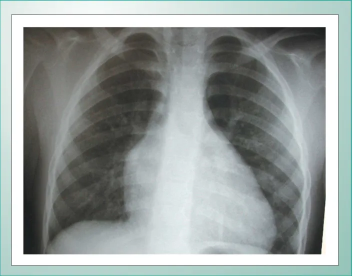
Fig. 1 -Chest X-ray showing the diminished pulmonary vascularity in the presence of enlarged right cavities. The straight mid-arch indicates right ventricular outflow tract obstruction.
Key words
infundibular pulmonary stenosis, perimembranous ventricular septal defect, Tetralogy of Fallot
Clinicoradiological Session
Edmar Atik CASE 1/2007 – A THREE-YEAR OLD CHILD WITH INFUNDIBULAR PULMONARY STENOSIS
Arq Bras Cardiol 2007; 88(1) : 98-99 Fig. 2 -Echocardiogram showing an anomalous muscle bundle in the right ventricular outflow tract, seen in a cross-section (A), causing obstruction with turbulence on the color flow map (B). RA: right atrium; Ao: aorta; PT: pulmonary trunk; RV: right ventricle; PV: pulmonary valve.
PT
RV
RV
PV
RA
rate of 90 bpm, blood pressure of 91/58-66 mmHg, and 96% oxygen arterial saturation. No cyanosis was detected. Neither the aorta nor the ictus cordis were palpable. No deformities were found in her thorax. Cardiac examination showed regular rate and rhythm, and a soft systolic ejection murmur was heard in the third, 2nd, 3rd and, 4thleft intercostal spaces.
Her liver was not palpable.
An electrocardiogram showed sinus rhythm and signs of right ventricular overload with preserved left potentials by Q wave in V4 to V6 and wide R waves in the same leads. The R wave amplitude in V1 was 17 mm. P-axis:+60°, QRS-axis:+90°, T-axis:+60°.
Radiographic examination
Radiographic findings included the following: mildly enlarged cardiac silhouette due to prominent right atrial and ventricular arches, a long ventricular arch and an elevated cardiac apex, a straight mid-arch, and reduced peripheraleduced peripheral pulmonary vascularity (Fig. 1).
Diagnostic impression
These findings are suggestive of right ventricular outflow tract obstruction.
Differential diagnosis
Pulmonary valve stenosis is usually accompanied by dilation
of the main and hilar pulmonary arteries, unless stenosis is severe to the point of not causing such post-stenotic dilations. It is worth noting that the RV outflow tract obstruction also does not cause pulmonary artery dilation.
Diagnostic confirmation
Clinical findings are consistent with the diagnosis of right ventricular outflow tract obstruction. The echocardiogram (Fig, 2) revealed a small, perimembranous ventricular septal defect measuring 4 mm in length and an anomalous right ventricular muscle bundle with intraventricular gradient of 84 mmHg. The aortic annulus diameter was 16 mm, and the pulmonary annulus’, 15 mm. Other measurements were the following: right ventricle,11 mm; diastolic left ventricle diameter, 33 mm; aorta and left atrium, 26 mm; and interventricular septum, 4 mm.
Management
During surgery, no ventricular septal defect was found.
There was a fibrous ostium infundibuli, which was widely resected. The right ventricular outflow tract had to be enlarged using bovine pericardium. The pulmonary valve was unremarkable.
The ventricular septal defect was thought to close spontaneously secondary to a progressive ventricular and RV outflow tract hypertrophy. This hypothesis is supported by the fact that in the radiography obtained two years earlier, the pulmonary vascularity was clearly larger.
