Pulmonary Haemodynamics in Sickle Cell
Disease Are Driven Predominantly by a
High-Output State Rather Than Elevated
Pulmonary Vascular Resistance: A Prospective
3-Dimensional Echocardiography/Doppler
Study
Sitali Mushemi-Blake1,2, Narbeh Melikian1,2, Emma Drasar3, Amit Bhan2, Alan Lunt4, Sujal R. Desai5, Anne Greenough4, Mark J. Monaghan1,2, Swee Lay Thein3,6, Ajay M. Shah1,2*
1King’s College London British Heart Foundation Centre, Cardiovascular Division, London, United Kingdom,2Department of Cardiology, King’s College Hospital, Denmark Hill, London, United Kingdom,
3Department of Haematological Medicine, King’s College Hospital, Denmark Hill, London, United Kingdom,
4Department of Paediatrics, King’s College Hospital, Denmark Hill, London, United Kingdom,5Department of Radiology, King’s College Hospital, Denmark Hill, London, United Kingdom,6NIH / National Institute of Heart, Lung and Blood Diseases, Sickle Cell Branch, Bethesda, Maryland, United States of America
*ajay.shah@kcl.ac.uk
Abstract
Aims
Patients with sickle cell disease have significant morbidity and mortality. Pulmonary hyper-tension is suggested to be an important contributor but its nature and severity in these patients and how best to non-invasively assess it are controversial. We hypothesised that a high-output state rather than primary pulmonary vascular pathology may be the major abnormality in sickle cell disease. This study aimed to evaluate the characteristics and severity of pulmonary hypertension in patients with sickle cell disease using detailed echocardiography.
Methods and Results
We undertook a prospective study in 122 consecutive stable outpatients with sickle cell dis-ease and 30 age, gender and ethnicity-matched healthy controls. Echocardiographic evalu-ation included 3D ventricular volumes, sphericity, tissue Doppler, and non-invasive
estimation of pulmonary vascular resistance. 36% of patients had a tricuspid regurgitant velocity2.5 m.s-1but only 2% had elevated pulmonary vascular resistance and the
preva-lence of right ventricular dysfunction was very low. Patients with raised tricuspid regurgitant velocity had significantly elevated biventricular volumes and globular left ventricular remod-elling, related primarily to anaemia. In a subgroup of patients who underwent cardiac cathe-terization, invasive pulmonary haemodynamics confirmed the echocardiographic findings. a11111
OPEN ACCESS
Citation:Mushemi-Blake S, Melikian N, Drasar E, Bhan A, Lunt A, Desai SR, et al. (2015) Pulmonary Haemodynamics in Sickle Cell Disease Are Driven Predominantly by a High-Output State Rather Than Elevated Pulmonary Vascular Resistance: A Prospective 3-Dimensional Echocardiography/ Doppler Study. PLoS ONE 10(8): e0135472. doi:10.1371/journal.pone.0135472
Editor:Philippe Connes, Université Claude Bernard Lyon 1, FRANCE
Received:May 11, 2015
Accepted:July 22, 2015
Published:August 13, 2015
Copyright:This is an open access article, free of all copyright, and may be freely reproduced, distributed, transmitted, modified, built upon, or otherwise used by anyone for any lawful purpose. The work is made available under theCreative Commons CC0public domain dedication.
Data Availability Statement:All relevant data are within the paper and its Supporting Information files.
Conclusions
Elevated cardiac output and left ventricular volume overload secondary to chronic anaemia may be the dominant factor responsible for abnormal cardiopulmonary haemodynamics in patients with sickle cell disease. 3D echocardiography with non-invasive estimation of pul-monary vascular resistance represents a valuable approach for initial evaluation of cardio-pulmonary haemodynamics in sickle cell disease.
Introduction
Sickle cell disease (SCD), prevalent in peoples of African descent, results from the presence of haemoglobin S (HbS) due to a genetic mutation in theβ-globin chain of haemoglobin. The abnormal HbS polymerizes under low oxygen conditions leading to the formation of irrevers-ibly sickled red blood cells that cause repeated episodes of vaso-occlusion and chronic anaemia, with multi-organ complications that impose substantial morbidity and reduce life expectancy [1]. With improved general medical care and reduction in infective complications, SCD has evolved into a chronic condition in which current treatment options are largely limited to blood transfusion and hydroxyurea [2].
Considerable attention has focused on pulmonary arterial hypertension as a potential cause of long-term morbidity and mortality in SCD [3–6]. It was proposed that chronic haemolysis leads to depletion of vasodilator nitric oxide in the microcirculation and induces an increase in pulmonary resistance that has long-term detrimental effects [4]. These authors used echocar-diographically-measured tricuspid valve regurgitation velocity (TRV) of2.5 m.s-1as a surro-gate marker of abnormally elevated pulmonary arterial pressure and reported that>30% of
patients had abnormal values and that this correlated with an increased likelihood of prema-ture death [4]. Other studies also found a high prevalence of TRV2.5 m.s-1in SCD [7,8]. However, this hypothesis has been challenged (6), and clinical trials of agents that target pul-monary arterial hypertension have been disappointing in SCD [9,10]. More recent studies involving systematic right heart catheterisation in patients with SCD and elevated TRV suggest a much lower prevalence of pulmonary hypertension [5,11]. In the largest study, Parent et al [5] found only 6% of patients to have pulmonary hypertension and this was most commonly post-capillary (venous) rather than pre-capillary (arterial). These authors concluded that sim-ple echocardiographic evaluation alone is of limited value for the detection of pulmonary hypertension in SCD. However, invasive and potentially repeated assessment by right heart catheterisation in all patients is impractical. Furthermore, the pathophysiology responsible for the high prevalence of elevated TRV in stable patients with SCD remains unclear.
Most screening studies in SCD to date have employed basic 2D Doppler echocardiography and have not taken advantage of techniques such as 3D imaging (which allows accurate volume estimation), tissue Doppler, strain analysis and non-invasive estimation of pulmonary vascular resistance (PVR). Cardiac output in SCD is often significantly elevated secondary to chronic anaemia and may confound interpretation of TRV. We hypothesized that elevated cardiac out-put rather than an elevated PVR may be the major driver of abnormal cardiopulmonary hae-modynamics in SCD and that the relative contributions of these factors can be assessed by comprehensive echocardiography. The primary aim of this study was to prospectively under-take comprehensive non-invasive evaluation of cardiopulmonary haemodynamics in a large population of outpatients with SCD and stable symptoms.
design, data collection, decision to publish, or preparation off the manuscript.
Methods
Study population
We studied 152 subjects, comprising 122 consecutive adult outpatients with stable SCD and 30 healthy controls matched for age, gender and ethnicity. Patients with a painful sickle crisis within the previous 6 weeks were excluded. The SCD genotype was haemoglobin SS in 82 (67%), haemoglobin SC in 22 (18%) and haemoglobin S-beta thalassaemia in 18 (15%). Con-trols were recruited from the general public and hospital staff. The study complied with the Declaration of Helsinki and was approved by the National Research Ethics Service London Dulwich Committee and the King’s College Hospital Directorate of Research and Develop-ment. All participants provided written informed consent.
Echocardiography
Transthoracic echocardiography was performed according to American Society of Echocardi-ography guidelines [12], using a Philips IE33 system with 2.5 MHz matrix array and stand-alone transducers. TRV was measured with continuous wave Doppler, using the highest value obtained in any of four standard views. Non-invasive estimation of PVR (i.e. PVRecho) was derived from TRV and the RV outflow tract time-velocity integral (PVRecho= 10 x TRV / RV outflow tract time-velocity integral), as previously described [13]. 2D colour-coded pulse wave tissue Doppler recordings were made at the level of the LV lateral mitral valve annulus and the RV free wall tricuspid valve annulus. The ratios of early diastolic LV inflow (E) to lateral mitral annulus velocity (E’), and RV inflow to tricuspid free wall annulus velocity, were derived as measures of LV and RV filling pressure respectively [14]. Tricuspid annular plane systolic excursion (TAPSE) was recorded as a measure of right ventricular systolic function [15]. Images were analysed off-line using Philips Xcelera software.
LV myocardial strain analysis was performed using standard grayscale 2D images in the short axis parasternal view at the level of the papillary muscle and in the 4-chamber apical view. Myocardial deformation was quantified at basal, mid and apical levels by speckle tracking analysis [16]. Strain quantification was performed using Philips QLab software v.9.
Full-volume 3D datasets were obtained from the apical 4-chamber window over 4 consecu-tive cardiac cycles and analyzed off-line [17]. 3D sphericity index was calculated as LV end-dia-stolic volume divided by volume of a sphere with the same diameter as the LV long-axis length; a higher index indicates a more globular LV. LV remodelling index was calculated as the ratio of LV mass to LV end-diastolic volume; it is usually unchanged in physiological hypertrophy, increased in pathological hypertrophy and decreased in heart failure [18].
Cardiac catheterisation and pulmonary assessment
A random subset of 18 patients with a TRV2.5 m.s-1underwent right heart catheterization, pulmonary high resolution computed tomography, and lung function testing, performed within a 2-week window for each patient. Measurements were made of pulmonary artery pres-sure (PAP), pulmonary capillary wedge prespres-sure (PCWP) and thermodilution cardiac output. Pulmonary vascular resistance (PVRRHC) was calculated in Wood Units. Computed tomogra-phy was performed on a dual detector helical scanner (HiSpeed NX/I, GE Medical Systems, Milwaukee, WI). Lung function tests were assessed according to American Thoracic Society/ European Thoracic Society guidelines [19].
Statistics. Data are presented as mean±SD or percentage as appropriate. Student's t-test was used to compare continuous variables and Chi square or Mann-Whitney U test to compare dichotomous variables. Associations between continuous variables were quantified by Pear-son’s correlation coefficients (two-tailed). Multivariable regression models with stepwise elimi-nation, based on a selection of clinical and laboratory variables, were created to determine the independent determinants of echocardiographic parameters. Standardized (beta) coefficients were calculated for covariates in models. Data analyses were performed using SPSS statistical software. P<0.05 was considered significant.
Results
Patient characteristics
All patients and controls were of African-Caribbean or African ethnic origin, with a majority of females. The SCD cohort had typical clinical characteristics of the disease, including multi-organ complications, chronic anaemia, reticulocytosis and mild hypoxaemia (Table 1). 62% of all SCD outpatients had at least 1 sickle-related complication. Blood pressure was significantly lower in patients with SCD.
Non-invasive assessment of pulmonary haemodynamics
Patients with SCD had significantly higher TRV and PVRechovalues than control subjects (Fig
1,Table 2). However, although 36% of patients had a TRV2.5 m.s-1and 12% had a TRV3
m.s-1, only 2% had a PVRecho>3 Wood units, which is the accepted cutoff for an abnormally
elevated PVR [20]. 14% of patients had a PVRechobetween 2–3 Wood units.
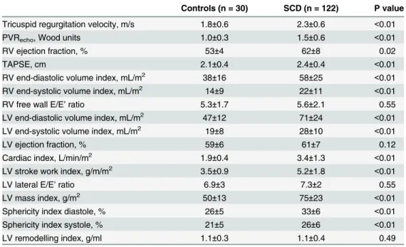
To assess the functional impact of potential pulmonary hypertension, we assessed RV struc-ture and function. Both RV ejection fraction and TAPSE were significantly higher in patients than controls, suggesting preserved or increased systolic function (Table 2). RV diastolic func-tion assessed by tricuspid E/E’was similar in patients and controls. The 3D RV end-diastolic and end-systolic volumes were significantly higher in SCD than controls (Fig 1,Table 2). These results suggest that the elevated TVR values are not related to a raised PVRechobut patients with SCD have an enlarged RV.
Left ventricular structure and function
Pulmonary haemodynamics may also be influenced by left-sided heart pressures and volumes, which can contribute to pulmonary venous hypertension. Patients with SCD had a significantly higher 3D cardiac index, LV end-diastolic and end-systolic volume index than controls (Fig 1,
Table 2). The main independent determinants of LV end-diastolic volume on multivariable
regression analysis were haemoglobin concentration (P = 0.04) and female gender (P = 0.01). LV mass index was also significantly higher in SCD. The 3D LV sphericity index was signifi-cantly higher in SCD (Table 2), indicating globular LV remodelling. Ejection fraction and LV remodelling index were unaltered in SCD. LV stroke work index, which controls for differences in afterload, was significantly higher in patients than controls. The LV E/E' ratio was similar in patients and controls.
Determinants of elevated TRV
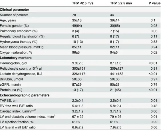
Table 3compares the characteristics of SCD patients with TRV2.5 m.s-1and<2.5 m.s-1.
Patients with elevated TRV had significantly lower haemoglobin levels and oxygen saturation, higher LDH levels and cardiac volumes, and were more likely to have suffered previous pulmo-nary embolism. RV systolic function assessed by TAPSE was more hyperdynamic in patients with TRV2.5 m.s-1.
We looked for association between TRV and various clinical, laboratory and echocardio-graphic parameters and then undertook multivariable regression analysis for independent determinants of TRV2.5 m.s-1. This showed that haemoglobin concentration and a history of pulmonary embolism were independent determinants of TRV2.5 m.s-1(S2 Table). Since very few patients had PVRecho3 Wood units, we undertook a similar analysis in those with PVRecho2 Wood units (22 patients; 18% of the total cohort). On multivariable regression analysis, the independent determinants of PVRecho2 Wood units were a history of pulmo-nary embolism and advancing age.
Invasive assessment of pulmonary haemodynamics
The findings so far suggest that elevated TRV2.5 m.s-1in patients with SCD is accompanied by increased cardiac volumes, likely due to chronic anaemia, but is rarely related to a raised
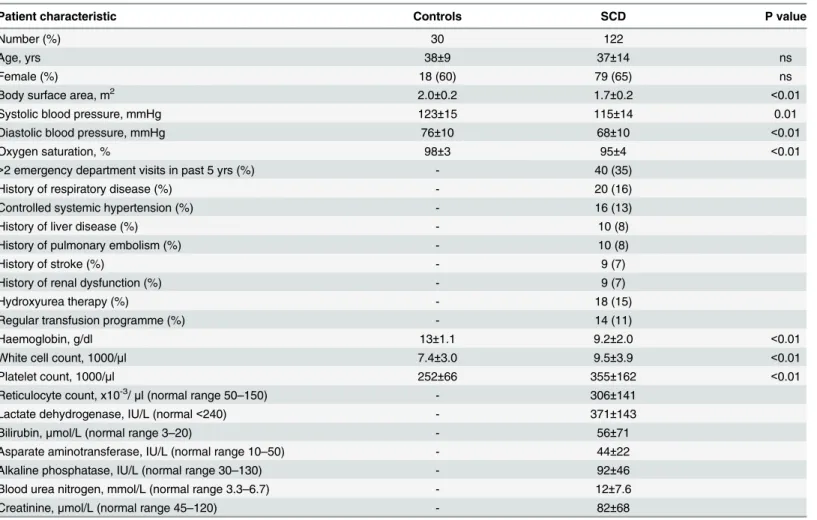
Table 1. Clinical characteristics of SCD patients compared with matched control subjects.
Patient characteristic Controls SCD P value
Number (%) 30 122
Age, yrs 38±9 37±14 ns
Female (%) 18 (60) 79 (65) ns
Body surface area, m2 2.0±0.2 1.7±0.2 <0.01
Systolic blood pressure, mmHg 123±15 115±14 0.01
Diastolic blood pressure, mmHg 76±10 68±10 <0.01
Oxygen saturation, % 98±3 95±4 <0.01
>2 emergency department visits in past 5 yrs (%) - 40 (35)
History of respiratory disease (%) - 20 (16)
Controlled systemic hypertension (%) - 16 (13)
History of liver disease (%) - 10 (8)
History of pulmonary embolism (%) - 10 (8)
History of stroke (%) - 9 (7)
History of renal dysfunction (%) - 9 (7)
Hydroxyurea therapy (%) - 18 (15)
Regular transfusion programme (%) - 14 (11)
Haemoglobin, g/dl 13±1.1 9.2±2.0 <0.01
White cell count, 1000/μl 7.4±3.0 9.5±3.9 <0.01
Platelet count, 1000/μl 252±66 355±162 <0.01
Reticulocyte count, x10-3/
μl (normal range 50–150) - 306±141
Lactate dehydrogenase, IU/L (normal<240) - 371±143
Bilirubin,μmol/L (normal range 3–20) - 56±71
Asparate aminotransferase, IU/L (normal range 10–50) - 44±22
Alkaline phosphatase, IU/L (normal range 30–130) - 92±46
Blood urea nitrogen, mmol/L (normal range 3.3–6.7) - 12±7.6
Creatinine,μmol/L (normal range 45–120) - 82±68
Fig 1. Scatter plots showing the distribution of values for echocardiographic parameters in SCD and control subjects.LVEDVI, LV end-diastolic volume index; RVEDVI, RV end-diastolic volume index.
doi:10.1371/journal.pone.0135472.g001
Table 2. Echocardiographic parameters in SCD outpatients.
Controls (n = 30) SCD (n = 122) P value
Tricuspid regurgitation velocity, m/s 1.8±0.6 2.3±0.6 <0.01
PVRecho, Wood units 1.0±0.3 1.5±0.6 <0.01
RV ejection fraction, % 53±4 62±8 0.02
TAPSE, cm 2.1±0.4 2.4±0.4 <0.01
RV end-diastolic volume index, mL/m2 38±16 58±25 <0.01 RV end-systolic volume index, mL/m2 14±9 22±11 <0.01
RV free wall E/E’ratio 5.3±1.7 5.6±2.1 0.55
LV end-diastolic volume index, mL/m2 47±12 71±24 <0.01 LV end-systolic volume index, mL/m2 19±8 28±10 <0.01
LV ejection fraction, % 59±6 61±7 0.12
Cardiac index, L/min/m2 1.9±0.4 3.4±1.3 <0.01
LV stroke work index, g/m/m2 3.5±0.9 5.2±1.8 <0.01
LV lateral E/E’ratio 6.9±3 7.3±2 0.55
LV mass index, g/m2 50±13 75±23 <0.01
Sphericity index diastole, % 26±5 33±6 <0.01
Sphericity index systole, % 21±5 26±6 <0.01
LV remodelling index, g/ml 1.1±0.3 1.1±0.4 0.49
PVRecho. To confirm this and to establish the contribution of elevated pulmonary venous pres-sures, we undertook right heart catheterization in 18 SCD patients with TRV2.5 m.s-1. Echo-cardiography was repeated on the day of catheterization; 2 patients had TRV<2.5 m.s-1on
this occasion. Patient clinical characteristics were similar to the overall group of 44 patients with TRV2.5 m.s-1.
S3 Tableshows the invasive haemodynamic and non-invasive data for individual patients.
The mean PAP was 22±7 mmHg (range 12–37 mmHg), mean PCWP 13±4 mmHg (range 6–20 mmHg) and mean cardiac output 8 L.min-1(range 4–12 L.min-1). 8 patients (44%) had PCWP15 mmHg, indicative of pulmonary venous hypertension. 13 patients (72%) had sig-nificant parenchymal lung disease on computed tomography, including the 1 patient who met diagnostic criteria for pulmonary arterial hypertension. All 15 patients who had lung function tests showed abnormal results.
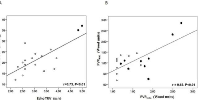
Fig 2Ashows the association between TRV and mean PAP. Although there was a significant
correlation, only 4 of 18 patients had a mean PAP25 mmHg, of whom 3 had a PCWP15 mmHg. Therefore, using TRV2.5 m.s-1greatly overestimated PAP and only 1 patient met diag-nostic criteria for pulmonary arterial hypertension.Fig 2Bshows the association between PVRecho and invasively measured PVR (PVRRHC). No patient had a PVRRHC>3 Wood units (diagnostic
cut-off for pulmonary arterial hypertension). The 2 patients with PVRRHCbetween 2–3 Wood units had PVRechovalues in the same range and had TRV>4 m.s-1. None of the 16 patients with
PVRecho<2 Wood units had a PVRRHCover this value; i.e. no patients with even physiologically
elevated PVR were missed by using a conservative PVRechocut-off of 2 Wood units.
Table 3. Clinical, laboratory and echocardiographic parameters for patients divided according to TRV <2.5 and2.5 m/s. eGFR, estimated glomerular filtration rate.
TRV<2.5 m/s TRV2.5 m/s P value
Clinical parameter
Number of patients 78 44
Age, years 35±13 39±14 0.1
Female gender (%) 49(64) 30(65) 0.93
Pulmonary embolism (%) 3 (4) 7 (15) 0.03
Regular blood transfusion (%) 6 (7) 8 (17) 0.11
Hydroxyurea therapy (%) 10 (13) 8 (17) 0.53
Mean blood pressure, mmHg 85±11 82±11 0.24
Oxygen saturation, % 96±3 94±5 0.02
Laboratory markers
Haemoglobin, g/dl 9.9±2.0 8.1±1.6 <0.01
Reticulocyte count, x10-3/
μl 303±151 309±127 0.81
Lactate dehydrogenase, IU/l 326±117 441±153 <0.01
Bilirubin,μmol/l 50±38 50±33 0.97
eGFR, ml/min 87±29 90±28 0.74
Proteinuria (%) 13 (17) 21 (45) <0.01
Echocardiographic parameters
TAPSE, cm 2.3±0.4 2.5±0.4 0.01
RV free wall E/E’ratio 5.4±1.8 5.8±2.4 0.43
Cardiac index, L/min/m2 3.2±1.2 3.7±1.2 0.06
LV end-diastolic volume index, ml/m2 67±22 79±26 0.01
LV ejection fraction, % 61±6 61±8 0.92
LV lateral wall E/E’ratio 6.9±2.2 7.9±2.5 0.06
Discussion
The main findings of this study in stable SCD outpatients are that: (a) although>35% of
patients have TRV2.5 m.s-1, there is little evidence of haemodynamically significant pulmo-nary hypertension as assessed by RV function and non-invasive PVRecho; (b) patients with raised TRV have elevated cardiac output, biventricular dilatation and 3D features of volume overload, likely to be related to chronic anaemia. Therefore, the high-output state of SCD rather than primary abnormalities of the pulmonary microvasculature may be the major driver of abnormal pulmonary haemodynamics. To our knowledge, this is the first prospective study to use comprehensive 2D and 3D echocardiography to evaluate the complex cardiopulmonary physiology of SCD outpatients.
The prevalence and role of pulmonary hypertension in the pathophysiology and outcome of SCD are controversial. Reports by Gladwin [4,21] and others [7,8], based primarily on TRV as a marker for pulmonary hypertension, suggested that this was a substantial problem and that targeting the pulmonary microvasculature might be of therapeutic value. It was proposed that chronic haemolysis impairs vascular nitric oxide bioactivity and leads to pulmonary vasocon-striction and pulmonary hypertension in SCD [4]. However, more recent studies that per-formed systematic cardiac catheterization indicate a much lower (<10%) prevalence of
pulmonary hypertension, with post-capillary hypertension predominating over arterial pulmo-nary hypertension [5,11]. However, it remains unclear whether SCD patients can usefully be evaluated by echocardiography and what the basis is for the elevated TRV values in a large pro-portion of patients.
Our study confirms the high prevalence of elevated TRV in unselected SCD outpatients but, consistent with previous work [5], we find a very low rate of pulmonary hypertension. Impor-tantly, our study used non-invasive methodology to assess patients and did not exclude any subjects on the basis of disease severity or complications. We found that very few patients had elevated PVRechoand, furthermore, there was no evidence of significant RV dysfunction as assessed by ejection fraction or TAPSE. In fact, SCD patients with TRV2.5 m.s-1had more
Fig 2. Correlation between echocardiographic estimates and invasive values of pulmonary haemodynamic parameters.A) TRV versus mean PAP; B) PVRechoversus PVRRHC. In A, solid symbols denote patients with PVRecho2 Wood units. In B, solid symbols denote patients with TRV3 m/s.
hyperdynamic RV function than those with lower TRV values indicating an absence of RV pressure overload. These findings were confirmed in the subgroup of patients with TRV2.5 m.s-1who underwent invasive assessment; only 1 patient met the diagnostic criteria for pulmo-nary arterial hypertension. It should be noted that the thermodilution method may underesti-mate cardiac output in patients with significant TR and high flow but this would, if anything, result in an overestimation of the PVR.
We then wished to establish the reasons for elevated TRV values in a high proportion of SCD patients. We found that haemoglobin concentration was an independent determinant of TRV2.5 m.s-1but markers of haemolysis (e.g. reticulocyte count, lactate dehydrogenase) were not independent predictors, arguing against haemolysis, nitric oxide consumption and consequent increase in PVR as a potential mechanism. Chronic anaemia in SCD is known to be accompanied by an increase in LV volumes and mass [21–23], and we found that patients with elevated TRV had higher LV and RV volumes than those with TRV<2.5 m.s-1. 3D
echo-cardiography not only allowed more accurate estimation of volumes than 2D echocardiogra-phy [17] but also enabled calculation of the LV sphericity index. This showed that patients with elevated TRV had globular LV remodelling (a higher sphericity index), suggestive of a vol-ume-overloaded LV due to the high-output state associated with chronic anaemia. We assessed whether LV systolic function was impaired in patients with enlarged ventricles, which could potentially promote adverse remodelling and dilatation. However, systolic function assessed by LV strain, ejection fraction and stroke work appeared well preserved, again pointing to a high-output state and volume overload as the major driver of ventricular dilatation. A high-high-output state and LV volume overload may contribute to the elevated TRV—which is used as a non-invasive estimate of PAP—through at least two mechanisms. Firstly, since PAP is directly dependent on the product of flow and microvascular resistance, an elevated cardiac output (flow)per semay lead to elevated pressures if PVR is unchanged. Secondly, LV volume over-load may lead to elevated pulmonary venous pressures and resistance. We found evidence for both mechanisms in that cardiac index was increased in SCD patients and a high proportion (44%) of those who underwent cardiac catheterization had an elevated PCWP. Taken together, these results strongly suggest that a high-output stateper seis the major driver of abnormal pulmonary haemodynamics, both through direct (flow) and indirect (LV overload) mecha-nisms. The majority of our patients (67%) had the haemoglobin SS genotype, with only 18% haemoglobin SC and 15% haemoglobin S-beta thalassaemia, and so no systematic comparison among genotypes was undertaken. However, the results in the haemoglobin SS group reflected those in the total cohort. The finding in multivariable analyses that haemoglobin concentration was the main independent determinant of both TRV and LVEDV suggests that any difference between genotypes may be related mainly to the haemoglobin concentration and associated severity of high-volume state.
In this study, we used a non-invasive echocardiography-based estimate of PVR (i.e. PVRecho) that has been validated in a non-SCD population [13]. This estimate, while simple to use, does not take filling pressures into account and so may be regarded more an estimate of total pulmo-nary resistance than pulmopulmo-nary arterial resistance [24]. This may explain why PVRechotended to overestimate PVRRHC, with SCD patients known to often have raised LV filling pressures [25]. However, such overestimation would reduce the likelihood of false negative results and indeed no patients with PVRRHC>2 Wood units were missed by the use of PVRecho. Therefore,
our results suggest that PVRechomay be a useful estimate of PVR in SCD.
Agents such as endothelin receptor antagonists and phosphodiesterase V inhibitors may be effective in pulmonary arterial hypertension where an increased PVR is the major pathogenic factor [20]. However, they would not be expected to be of benefit when the PVR is relatively low. In theory, therapies that target chronic anaemia, hypoxia, pulmonary venous congestion and pulmonary embolism (such as hydroxyurea, domiciliary oxygen, diuretics and anticoagula-tion) may be more beneficial, although randomised clinical trials to test this are required.
The evaluation of cardiopulmonary haemodynamics and its contribution to disease patho-physiology and outcome in SCD is complex. Invasive right heart catheterization remains the gold-standard for accurate measurement of pulmonary vascular pressures and resistance, but comprehensive non-invasive evaluation by echocardiography can identify a large proportion of patients in whom such invasive assessment may be unnecessary. Our study indicates that 3D analysis of ventricular volumes and sphericity, cardiac index, analysis of RV function, and esti-mation of PVRechoare especially useful in this population. A limitation of our study is that we did not have detailed data on biomarkers (e.g. NT-proBNP) or objective functional capacity in the study population, which would be valuable in the overall evaluation of these patients. In conclusion, this study suggests that a dominant factor that contributes to abnormal pulmonary haemodynamics in stable SCD is the elevated cardiac output and volume overload state associ-ated with chronic anaemia.
Supporting Information
S1 Table. LV strain.
(DOCX)
S2 Table. Independent determinants of TRV2.5 m/s and PVRecho2 Wood units.
Vari-ables included in the model were: age, gender, history of pulmonary embolus, haemoglobin concentration, LDH level, proteinuria, and cardiac index.
(DOCX)
S3 Table. Individual invasive pulmonary hemodynamic and non-invasive parameters in 18 SCD patients.TPG, transpulmonary gradient, ILD, interstitial lung disease.
(DOCX)
Author Contributions
Conceived and designed the experiments: AMS SLT. Performed the experiments: SMB NM ED AB AL SRD AMS. Analyzed the data: SMB NM AB AL SRD AG MJM AMS. Contributed reagents/materials/analysis tools: SRD AG MJM SLT AMS. Wrote the paper: SMB NM AL SRD AG MJM SLT AMS.
References
1. Bunn HF. Pathogenesis and treatment of sickle cell disease. N Engl J Med. 1997; 337: 762–769. PMID:
9287233
2. Prabhakar H, Haywood C Jr, Molokie R. Sickle cell disease in the United States: looking back and for-ward at 100 years of progress in management and survival. Am J Hematol. 2010; 85: 346–353. doi:10. 1002/ajh.21676PMID:20425797
3. Sutton LL, Castro O, Cross DJ, Spencer JE, Lewis JF. Pulmonary hypertension in sickle cell disease. Am J Cardiol. 1994; 74: 626–628. PMID:8074054
4. Gladwin MT, Sachdev V, Jison ML, Shizukuda Y, Plehn JF, Minter K, et al. Pulmonary hypertension as a risk factor for death in patients with sickle cell disease. N Eng J Med. 2004; 350: 886–895.
6. Bunn HF, Nathan DG, Dover GJ, Hebbel RP, Platt OS, Rosse WF, et al. Pulmonary hypertension and nitric oxide depletion in sickle cell disease. Blood. 2010; 116: 687–692. doi: 10.1182/blood-2010-02-268193PMID:20395414
7. Ataga KI, Moore CG, Jones S, Olajide O, Strayhorn D, Hinderliter A, et al. Pulmonary hypertension in patients with sickle cell disease: a longitudinal study. Br J Haematol. 2006; 134: 109–115. PMID:
16803576
8. De Castro LM, Jonassaint JC, Graham FL, Ashley-Koch A, Telen MJ. Pulmonary hypertension associ-ated with sickle cell disease: clinical and laboratory endpoints and disease outcomes. Am J Hematol. 2008; 83: 19–25. PMID:17724699
9. Barst RJ, Mubarak KK, Machado RF, Ataga KI, Benza RL, Castro O, et al. Exercise capacity and hae-modynamics in patients with sickle cell disease with pulmonary hypertension treated with bosentan: results of the ASSET studies. Br J Haematol. 2010; 149: 426–435. doi:10.1111/j.1365-2141.2010. 08097.xPMID:20175775
10. Machado RF, Barst RJ, Yovetich NA, Hassell KL, Kato GJ, Gordeuk VR, et al. Hospitalization for pain in patients with sickle cell disease treated with sildenafil for elevated TRV and low exercise capacity. Blood. 2011; 118: 855–864. doi:10.1182/blood-2010-09-306167PMID:21527519
11. Fonseca GHH, Souza R, Salemi VMC, Jardin CVP, Gualandro SFM. Pulmonary hypertension diag-nosed by right heart catheterisation in sickle cell disease. Eur Respir J. 2012; 39: 112–118. doi:10. 1183/09031936.00134410PMID:21778170
12. Lang RM, Bierig M, Devereux RB, Flachskampf FA, Foster E, Pellikka PA, et al. Recommendation for chamber quantification: a report from the American Society of Echocardiography’s Guidelines and Standards Committee and the Chamber Quantification Writing Group, developed in conjunction with the European Association of Echocardiography, a branch of the European Society of Cardiology. J Am Soc Echocardiogr. 2005; 18: 1440–1463. PMID:16376782
13. Abbas AE, Fortuin FD, Schiller NB, Appleton CP, Moreno CA, Lester SJ. A simple method for non inva-sive estimation of pulmonary vascular resistance. J Am Coll Cardiol. 2003; 41: 1021–1027. PMID:
12651052
14. Nagueh SF, Appelton CP, Gillebert TC, Marino PN, Oh JK, Smiseth OA, et al. Recommendations for the evaluation of left ventricular diastolic function by echocardiography. J Am Soc Echocardiogr. 2009; 22: 107–133. doi:10.1016/j.echo.2008.11.023PMID:19187853
15. Saxena N, Rajagopalan N, Edelman K, Lopez-Candales A. Tricuspid annular systolic velocity: a useful measurement in determining right ventricular systolic function regardless of pulmonary artery pres-sures. Echocardiography. 2006; 23: 750–755. PMID:16999693
16. Mor-Avi V, Lang RM, Badano LP, Belohlavek M, Cardim NM, Derumeaux G, et al. Current and evolving echocardiographic techniques for the quantitative evaluation of cardiac mechanics: ASE/EAE consen-sus statement on methodology and indications endorsed by the Japanese society of echocardiography. Eur J Echocardiogr. 2011; 12: 167–205. doi:10.1093/ejechocard/jer021PMID:21385887
17. Jenkins C, Bricknell K, Hanekom L, Marwick TH. Reproducibility and accuracy of echocardiographic measurements of left ventricular parameters using real-time three-dimensional echocardiography. J Am Coll Cardiol. 2004; 44: 878–886. PMID:15312875
18. De Castro S, Caselli S, Maron M, Pelliccia A, Cavarretta E, Maddukuri P, et al. Left ventricular remodel-ling index (LVRI) in various pathophysiological conditions: a real-time three-dimensional echocardio-graphic study. Heart. 2007; 93: 205–209. PMID:16914482
19. Laszlo G. Standardisation of lung function testing; helpful guidance from the ATS / ERS Task Force. Thorax. 2006; 61: 744–746. PMID:16936234
20. McLaughlin VV, Archer SL, Badesch DB, Barst RJ, Farber HW, Lindner JR, et al. ACCF/AHA 2009 expert consensus document on pulmonary hypertension: a report of the American College of Cardiol-ogy Foundation Task Force on Expert Consensus Documents and the American Heart Association developed in collaboration with the American College of Chest Physicians; American Thoracic Society, Inc.; and the Pulmonary Hypertension Association. J Am Coll Cardiol. 2009; 53: 1573–1619. doi:10. 1016/j.jacc.2009.01.004PMID:19389575
21. Gladwin MT, Sachdev V. Cardiovascular abnormalities in sickle cell disease. J Am Coll Cardiol. 2012; 59: 1123–1133. doi:10.1016/j.jacc.2011.10.900PMID:22440212
22. Gerry JL, Baird MG, Fortuin NJ. Evaluation of left ventricular function in patients with sickle cell anemia. Am J Med. 1976; 60: 968–972. PMID:937358
23. Covitz W, Espeland M, Gallagher D, Hellenbrand W, Leff S, Talner M. The heart in sickle cell anemia. The Cooperative Study of Sickle Cell Disease (CSSCD). Chest. 1995; 108: 1214–1219. PMID:7587419
25. Sachdev V, Kato GJ, Gibbs JS, Barst RJ, Machado RF, Nouraie M, et al. Echocardiographic markers of elevated pulmonary pressure and left ventricular diastolic dysfunction are associated with exercise intolerance in adults and adolescents with homozygous sickle cell anemia in the United States and United Kingdom. Circulation. 2011; 124: 1452–1460. doi:10.1161/CIRCULATIONAHA.111.032920