ARTICLE DOI: 10.1590/0004-282X20130063
Ulnar sensory-motor amplitude ratio:
a new tool to differentiate
ganglionopathy from polyneuropathy
Razão de amplitude sensitivo-motora ulnar: novo parâmetro
para diferenciar ganglionopatia de polineuropatia
Raphael Ubirajara Garcia, João Adilson Gama Ricardo, Cassiana Abreu Horta, Solange Garcia Garibaldi, Anamarli Nucci, Marcondes Cavalcante França Junior
Ganglionopathies (GNP), also known as sensory neuronop-athies, are a group of conditions characterized by primary and selective damage to the dorsal root ganglia (DRG) of the spinal cord and sensory nuclei of the brainstem1,2. he etiologies are
di-verse and include immune-mediated diseases, vitamin deicien-cies, drug toxicity, paraneoplastic syndromes and genetic causes, but many patients are yet deined as idiopathic1,2. he clinical
presentation is characterized by difuse, often asymmetric, sen-sory deicits and marked ataxia due to loss of proprioception1,2.
In neurological practice, it is important to diferenti-ate GNP from polyneuropathies (PNP) because the etiolo-gies, therapeutic strategies and prognosis are often diverse3.
Clinically, GNP can be distinguished from PNP due to a pure-ly sensory dysfunction and the absence of length-dependent gradient of involvement. Often it is not possible to deine a clear pattern of symmetry or predominant distal involvement (either by clinical or electrophysiological criteria), making it diicult to distinguish a GNP from a sensory PNP.
Department of Neurology, School of Medicine, University of Campinas (UNICAMP), Campinas SP, Brazil.
Correspondence: Marcondes Cavalcanti França Junior; Rua Tessália Vieira de Camargo 126 / Cidade Universitária “Zeferino Vaz”; 13083-887 Campinas SP - Brasil; E-mail: [email protected]
Conflict of interest: There is no conflict of interest to declare.
Received 25 September 2012; Received in final form 08 March 2013; Accepted 15 March 2013.
ABSTRACT
The objective of this study was to evaluate if the ratio of ulnar sensory nerve action potential (SNAP) over compound muscle action potential
(CMAP) amplitudes (USMAR) would help in the distinction between ganglionopathy (GNP) and polyneuropathy (PNP). Methods: We reviewed
the nerve conductions studies and electromyography (EMG) of 18 GNP patients, 33 diabetic PNP patients and 56 controls. GNP was defined by simultaneous nerve conduction studies (NCS) and magnetic resonance imaging (MRI) abnormalities. PNP was defined by usual clinical and NCS criteria. We used ANOVA with post-hoc Tukey test and ROC curve analysis to compare ulnar SNAP and CMAP, as well as USMAR in the groups. Results: Ulnar CMAP amplitudes were similar between GNP x PNP x Controls (p=0.253), but ulnar SNAP amplitudes (1.6±3.2 x 11.9±9.1 x 45.7±24.7) and USMAR values (0.3±0.3 x 1.5±0.9 x 4.6±2.2) were significantly different. A USMAR threshold of 0.71 was able to dif-ferentiate GNP and PNP (94.4% sensitivity and 90.9% specificity). Conclusions: USMAR is a practical and reliable tool for the differentiation between GNP and PNP.
Key words: clinical neurophysiology, ganglionopathy, polyneuropathy, sensory neuronopathy, ulnar nerve.
RESUMO
O objetivo deste estudo foi avaliar se a razão entre as amplitudes dos potenciais de ação sensitivo (SNAP) e motor (CMAP) do nervo ulnar (USMAR) auxiliaria na distinção entre ganglionopatia (GNP) e polineuropatia (PNP). Métodos: Revisamos os estudos de neurocondução e ele-tromiografia de 18 pacientes com GNP, 33 com PNP diabética e 56 controles. GNP foi definida pela presença simultânea de anormalidades na neurocondução e na ressonância magnética cervical. PNP foi definida por critérios clínicos e neurofisiológicos usuais. Usamos o teste ANOVA
com Tukey post-hoc e análise da curva ROC para comparar o SNAP e CMAP ulnares, assim como o USMAR entre os grupos. Resultados: As
amplitudes dos CMAPs ulnares foram similares entre GNP x PNP x Controles (p=0,253), mas as amplitudes dos SNAPs ulnares (1,6±3,2 x
11,9±9,1 x 45,7±24,7) e os valores de USMAR (0,3±0,3 x 1,5±0,9 x 4,6±2,2) foram significativamente diferentes. Um corte de 0,71 para a US-MAR foi capaz de diferenciar GNP de PNP (sensibilidade de 94,4% e especificidade de 90,9%). Conclusões: A USMAR é um parâmetro útil e confiável para o diagnóstico diferencial entre GNP e PNP.
Nerve conduction studies (NCS) are able to evaluate sepa-rately peripheral motor and sensory ibers. his helps to de-termine the relative proportion of sensory over motor impair-ment in peripheral nervous system diseases, and might prove useful to separate GNP from PNP4. In this setting, we
hypoth-esized that the ratio of the ulnar sensory nerve action potential (SNAP) amplitude divided by its compound muscle action po-tential (CMAP) amplitude would be a good parameter to assist in this diferentiation. We have then proceeded with this inves-tigation by reviewing and comparing NCS in a sample of pa-tients with GNP, length-dependent PNP and healthy controls.
METHODS
Subjects
We reviewed the electrodiagnostic studies of 107 subjects: 18 patients with GNP, 33 patients with PNP, and 56 normal controls. All subjects were evaluated by clinical neurophysi-ologists from the Department of Neurology of the University of Campinas (UNICAMP) between 1998 and 2011. he study was approved by our institution Ethics Committee, and a written informed consent was obtained from all participants.
Inclusion criteria
GNP
he diagnosis of GNP was set when we could demon-strate simultaneous damage both to central and peripheral extensions of DRG5,6. his was established by a combination
of electrophysiological and spinal cord magnetic resonance imaging (MRI) criteria: (1) NCS showed widespread reduc-tion of SNAP amplitudes (in a sural nerve and at least one other sensory nerve of the upper limbs) combined with nor-mal sensory conduction velocities, motor NCS and elec-tromyography (EMG); (2) cervical spinal cord MRI showed hyperintense T2 lesions in the dorsal funiculi. Each GNP pa-tient presented sensory ataxia and preserved motor strength.
PNP
he diagnosis of PNP was deined by NCS and clinical pre-sentation. he NCS required at least one abnormality (potential amplitude, conduction velocity or distal latency) on at least two of the following nerves: median motor, peroneal motor, median sensory and sural. he clinical presentation required at least one abnormality along muscle strength, tendon relexes, distal sen-sation or autonomic dysfunction judged to be due to diabetic polyneuropathy7,8. All patients in this group had diabetes
melli-tus, but no other possible etiologies for PNP. hese were exclud-ed through anamnesis and extensive laboratory work-up8.
Control group
his group included only asymptomatic subjects with normal neurological examination, NCS and EMG8.
Exclusion criteria
Patients with clinical or electrodiagnostic evidence of brachial plexopathy, C8-T1 radiculopathy or ulnar mononeu-ropathy were not included in our study9.
Electrophysiology
NCS and EMG were performed using Nihon Kohden de-vices model MEB-9200J or Neuropack 2. All subjects had skin temperature maintained above 30ºC.
Ulnar sensory conduction studies were performed anti-dromically, with ring surface electrodes recording from the right ifth inger and stimulation at the right wrist at a distance of 100–140 mm from the active recording electrode. Stimulations were repeated 3–10 times to assure supramaximal stimulation, and the highest possible SNAP amplitude was registered.
Ulnar motor conduction studies were performed by re-cording with surface electrodes on the abductor digiti min-imi muscle of the right hand and stimulating at the wrist, 40–70 mm away from the active recording surface electrode. Stimulations were repeated 3–10 times to assure supramaxi-mal stimulation, and the highest CMAP amplitude was re-corded. Responses obtained by more proximal stimulations were not considered in the study.
Additional NCS and EMG were performed to assess mo-tor impairment or length-dependent gradient of involve-ment according to inclusion and exclusion criteria described above. hose additional studies are not reported here.
Ulnar sensory-motor amplitude ratio (USMAR) was cal-culated by dividing the ulnar SNAP amplitude (uV) by the distal ulnar CMAP amplitude (mV) for each subject.
Statistical analysis
Demographic and NCS data of patients and controls are detailed with descriptive statistics. We compared the ages and the gender distribution in the three groups using ANOVA and Pearson’s chi-square tests, respectively. Ulnar SNAPs, ulnar CMAPs and USMAR values were compared in the GNP, PNP and control groups using ANOVA, with post-hoc Tukey test for the USMAR values. We used a ROC curve to assess the usefulness of USMAR in the diferentiation be-tween GNP and PNP. Level of signiicance was set at α=0.05 for all comparisons. Statistical analyses were performed with SYSTAT v9.0 (San Jose, CA) and SPSS v17 (Chicago, IL) softwares.
RESULTS
Group characteristics
SNAP, CMAP and USMAR distribution
No signiicant diference was observed regarding the CMAP amplitudes of the ulnar nerve (p=0.253). We found signiicant diferences between ulnar SNAP amplitudes and USMAR values among each of the three groups (p<0.001 and p<0.001, respectively) (Table). As expected, the lowest mean USMAR was observed in the GNP group, and the highest in the control group (Fig 1).
ROC curve
he area under the ROC curve was 0.929, showing that USMAR was able to diferentiate both groups (Fig 2). A cutof of 0.71 for USMAR presented the best proile to dis-criminate between GNP and PNP (94.4% sensitivity and 90.9% speciicity).
DISCUSSION
GNP are a distinctive group of peripheral nervous system diseases and may be the irst manifestation of systemic dis-orders, such as cancer and Sjögren’s syndrome10,11. In clinical
practice, GNP must be diferentiated from sensory PNP, but this is not always a simple task. he hallmarks of GNP — dis-proportionate sensory involvement and multifocal distribu-tion of deicits — are often diicult to determine either by clinical or electrophysiological criteria. Sometimes, additional investigation such as spinal cord MRI, skin biopsy with epider-mal nerve iber density evaluation or even dorsal root ganglia biopsy is needed to reach the correct diagnosis6,12-14. Although
valuable, some of these tests are expensive, invasive and time-consuming. herefore, other diagnostic tools are needed to dif-ferentiate GNP and PNP.
Camdessanché et al. investigated whether SNAP ampli-tudes of the median, ulnar, radial, sural and supericial pe-roneal nerves would individually enable the distinction be-tween GNP and PNP3. hey found that nerves in the upper
limbs are signiicantly more compromised in GNP, but using diferent thresholds to separate GNP and PNP sensitivity and speciicity values ranged between 70 and 85%3. his
motivat-ed us to investigate other parameters derivmotivat-ed from NCS that might work better.
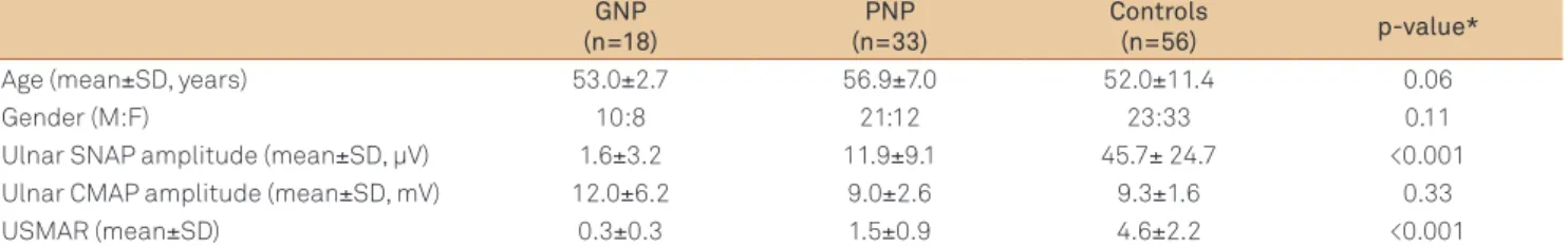
Table. Demographic and neurophysiological data of the subjects included in the study. GNP
(n=18)
PNP (n=33)
Controls
(n=56) p-value*
Age (mean±SD, years) 53.0±2.7 56.9±7.0 52.0±11.4 0.06
Gender (M:F) 10:8 21:12 23:33 0.11
Ulnar SNAP amplitude (mean±SD, µV) 1.6±3.2 11.9±9.1 45.7± 24.7 <0.001
Ulnar CMAP amplitude (mean±SD, mV) 12.0±6.2 9.0±2.6 9.3±1.6 0.33
USMAR (mean±SD) 0.3±0.3 1.5±0.9 4.6±2.2 <0.001
*ANOVA p-values; GNP: ganglionopathies; PNP: polyneuropathies; SD: standard deviation; M: male; F: female; SNAP: sensory nerve action potential; CMAP: compound muscle action potential; USMAR: ulnar sensory-motor amplitude ratio.
Fig 2. ROC curve for the determination of threshold differentiating ganglionopathy from polyneuropathy.
ROC Curve
1 - Specificity
Sensitivity
1.0
0.8
0.6
0.4
0.2
0.0
0.0 0.2 0.4 0.6 0.8 1.0
Fig 1. Box and Whiskers Plot showing the distribution of ulnar sensory-motor amplitude ratio values in the three groups.
10
8
6
4
2
0
1 2 3
Groups
USMAR
Groups
1. Controls
2. Polyneuropathy
3. Ganglionopathy
1. Kuntzer T, Antoine JC, Steck AJ. Clinical features and pathophysiological basis of sensory neuronopathies (ganglionopathies). Muscle Nerve 2004;30:255-268.
2. Damasceno A, França MC Jr, Nucci A. Chronic acquired sensory neuron diseases. Eur J Neurol 2008;15:1400-1405.
3. Camdessanché JP, Jousserand G, Ferraud K, et al. The pattern and diagnostic criteria of sensory neuronopathy: a case-control study. Brain 2009;132:1723-1733.
4. England JD, Gronseth GS, Franklin G, et al. Distal symmetric polyneuropathy: a definition for clinical research: report of the American Academy of Neurology, the American Association of Electrodiagnostic Medicine, and the American Academy of Physical Medicine and Rehabilitation. Neurology 2005;64:199-207.
5. Lauria G, Pareyson D, Sghirlanzoni A. Neurophysiological diagnosis of acquired sensory ganglionopathies. Eur Neurol 2003;50:146-152. 6. França MC Jr, D’Abreu A, Zanardi VA, et al. MRI shows dorsal lesions
and spinal cord atrophy in chronic sensory neuronopathies. J Neuroimaging 2008;18:168-172.
7. Dyck PJ. Detection, characterization, and staging of polyneuropathy: assessed in diabetics. Muscle Nerve 1988;11:21-32.
8. Garibaldi SG. Contribuição da imunohistoquímica cutânea na avaliação das fibras nervosas no diabete melito tipo 2. [dissertation]. Campinas: Faculty of Medical Sciences, Universidade Estadual de Campinas (UNICAMP), 2001.
9. Preston CP, Shapiro BE. Electromyography and Neuromuscular Disorders: Clinical-Electrophysiologic Correlations. 2nd ed. Newton: Butterworth-Heinemann; 2005.
10. Horwich MS, Cho L, Porro RS, Posner JB. Subacute sensory neuropathy: a remote effect of carcinoma. Ann Neurol 1977; 2:7-19.
11. Griffin JW, Cornblath DR, Alexander E, et al. Ataxic sensory neuropathy and dorsal root ganglionitis associated with Sjögren’s syndrome. Ann Neurol 1990;27:304-315.
12. Lauria G, Pareyson D, Grisoli M, Sghirlanzoni A. Clinical and magnetic resonance imaging findings in chronic sensory ganglionopathies. Ann Neurol 2000;47:104-109.
13. Lauria G, Sghirlanzoni A, Lombardi R, Pareyson D. Epidermal innervation in sensory ganglionopathies: clinical and neurophysiological correlations. Muscle Nerve 2001;24:1034-1039. 14. Colli BO, Carlotti CG Jr, Assirati JA Jr, et al. Dorsal root ganglionectomy
for the diagnosis of sensory neuropathies. Surgical technique and results. Surg Neurol 2008;69:266-273.
15. Rutkove SB, Kothari MJ, Raynor EM, Levy ML, Fadic R, Nardin RA. Sural/Radial amplitude ratio in the diagnosis of mild axonal polyneuropathy. Muscle Nerve 1997;20:1236-1241.
16. Overbeek BUH, van Alfen N, Bor JA, Zwarts MJ. Sural/Radial amplitude ratio: reference values on healthy subjects. Muscle Nerve 2005;32:613-618.
References
Severe involvement of distal sensory nerves of the legs (sural and superficial peroneal) is a frequent finding both in GNP and PNP, sometimes with unobtainable sural SNAPs on more severe cases3. This limits the usefulness of
indices that rely upon SNAP amplitudes of these nerves to differentiate GNP and PNP, such as the sural/radial ampli-tude ratio (SRAR)15,16. In our study, we have thus chosen
to investigate an upper limb nerve. Although the median nerve could have been used for the same purpose, the ul-nar nerve was preferred due to a lower rate of entrapment mononeuropathies17-19. Radial nerve was not chosen
ei-ther, because motor NCS of this nerve are not routinely performed. To assess the proportion of sensory over motor involvement, not covered by the previous indices, a com-parison of SNAP and CMAP amplitudes of the same nerve through a ratio was an intuitive choice. Furthermore, such ratios compare different nerves in the same patient, which possibly reduces variability due to aging, as discussed by Rutkove et al.15.
Numerical indices based on electrodiagnostic studies have already been proposed for similar situations. Cocito et al. elaborated the Terminal Latency Index (TLI) and found that low values (<0.26 for the median nerve and <0.33 for the ulnar nerve) suggested anti-MAG neuropathy instead of classical chronic inlammatory demyelinating polyradiculoneuropa-thy (CIDP)20. Regarding common PNP, Rutkove et al. assessed
the expected length-dependent gradient of involvement with their SRAR, for which values lower than 0.40 were considered as a good parameter for the diagnosis of mild axonal PNP15.
In a further study, the threshold value to separate healthy controls and patients with neuropathy was 0.3616.
As expected, our results showed lower USMAR values for the GNP patients when compared to those of diabetic PNP pa-tients. Statistical analysis revealed that a USMAR value equal to or lower than 0.71 was able to diferentiate GNP from dia-betic PNP with a good combination of sensitivity and speciici-ty (94.4 and 90.9%, respectively), conirmed by ROC curve anal-ysis. hese values indeed proved better than isolated SNAP values of upper limb nerves to discriminate GNP and PNP3.
We have chosen diabetes mellitus as the single etiology for the PNP group because this is the most frequent and the best studied model of length-dependent PNP21,22. By
choos-ing a unique etiology, the PNP group was made more homo-geneous, which helped in the comparison with GNP. In ad-dition, there is no description of GNP related to diabetes. Despite this, we acknowledge that further prospective stud-ies with other etiologstud-ies for PNP are needed for a wider val-idation of USMAR. Another limitation, related to the study design, is due to the asymmetry often found in GNP, so that our research could be improved with bilateral calculation of the USMAR. We also emphasize that a complete ulnar elec-trodiagnostic evaluation should be performed before the USMAR calculation in order to exclude focal mononeuropa-thies, since these might obviously inluence the result.
17. de Krom MC, Knipschild PG, Kester AD, Thijs CT, Boekkooi PF, Spaans F. Carpal tunnel syndrome: prevalence in the general population. J Clin Epidemiol 1992;45:373-376.
18. McPherson SA, Meals RA. Cubital tunnel syndrome. Orthop Clin North Am 1992;23:111-123.
19. Latinovic R, Gulliford MC, Hughes RA. Incidence of common compressive neuropathies in primary care. J Neurol Neurosurg Psychiatry 2006;77:263-265.
20. Cocito D, Isoardo G, Ciaramitaro P, et al. Terminal latency index in polyneuropathy with IgM paraproteinemia and anti-MAG antibody. Muscle Nerve 2001;24:1278-1282.
21. Sumner CJ, Sheth S, Griffin JW, Cornblath DR, Polydefkis M. The spectrum of neuropathy in diabetes and impaired glucose tolerance. Neurology 2003;60:108-111.