308
LETTERS
Penetrating cervical spine injury and spinal
cord intramedullary abscess
Trauma raquimedular penetrante e abscesso medular
Regis Tavares da Silva1, Henrique Caetano de Souza1, Ricardo de Amoreira Gepp1, Giovani Rodrigues
Batista2, Thomas Anthony Horan3, Paulo Cesar Rocha Oliveira2
SARAH Network of Rehabilitation Hospitals, Brasília DF, Brazil:
1 Neurosurgeon; 2 Radiologist; 3 Surgeon.
Correspondence: Paulo Cesar Rocha Oliveira; Diagnostic Imaging Department, Hospital SARAH – Brasília; SARAH Network of Rehabilitation Hospitals; SMHS Qd 501 / Bl A; 70335-901 Brasilia DF - Brasil; E-mail: [email protected]
Conflict of interest: There is no conflict of interest to declare.
Received 07 October 2011; Received in final form 28 November 2011; Accepted 05 December 2011
Penetrating spinal injuries by wooden foreign body (WFB) is rare1. We present a 52 year-old man with a cer-vical spine injury by a WFB, identified 172 days after the accident, with an intramedullary abscess. Surgical in-tervention and antibiotic treatment led to neurological improvement.
CASE
A 52 year-old man had fallen from a horse on May 29, in 2008, with a penetrating injury from a piece of wood be-low the angle of the left mandible causing prompt change in sensibility and strength in his left side. He underwent a supericial exploration and suture in the local hospital. Neurological deicits evolved over time to loss of indepen-dent gait and severe functional limitations of the upper limbs, particularly the left.
He was admitted on October 13, in 2008, without signs of inlammation, dehiscence, or cerebrospinal luid (CSF) istula in the neck. Neurological examination showed tetra-paresis, predominantly on the left side, and hyperrelexia in lower limbs with preserved vibration and position sensation in all four limbs. He was only able to walk with great dii-culty if supported by two helpers. Bladder and bowel func-tions were normal.
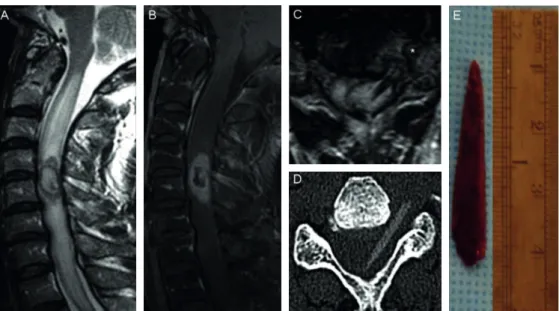
he magnetic resonance image (MRI) revealed spinal cord edema and a heterogeneous intramedullary lesion at C4-C5 level, with ring like post-contrast enhancement, con-sistent with an abscess (Fig A and B). here was a hypoin-tense linear image within the left C4-C5 intervertebral fo-ramen with intramedullary extension and hyperdense on computed tomography, measuring 3.0 cm, consistent with a WFB. It transixed the ipsilateral vertebral artery, which was obliterated (Fig C and D).
The WFB was surgically removed (Fig E). Purulent mat-ter was drained from location, where the WFB had been lodged. Lab work showed the presence of Staphilococcus epidermiditis, and the patient was treated with oxacillin. At the latest outpatient follow-up visit on January 18th, 2011, he was independent for activities of daily living, walking without any aid, with intact sphincter control.
DISCUSSION
Non-missile penetrating spinal injuries are more com-monly caused by stabs2. WFB is rarely reported as the cause of this type of injury1. Since the wood is an optimal source of infection due its porous, soft, organic, and veg-etable nature, spinal infectious complications are prone to occur1,3. Foreign body reaction, another inflamma-tory complication, may lead to long-term neurological worsening1.
CT and MRI are essential to evaluate spinal cord and the presence of a foreign body in penetrating spinal inju-ries. The density of wood on CT is highly variable and de-pends on its type and hydration status, as well as on the length of stay in the body4. This latter factor may explain the increased density of the wooden fragment in this pa-tient. MRI is mandatory in non-missile penetrating spinal injuries for the diagnosis of associated lesions5, whenev-er a metallic foreign body has been excluded or removed2. Even when CT is negative, MRI should be performed to ex-clude retained foreign bodies1.
309
LETTERS
1. Gul S, Dusak A, Songur M, Kalayci M, Acikgoz B. Penetrating spinal injury with a wooden fragment: a case report and review of the literature. Spine 2010;35:1534-1536.
2. Jacobsohn M, Semple P, Dunn R, Candy S. Stab injuries to the spinal cord: a retrospective study on clinical findings and magnetic resonance imaging changes. Neurosurgery 2007;61: 1262-1266.
3. Piqueras C, Martínez-Lage JF, Almagro MJ, Ros De San Pedro J, Torres
Tortosa P, Herrera A. Cauda equina-penetrating injury in a child. Case report. J Neurosurg 2006;104:279-281.
4. Ginsberg LE, Williams DW, Mathews VP. CT in penetrating craniocervical injury by wooden foreign bodies: reminder of a pitfall. AJNR Am J Neuroradiol 1993;14:892-895.
5. Pal D, Timothy J, Marks P. Penetrating spinal injury with wooden fragments causing cauda equina syndrome: case report and literature review. Eur Spine J 2006;15:574-577.
References