Letters to the Editor
Radiol Bras. 2016 Nov/Dez;49(6):406–413
408
http://dx.doi.org/10.1590/0100-3984.2015.0190 There is a broad spectrum of diagnoses of bilateral lesions in
the basal ganglia in the pediatric population. The main causes cited are hypoxic-ischemic encephalopathy; hypoglycemia; en-cephalitis; inborn errors of metabolism; water and electrolyte turbances; carbon monoxide poisoning; and demyelinating dis-orders. The correlation with clinical and laboratory data is funda-mental for making the definitive diagnosis(7,12,13).
In conclusion, the possibility of acute or chronic kernicterus should be considered when clinical symptoms, biochemical data, and MRI findings are suggestive of the disease, the chronic pre-sentation and permanent, irreversible profile being promoted by bilirubin neurotoxicity.
REFERENCES
1. Alfenas R, Niemeyer B, Bahia PRV, et al. Parry-Romberg syndrome: findings in advanced magnetic resonance imaging sequences – case report. Radiol Bras. 2014;47:186–8.
2. Bimbato EM, Carvalho AG, Reis F. Toxic and metabolic encephalopa-thies: iconographic essay. Radiol Bras. 2015;48:121–5.
3. Castro FD, Reis F, Guerra JGG. Intraventricular mass lesions at mag-netic resonance imaging: iconographic essay – part 1. Radiol Bras. 2014;47:176–81.
4. Ono SE, Carvalho Neto A, Gasparetto EL, et al. X-linked adrenoleukodys-trophy: correlation between Loes score and diffusion tensor imaging parameters. Radiol Bras. 2014;47:342–9.
5. Barbosa JHO, Santos AC, Salmon CEG. Susceptibility weighted imag-ing: differentiating between calcification and hemosiderin. Radiol Bras. 2015;48:93–100.
Bruno Niemeyer de Freitas Ribeiro1, Gabriela de Almeida Lima1, Nina Ventura1, Emerson Leandro Gasparetto1, Edson Marchiori2
1. Instituto Estadual do Cérebro Paulo Niemeyer, Rio de Janeiro, RJ, Brazil. 2. Universidade Federal do Rio de Janeiro (UFRJ), Rio de Janeiro, RJ, Brazil. Mailing address: Dr. Bruno Niemeyer de Freitas Ribeiro. Instituto Estadual do Cérebro Paulo Niemeyer – Departamento de Radiologia. Rua do Rezende, 156, Centro. Rio de Janeiro, RJ, Brazil, 20231-092. E-mail: [email protected].
6. Turkel SB, Miller CA, Guttenberg ME, et al. A clinical pathologic re-appraisal of kernicterus. Pediatrics. 1982;69:267–72.
7. Parashari UC, Singh R, Yadav R, et al. Changes in the globus pallidus in chronic kernicterus. J Pediatr Neurosci. 2009;4:117–9.
8. Perlstein MA. The late clinical syndrome of posticteric encephalopathy. Pediatr Clin North Am. 1960;7:665–87.
9. Martich-Kriss V, Kollias SS, Ball WS Jr. MR findings in kernicterus. AJNR Am J Neuroradiol. 1995;16(4 Suppl):819–21.
10. Coskun A, Yikilmaz A, Kumandas S, et al. Hyperintense globus pallidus on T1-weighted MR imaging in acute kernicterus: is it common or rare? Eur Radiol. 2005;15:1263–7.
11. Govaert P, Lequin M, Swarte R, et al. Changes in globus pallidus with (pre)term kernicterus. Pediatrics. 2003;112(6 Pt 1):1256–63. 12. Hegde AN, Mohan S, Lath N, et al. Differential diagnosis for bilateral
abnormalities of the basal ganglia and thalamus. Radiographics. 2011; 31:5–30.
13. Khanna PC, Iver RS, Chaturvedi A, et al. Imaging bithalamic pathol-ogy in the pediatric brain: demystifying a diagnostic conundrum. AJR Am J Roentgenol. 2011;197:1449–59.
Renal lymphangiectasia: know it in order to diagnose it
Dear Editor,
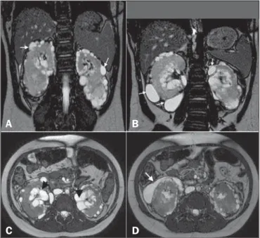
Here, we report the case of a 9-year-old girl with hyperpar-athyroidism. Ultrasound showed renal cysts and increased echo-genicity of the parenchyma in both kidneys. The diagnostic hy-pothesis was hyperparathyroidism secondary to chronic/polycystic kidney disease. The patient presented with gradually worsening kidney function and hypertension, and new imaging scans were requested. The ultrasound showed anechoic, multiloculated im-ages in the pyelocaliceal region of both kidneys, and perirenal, subcapsular cysts. A computed tomography (CT) scan was ac-quired, although no contrast agent was used, which precluded an accurate characterization. Nevertheless, the CT scan revealed changes similar to those observed on ultrasound. We also per-formed magnetic resonance imaging (MRI), which showed pyelocaliceal, perirenal cysts, with altered intensity of the signal of the renal parenchyma and loss of corticomedullary differentiation (Figure 1A), confirming, in conjunction with the clinical and bio-chemical data, the diagnosis of renal lymphangiectasia (RL).
RL is a rare benign disease that occurs because of miscom-munication between the renal lymphatic drainage system and the retroperitoneal lymphatic system(1). As a result, there is accumu-lation of lymph in the renal lymph ducts, making them ectatic and forming simple or multiloculated, typically asymmetric and bilateral, collections in the pyelocaliceal, perinephric, or parenchy-mal regions, although, in some cases, only a part of one kidney is affected (Figure 1—B,C). There is no predilection for a given gender or age group. As of 2005, only 40 cases had been de-scribed(1,2).
In most cases, RL is an incidental finding, with or without signs and symptoms of pain, increased abdominal volume, he-maturia, ascites, edema of the lower limbs, hypertension, erythro-cytosis with renal vein thrombosis, and, rarely, chyluria(3). Such manifestations can be explained by the distention of the renal
fascia and compression of the renal parenchyma by cysts, fistuli-zation to the pelvic cavity, and changes in the renin-angiotensin system(2–4). In rare cases, chronic kidney disease has been re-ported(5). To our knowledge, there have been no specific reports of clinical evolution to hyperparathyroidism, although a relation-ship with chronic kidney disease can be assumed.
A CT scan can reveal expansive perirenal formations with fluid attenuation, bounded by the renal fascia, that conform to
Figure 1. A: Coronal T2-weighted MRI sequence showing a loss of corticomedullary differentiation in both kidneys and multiple cystic lesions, with thin walls, located in the cortex (arrows). B: Cystic formations in the subcapsular cortex (arrows). C:
Axial T2-weighted MRI sequence showing cysts located in the renal sinuses (ar-rowheads) and perinephric spaces, simulating pelvic dilatation. D: The same images simulating cystic collections in the subcapsular cortex (arrow).
A
B
Letters to the Editor
Radiol Bras. 2016 Nov/Dez;49(6):406–413
409
http://dx.doi.org/10.1590/0100-3984.2015.0025 (and do not invade) the adjacent structures. Those formations
can compress the kidney cortex, expand the sinus and distort the calyceal system. In some cases, small, predominantly peripheral, hypodense collections can be seen, with attenuation values of 0–15 HU(3). There may be thickening of the renal fasciae and retroperitoneal collections crossing the midline at the level of the renal hilum. After the administration of iodine contrast, there is no enhancement of the collections or of the walls of the cystic formations(6). In MRI, the cysts exhibit a low signal on T1-weighted sequences—although the signal strength can be in-creased if there is bleeding(6)—and a more intense signal on T2-weighted sequences, without enhancement. In addition, RL can be diagnosed on MRI scans by identifying perirenal lymphatic collections with inversion of the corticomedullary signal inten-sity(1,4), as depicted in Figure 1—B,C,D.
To suggest a diagnosis of RL, as well as to devise a treatment strategy and to prevent complications, it is essential to understand the radiological aspects of the disease and to differentiate it from other conditions that mimic cystic kidney disease. Although the combination of RL and renal failure is rare, knowledge of that association is also important to prevent comorbid conditions that can evolve with this complication, such as obesity and high blood pressure.
Andréa Farias de Melo Leite1, Bruna Venturieri1, Rosana Gonçalves de Araújo1, Eduardo Just Costa e Silva1, Jorge Elias Junior2
1. Instituto de Medicina Integral Professor Fernando Figueira de Per-nambuco (IMIP), Recife, PE, Brazil. 2. Faculdade de Medicina de Ri-beirão Preto da Universidade de São Paulo (FMRP-USP), RiRi-beirão Preto, SP, Brazil. Mailing address: Dra. Andréa Farias de Melo Leite. Rua Laura Campelo, 130, Torre. Recife, PE, Brazil, 50710-270. E-mail: [email protected].
REFERENCES
1. Rastoji R, Rastogi V. Computed tomographic scan in the diagnosis of bilateral renal lymphangiectasia. Saudi J Kidney Dis Transpl. 2008;19: 976–9.
2. Ashraf K, Raza SS, Ashraf O, et al. Renal lymphangiectasia. Br J Radiol. 2007;80:e117–8.
3. Vega J, Santamarina M. Linfangiectasia renal unilateral. Caso clínico. Rev Méd Chile. 2012;140:1312–5.
4. Restrepo JM, Amaya JEL, Sepúlveda NA, et al. Renal lymphangiectasia: MDCT and MRI findings. Rev Colomb Radiol. 2011;22:1–8. 5. Ueda S, Yanagida H, Sugimoto K, et al. Chronic renal insufficiency in a
boy with cystic renal lymphangiectasia: morphological findings and long-term follow-up. Clin Nephrol. 2007;68:416–21.
6. Vasconcelos RA, Pereira ES, Bauab Jr T, et al. Renal lymphangiectasia: incidental finding at multislice computed tomography and literature re-view. Radiol Bras. 2012;45:178–80.
Primary undifferentiated sarcoma in the thorax: a rare diagnosis in young patients
Dear Editor,
A 30-year-old man was admitted to the thoracic surgery de-partment of a tertiary hospital for investigation of a thoracic mass. Having previously received treatment for pneumonia, he presented with a two-week history of progressively increasing pain in the right hemithorax and right flank, between the anterior axillary line and midaxillary line. On clinical examination, there was an absence of breath sounds in the right hemithorax.
Computed tomography (CT) of the chest showed an exten-sive, heterogeneous, mostly solid mass in right thoracic region (Figure 1), with areas of inner content of low attenuation (21–26 Hounsfield units) and foci of bleeding, without intervening calci-fications and without osteolysis of the rib. Laboratory tests pro-duced results within the limits of normality. The patient under-went percutaneous biopsy, and the pathology examination revealed undifferentiated sarcoma (Figure 2).
Sarcomas represent a heterogeneous group of tumors derived from mesenchymal cells(1–3). They account for 1% of all neo-plasms and occur mainly in the extremities (in 60% of cases), gas-trointestinal tract (in 25%), retroperitoneal space (in 20%), and the head and neck region (in 4.1%). Primary sarcomas of the tho-rax are exceptionally rare, accounting for only 0.2% of lung can-cers and only 5% of all the thoracic neoplasms. Such sarcomas can involve the lungs, mediastinum, pleura, and, mainly, the chest wall. The presence of sarcoma in any other part of the body must be ruled out, because metastasis to the chest is much more com-mon than is primary sarcoma of the thorax(4–7).
The most common histological types of primary sarcomas are angiomyosarcoma, leiomyosarcoma, rhabdomyosarcoma, and sarcomatoid mesothelioma(8). In the chest wall, the most com-mon primary sarcomas are Ewing’s sarcoma, primitive neuroec-todermal tumor, malignant fibrous histiocytoma, chondrosar-coma, osteosarchondrosar-coma, synovial sarchondrosar-coma, and fibrosarcoma(8). Ra-diologically, these tumors typically present as large, heterogeneous masses. However, their appearance can vary from an intrabronchial
Figure 1. CT scan showing a primary sarcoma in the right hemithorax. A: CT scout image showing opacification of the right hemithorax. B: Coronal CT reconstruction with heterogeneous enhancement (arrow). C: Axial CT slice showing contralateral mediastinal deviation.