Volume 1 | Issue 6 Optometry & Visual Performance 213
Article
4
Waardenburg Syndrome:
A Report of Two Familial Case Series
Safal Khanal, B.Optom, Southwestern University, Cebu City, PhilippinesPragati Gautam, MD, BP Koirala Lions Center for Ophthalmic Studies, Institute of Medicine, Maharajgunj, Kathmandu, Nepal
Nabin Paudel, B.Optom, University of Auckland, Auckland, New Zealand
ABSTRACT
Background: Waardenburg syndrome is a rare autosomally-inherited developmental disorder characterized by sensorineural deafness in association with pigmentary anomalies comprising various ocular features including dystopia canthorum, iris heterochromia, eyebrow lare, and fundus alterations. It is a congenital non-progressive genetic disorder that has been found to result in hearing loss, reduced vision, reduced self esteem, problems related to appearance, and decreased intellectual functioning.
Case Reports: We report two familial case series that presented with the characteristic ocular indings and the systemic features of Waardenburg syndrome. he irst series comprised a 32-year-old father with his two sons aged nine and six years. Two female siblings, aged 10 and eight years, both with cochlear implants, were included in the second series.
Conclusion: Waardenburg syndrome manifests diferently with dissimilar genetic penetration even within the same family. Some individuals will require no treatment, while others may need treatment or surgery for other abnormalities. Appropriate measures can be undertaken to negotiate the disabilities resulting from the ocular conditions associated with this syndrome.
Keywords: albinotic fundus, dystopia canthorum, heterochromia iridis, sensorineural deafness, Waardenburg syndrome
Introduction
Waardenburg syndrome (WS) is a rare autosomally-inherited developmental disorder characterized by sensorineural deafness in association with pigmentary anomalies and defects of neural crest-derived tissues.1 WS is named after a Dutch ophthalmologist, P. J. Waardenburg, who described a syndrome comprising six distinctive features: lateral displacement of the medial canthi and lacrimal punctae, broad and high nasal root, hypertrichosis of the medial part of the eyebrows, partial or total heterochromia iridis, white forelock, and congenital deaf mutism.2 his condition is caused by the physical absence of melanocytes in the skin, hair, and eyes. WS equally afects both sexes and all races.1,3 Its prevalence was estimated by Waardenburg to be 1/42000 of the population.2 Based on the clinical presentations, four subtypes were subsequently described.3,4
• Type I WS (WS1) consists of dystopia canthorum and broad nasal root.
• Type II WS (WS2) lacks the dystopia canthorum. • Type III WS (WS3) (Klein-Waardenburg syndrome),
a severe form of WS1, is associated with upper limb defects.
• Type IV WS (WS4) (Shah-Waardenburg syndrome) is characterised by Hirschsprung disease.
he clinical variability of WS is attributed to the diferent penetrance and expression of the responsible genes, just as in other genetic syndromes.
Waardenburg syndrome is a rare nonprogressive congeni-tal genetic disorder. he diagnostic criteria for WS1 was proposed by the Waardenburg Consortium in 1992.5 he individuals must have two major or one major and two minor criteria to be diagnosed as WS1 (Table 1).
Multiple genes have been implicated in the syndrome. Abnormalities in the paired box gene 3 (PAX3 gene) account for most of the WS1 and WS3 patients. Microphthalmia associated transcription factor (MITF) gene abnormality is
Table 1: Diagnostic criteria for WS as proposed by the Waardenburg Consortium.
Major Criteria
Congenital sensorineural hearing loss
Pigmentary disturbances of iris: Complete heterochromia iridis, partial segmental heterochromia iridis, hypoplastic blue irides White forelock
Dystopia canthorum Affected irst degree relative
Minor Criteria
Congenital leukoderma: several areas of hypopigmented skin Medial eyebrow lare (synophrys)
214 Optometry & Visual Performance Volume 1 | Issue 6
responsible for WS2. WS4 is heterogeneous, with reported mutations in endothelin 3 (EDN3), in its receptor endothelin receptor type B (EDNRB), or in SRY-sex determining region Y-box 10 (SOX10).6
We report two case series of classical features of Waardenburg syndrome; one occurred within the same family covering two generations and the second involves two sisters.
Case report
Familial case series 1 (WS type I)
Our irst patient, a 32-year-old male, presented to our Outpatient Department with blurry vision OD since childhood and was found to have partial heterochromia of iris OU. He had congenital hearing loss and lack of speech development. he physical examination revealed no abnormal pigmentation of the hair but hypopigmented patches on the skin. Presenting visual acuity was 6/24 in OD and 6/6 in OS, which improved to 6/12 OD with +1.50 D glasses. On further examination, ptosis of the right upper lid was noted with palpebral issure height of 10mm, marginal relex distance from upper lid of 1mm, and the levator muscle function of 8mm. Pupils were round, regular, and reactive to light with both direct and consensual relex intact. Fundus was albinotic in both eyes. Dystopia canthorum (lateral displacement of medial canthus) was noted, but there was no bilateral ptosis. he patient was counselled thoroughly regarding the genetic condition and was advised to undergo a trial of patching of the left eye. He was also made aware of the surgical correction to improve the ptosis but as the patient was not quite ready, the reattachment and strengthening of levator was planned as a possible surgical option in subsequent visits (Figure 1).
he second patient was the irst child of the patient above. A nine-year-old boy, he was born of non-consanguineous parents who reported that he was mute and deaf from birth. Prenatal, antenatal, and postnatal histories were all uneventful. Birth history reported full term normal vaginal delivery. Immunization was reported to be complete. On ocular examination, unaided binocular visual acuity was 3/2.4 with the Kay Picture test at 3 metres. One diopter of hyperopia was present in both eyes. Further examination revealed iris heterochromia (segmental OD, complete OS), dystopia canthorum, telecanthus, and pseudoexotropia, along with hypopigmented patches on the face (Figure 2). Fundi of both eyes were albinotic with hypopigmentation. Profound bilateral
sensorineural hearing loss was noted with no deformity of joints or neurological abnormalities. he child, as well as his father, was counselled regarding the ocular indings and that he can live a normal life compared to his peers. Grey tinted spectacles were advised for the child based on his preference.
Our third patient was a 6-year-old child born of the same parents who was also reported to have congenital hearing loss and mutism. Birth history and immunization history were all uneventful and similar to his older brother. Delivery was normal and he was born at term. Ocular examination revealed unaided visual acuity of 3/3.8 OU when tested with the Kay Picture Test with 0.50 D of hyperopia. On further examination, partial iris heterochromia OU, dystopia canthorum, premature graying of hair, hypopigmented patches on face, telecanthus, pseudoexotropia, and bilateral albinotic fundi were subsequently noted (Figure 3). his child was also counselled adequately and grey tinted spectacles were advised to reduce light sensitivity.
Familial case series 2 (WS2)
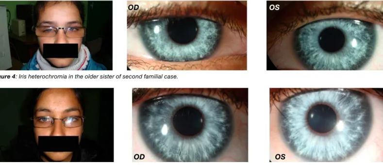
he irst case in this series was a 10-year-old girl with a history of congenital bilateral sensorineural deafness who had undergone cochlear implant at the age of three and had undergone speech therapy. Birth and immunization history were all uneventful with normal full term home delivery. Best corrected visual acuity was 6/9 with +2.50-2.50×140 OD and 6/6 with +1.50-1.00×050 OS. Further examination revealed complete iris heterochromia, premature graying of hair, and
Figure 1: Right upper lid ptosis and partial iris heterochromia in the father of first familial case.
Figure 2: Iris heterochromia, hypopigmented patches, and pseudoexo-tropia in older child of first familial case.
Figure 3: Partial iris heterochromia and premature graying of hair in the younger child of the first familial case.
Volume 1 | Issue 6 Optometry & Visual Performance 215
bilateral albinotic fundus (Figure 4). he child was managed with appropriate genetic counselling and full correction of refractive error. She was also advised to wear grey tinted spectacles to reduce photosensitivity. Furthermore, a constant part time patching trial, OD/OS:1/6, was initiated to improve the shallow amblyopia present in the right eye.
he second case was her younger sister, an eight-year-old girl with a similar history of cochlear implant at the age of two. She also had undergone speech therapy. Ocular examination revealed isoametropic amblyopia in both eyes, with best corrected visual acuity of 6/12 with +6.00-3.00 ×180. Complete heterochromia, bilateral hypopigmented fundus, and graying of hair was noted on further examination (Figure 5). Both the parents of these children were normal, but their fraternal aunt had a similar history. he child was fully corrected for refractive error with grey tinted spectacles after appropriate cycloplegic refraction. She had no problem adaptating to the prescription, as she had been wearing prescription spectacles since three years of age. Contact lenses as a possible alternative to spectacles were presented to the patient, but this was rejected on account of greater care required and also partly due to inancial problems. Part time, daily alternating patching was initiated, and appropriate genetic counselling was also performed.
Discussion
Waardenburg syndrome comprises both ocular and non-ocular features. Ocular features include dystopia canthorum, iris heterochromia, eyebrow lare, and fundus alterations. Dystopia presents with bilateral ptosis, and there may be hypertelorism. Synophrys, laring or fanning out of the eyebrow hair medially, has also been reported.2,4,5,7,8 Iris heterochromia is one of the major features which may be segmental or complete.4,5,7 Segmental heterochromia gives rise to iris bicolour with a clearly demarcated radial segmental
distribution pattern of two colours.5 In some cases characteristic brilliant blue or sapphire eyes may also be seen.4,5,8 Although hypopigmentation in the fundus is not considered as a major or minor criterion, it has been reported to be an important aspect of this syndrome.9,10 Müllner-Eidenböck9 suggested an ipsilateral correlation between iris and fundus pigmentation, while Goldberg10 also showed the heterogenicity in the fundus pigmentation corresponding with that of the iris. Strabismus, usually convergent, has also been reported.8
Hearing loss, hair hypopigmentation, and congenital leukoderma comprise the non-ocular features in WS. Hearing loss is one of the major non-ocular features, its type being mainly congenital, nonprogressive, unilateral or bilateral, and sensorineural.4,5,8
Waardenburg syndrome is reported to account for two percent of congenital deafness.6 his clinical inding is not a universal feature of WS, but penetrance of sensorineural hearing loss has been observed to be 69% in WS1 and 87% in WS2 after excluding probands ascertained through their hearing loss.1 Abnormality in hair pigmentation is another characteristic feature. A white forelock is usually described. It can be present at birth and then disappear later in life with reappearance in teens or adulthood, or it may appear for the irst time at any age.4,5,8,11 Farrer et al. also suggested the presence of premature graying of hair, with predominately scalp hair becoming white before the age of 30 years.5 Skin may also be hypopigmented, usually on face, trunk, and limbs, with or without associated white forelock.11 here may also be broad high nasal root and/or hypoplasia of alae nasi.1,4,5,8
Other rare features include neural tube defects, sprengel shoulder (congenital upward scapular displacement), cleft lip or palate, congenital heart abnormalities, Hirschsprung disease (primarily in cases of WS4), contractures, and limb muscle hypoplasia (in patients with WS3).4,5
Figure 4: Iris heterochromia in the older sister of second familial case.
Figure 5: Iris heterochromia in the younger sister of second familial case.
OD OS
216 Optometry & Visual Performance Volume 1 | Issue 6
he online version of this article contains digital enhancements. Our cases in the irst familial case series all showed bilateral
profound sensorineural hearing loss associated with complete or segmental iris heterochromia, dystopia canthorum, and broad nasal root. here were hypopigmented patches on the faces of the children with one showing premature graying of hair as well. he presence of an afected irst degree relative further emphasizes the autosomal dominant mode of transmission in WS1. he cases in the second familial case series showed profound bilateral sensorineural deafness with the history of cochlear implant, premature greying of hair, and complete iris heterochromia in one and segmental in the other. he exception of dystopia canthorum classiies them as WS2 with an autosomal recessive mode of transmission as their parents were healthy subjects.
Although the current management of this syndrome has been solely focused on addressing sensorineural hearing loss by cochlear implants if detected at an early age, the occurence of reduced vision, problems related to the ocular appearance, as well as increased sensitivity to light have demanded some attention towards management of such conditions. Full correction of refractive error from early childhood combined with appropriate patching therapies when amblyogenic factors are present combat reduced vision and prevent the development or permit the remediation of possible amblyopia. Tinted spectacles and contact lenses have also been useful options to those who are quite sensitive regarding the appearance of their eyes as well as those who are photosensitive. It has always been a matter of debate regarding the colour of the tints to be advised, but it is best to demonstrate various options and select the patient’s choice. All of the patients in our case series preferred the grey tint over amber ones and were quite satisied with the outcome when tested outdoors. Further, evaluation of patients’ visual needs can be addressed with a low vision evaluation if residual amblyopia and reduced vision result in signiicant visual impairment. he association of unilateral ptosis can be managed with a trial of patching in the better eye or with possible surgical corrections.
Waardenburg syndrome manifests diferently in every individual, even within the same family. Some individuals will require no treatment, while others may need treatment or surgery for other abnormalities. No special diet or activity restrictions are needed.1,12 Waardenburg syndrome does not usually afect the brain. Folic acid supplementation in pregnancy has been recommended for women at increased risk of having a child with WS.13 Inheritance of types 3 and 4 are more complex, but genetic counselling can help assess the risk of passing WS on to a child.12-15
Whereas pigmentation abnormalities do not herald any survival problem, hearing loss seems to be the most important prognostic factor because of impairment of life quality and poor cognitive abilities in WS1. he earlier it is diagnosed the better chance for cochlear implantation to improve speech discrimination and spoken language as in our second case series.11
Conclusion
Early diagnosis and improvement of the hearing defects are important for the psychological development of children
with this disease and help to reduce the sense of isolation. Apart from dedicating the focus of management only upon hearing loss, appropriate measures can be undertaken to negotiate the disabilities resulting from the associated ocular conditions. Appropriate refractive correction with management of amblyopia, tinted spectacles, and contact lenses to address photosensitivity and ptosis improvement form the core of ocular management regarding WS. Lack of resources in some societies may add to the physical and psychological obstacles faced by persons with WS. Tolerance and understanding of persons with WS will help support their integration into society.
References
1. Tagra S, Talwar AK, Walia RLS, Sidhu P. Waardenburg syndrome. Indian J Dermatol Venereol Leprol 2006;72(4):326.
2. Waardenburg PJ. A new syndrome combining developmental anomalies of the eyelids, eyebrows and nose root with pigmentary defects of the iris and head hair and with congenital deafness. Am J Hum Genet 1951;3:195-253.
3. Krishtul A, Galadari I. Waardenburg syndrome: Case report. Int J Dermatol 2003;42:651-2.
4. Read AP, Newton VE. Waardenburg syndrome. J Med Genet 1997;34:656-65. 5. Farrer LA, Grundfast KM, Amos J, Arnos KS, et al. Waardenburg syndrome
(WS) type 1 is caused by defects at multiple loci, one of which is near ALPP on chromosome 2: First report of the WS Consortium. Am J Hum Genet 1992;50:902-13.
6. Yang S, Cao J, Zhang R, Liu L, et al. Nonsense mutations in the PAX3 gene cause Waardenburg syndrome type 1 in two Chinese patients. Chin Med J 2007;120:46-9.
7. Liu XZ, Newton VE, Read AP. Waardenburg syndrome type 2: Phenotypic indings and diagnostic criteria. Am J Med Genet 1995;55:95-100.
8. Delleman JW, Hageman MJ. Heterogeneity in Waardenburg Syndrome. Am J Hum Genet 1977;29:468-85.
9. Müllner-Eidenböck A, Moser E, Frisch H, Read AP. Waardenburg syndrome type 2 in a Turkish family: implications for the importance of the pattern of fundus pigmentation. Br J Ophthalmol 2001;85:1384-6.
10. Goldberg MF. Waardenberg’s syndrome with fundus and other anomalies. Arch Ophthalmol 1966;76:797-810.
11. Arias S, Mota M. Apparent non-penetrance for dystopia in Waardenburg syndrome type 1 with some hints on the diagnosis of dystopia canthorum. J Genet Hum 1978;26:101-31.
12. Moore SW, Johnson AG. Hirschsprung’s disease: Genetic and functional associations of Down’s and Waardenburg syndromes. Semin Pediatr Surg 1998;7:156-61.
13. Karaman A, Aliagaoglu C. Waardenburg syndrome. Dermatol Online 2006; 12(3):21.
14. Verheij JB, Sival DA, van der Hoeven JH, Vos YJ, et al. Shah-Waardenburg syndrome and PCWH associated with SOX10 mutations: A case report and review of the literature. Eur J Paediatr Neurol 2006;10:11-7.
15. Pingault V, Bondurand N, Kuhlbrodt K. SOX10 mutations in patients with Waardenburg-Hirschsprung disease. NatGenet 1998;18:171-3.
Correspondence regarding this article should be emailed to safaladarsha09@ gmail.com or sent to Safal Khanal, Southwestern University Urgello Street, Cebu City, Philippines 639334557724. All statements are the authors’ personal opinions and may not reflect the opinions of the representative organizations, ACBO, COVD, or OEPF, Optometry & Visual Performance, or any institution or organization with which the author may be affiliated. Permission to use reprints of this article must be obtained from the editor. Copyright 2013 Optometric Extension Program Foundation. Online access is available at www.acbo.org.au, www.covd.org, and www.oepf.org.