Effect of High-Flux Dialysis on Circulating
FGF-23 Levels in End-Stage Renal Disease
Patients: Results from a Randomized Trial
Andreas Schneider1*, Markus P. Schneider2, Detlef H. Krieter1, Bernd Genser3,4, Hubert Scharnagl5, Tatjana Stojakovic5, Christoph Wanner1, Christiane Drechsler1
1Department of Medicine, Division of Nephrology, University Hospital, Würzburg, Germany,2Department of Nephrology and Hypertension, University Hospital, Erlangen-Nürnberg, Germany,3Mannheim Institute of Public Health, Social and Preventive Medicine, Medical Faculty, Mannheim, Germany,4BGStats
Consulting, Vienna, Austria,5Medical University of Graz, Clinical Institute of Medical and Chemical Laboratory Diagnostics, Graz, Austria
Abstract
Background
In patients undergoing maintenance hemodialysis (HD), increased levels of circulating fibro-blast growth factor-23 (FGF-23) are independently associated with cardiovascular events and mortality. Interventional strategies aiming to reduce levels of FGF-23 in HD patients are of particular interest. The purpose of the current study was to compare the impact of high-flux versus low-high-flux HD on circulating FGF-23 levels.
Methods
We conducted a post-hoc analysis of the MINOXIS study, including 127 dialysis patients randomized to low-flux (n = 62) and high-flux (n = 65) HD for 52 weeks. Patients with valid measures for FGF-23 investigated baseline and after 52 weeks were included.
Results
Compared to baseline, a significant increase in FGF-23 levels after one year of low-flux HD was observed (Delta plasma FGF-23: +4026 RU/ml; p<0.001). In contrast, FGF-23 levels remained stable in the high flux group (Delta plasma FGF-23: +373 RU/ml, p = 0.70). The adjusted difference of the absolute change in FGF-23 levels between the two treatment groups was statistically significant (p<0.01).
Conclusions
Over a period of 12 months, high-flux HD was associated with stable FGF-23 levels, where-as the low-flux HD group showed an increwhere-ase of FGF-23. However, the implications of the different FGF 23 time-trends in patients on high flux dialysis, as compared to the control group, remain to be explored in specifically designed clinical trials.
OPEN ACCESS
Citation:Schneider A, Schneider MP, Krieter DH, Genser B, Scharnagl H, Stojakovic T, et al. (2015) Effect of High-Flux Dialysis on Circulating FGF-23 Levels in End-Stage Renal Disease Patients: Results from a Randomized Trial. PLoS ONE 10(5): e0128079. doi:10.1371/journal.pone.0128079
Academic Editor:Carmine Zoccali, L' Istituto di Biomedicina ed Immunologia Molecolare, Consiglio Nazionale delle Ricerche, ITALY
Received:August 13, 2014
Accepted:April 21, 2015
Published:May 29, 2015
Copyright:© 2015 Schneider et al. This is an open access article distributed under the terms of the Creative Commons Attribution License, which permits unrestricted use, distribution, and reproduction in any medium, provided the original author and source are credited.
Data Availability Statement:All relevant data are within the paper and its Supporting Information files.
Trial Registration
German Clinical Trials Register (DRKS)DRKS00007612.
Introduction
Despite efforts to improve outcomes in patients with end-stage renal disease (ESRD), mortality remains excessively high [1]. Cardiac and vascular events are the predominant causes of death
[1,2]. Recently, non-traditional risk factors and specific biomarkers were identified that
distin-guish ESRD patients at high risk for CVD and mortality.
Fibroblast-growth-factor-23 (FGF-23) is a 251 amino-acid peptide synthesized by osteo-cytes and osteoblasts, and is involved in the regulation of phosphate homeostasis [3,4]. In chronic kidney disease (CKD), FGF-23 increases early in the course of the deterioration of kid-ney function, and has been proposed to be a physiological response that protects the organism from the adverse effects of phosphate retention by facilitating phosphate excretion [5]. On the other hand, elevated FGF-23 levels were found to be independently associated with mortality in ESRD patients as well as in patients with advanced CKD [6,7]. Thus, in view of the high car-diovascular risk in CKD and ESRD, strategies that are able to reduce FGF-23 levels are of par-ticular interest. During the past two decades, more permeable high-flux dialysis membranes, which are able to eliminate low-molecular weight toxins up to 50.000 Dalton [8] have been de-veloped. This is of clinical interest as FGF-23 is a free circulating peptide in the blood with a molecular weight of 32.000 Dalton [4,9] which might be removed by high-flux hemodialysis (HD).
In the MINOXIS study, maintenance HD patients were randomized to 52 weeks treatment with either low-flux or high-flux HD with the primary endpoint being parameters of anemia [10]. In the present post-hoc analysis of the MINOXIS data, effects of high-flux versus low-flux HD on FGF-23 serum levels were compared.
Methods
The study was performed in accordance with the ethical principles of the declaration of Hel-sinki. Written informed consent was obtained from each participant before entry into the study. The study was approved by the Freiburger Ethik-Kommission International (FEKI Nr.: 04/1952) and was conducted according to“good clinical practice”(GCP) guidelines. The study is registered in the„German Clinical Trials Register (DRKS)”(trial registry number
DRKS00007612).
Study design
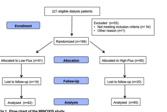
The MINOXIS study design, main outcome findings, and baseline data have been described previously [10]. In summary, MINOXIS was a prospective randomized controlled trial recruit-ing 166 patients with ESRD on maintenance HD (Fig 1). Included patients had to be18 years of age and receiving treatment for anemia with the erythropoietin stimulating agent (ESA) darbepoetin-alfa for at least 6 months. Incident patients undergoing HD for at least 1 or at most 3 months (recently diagnosed) had to be treated by low-flux dialysers. Prevalent pa-tients undergoing HD for more than 3 months had to be treated with low-flux dialysers for at least 3 months before inclusion in the study. Exclusion criteria were serious comorbidities with a life expectancy of less than 2 years, single-needle HD, use of temporary or permanent
roles of these authors are articulated in the‘author contributions’section.
catheters, planned kidney transplantation or pregnancy. Patients initially underwent a run-in period of 20 weeks, in which they received HD treatment with low-flux dialysers. Upon com-pletion of week 20, patients were randomly assigned to one of the two treatment groups (low-flux or high-(low-flux dialysis, 1:1 ratio). To be eligible for randomization, patients had to have a high-sensitive C-reactive protein (hsCRP) level of<50 mg/l and a hemoglobin concentration within the target range of 10.0–13.0 g/dl. After randomization, patients were followed-up for 52 weeks (main study period). The study had a total duration of 2 years and 5 months. As re-gards the main outcome parameters, high flux dialysis had no superior effects on hemoglobin levels or markers of inflammation, oxidative stress, and nutritional status [10].
Study dialyzers and HD treatment
In the main study, synthetic polysulfone dialysers (Fresenius Medical Care Deutschland GmbH, Bad Homburg) were used exclusively. For patients on low-flux dialysis, surface areas between 1.3 and 2.4 m2were used (Fresenius Hemoflow F6HPS to F10HPS, steam sterile). High-flux dialysis was administrated with surface areas between 1.0 and 2.4 m2(Fresenius FX50 to FX100, steam sterile). All patients had received HD before inclusion in the study es-sentially via an arterio-venous fistula (low-flux 91.9%, high-flux 89.2%). HD treatments were performed three times per week, each with a duration of at least 3 hours.
Data collection
Information on demographic characteristics, such as age, sex and, smoking status, were ob-tained. Comorbidities including coronary heart disease (CHD), myocardial infarction as well as duration of diabetes and HD treatment were reported by the patients`nephrologists. Blood samples for routine analyses (e.g. albumin, C-reactive protein, phosphate, calcium, haemoglo-bin etc.) were measured at local laboratories.
Fig 1. Flow chart of the MINOXIS study.
At the time of randomization (Month 0) and at completion of the study (Month +12), addi-tional blood samples were drawn for FGF-23 and bone-specific alkaline phophatase (BAP). These blood samples were frozen at -20°C. Serum FGF-23 (C-term) was measured by a second generation two-site enzyme-linked immunosorbent assay (ELISA, Immuntopics International, Can Clemente, CA, US). Serum BAP was measured using a sandwich ELISA (Immundiagnos-tic GmbH, Bensheim, Germany). The coefficient of variation (CV) were as follows: (i) FGF-23: intra-assay CV 3.4%, inter-assay CV 5.1%; (ii) BAP: intra-assay CV 3.5%, inter-assay CV 5.4%. All blood samples in our study were drawn pre-dialysis.
Statistical analysis
We calculated descriptive statistics (means and standard deviation, or median and interquartile range) for continuous variables, and frequency tables and percentages for categorical variables. The primary endpoint of this analysis was the difference between the high-flux and low-flux group in the change of FGF-23 concentrations between baseline (month 0) and end of follow-up (month 12) measurements. As secondary endpoints we analyzed the concentration of spe-cific biomarkers of mineral and bone metabolism, namely PTH, BAP, calcium, phosphate and 25-hydroxyvitamin D levels. Similar to calculations for the primary endpoint, the intra-indi-vidual changes of PTH, BAP, calcium, phosphate and 25-hydroxyvitamin D concentrations were determined and compared between the two treatment groups. Distributions of absolute differences for all outcome variables were approximately normal distributed, thus all statistical analyses for absolute differences were conducted on the original scale. We used unpaired t-tests to compare the absolute change between the two groups. For multivariate analysis we fit-ted two different regression models for the absolute difference: i) model A aimed to adjust for regression to the mean by including the baseline measurement as covariate and ii) model B ad-ditionally adjusting for a set of potential confounding variables (namely age, sex, C-reactive protein, albumin, calcium, phosphate, 25-hydroxyvitamin D and PTH). For both models we calculated the predicted marginal means of the absolute change evaluated at the mean of the covariates. We considered the full multivariate model B as the core model to test the hypothesis whether there was a difference between the two groups. In addition, we modelled the linear change between baseline and 12 months by a multivariate mixed effect models adjusted for the same covariates as in model B. All statistical analyses were conducted using the statistical soft-ware package STATA (StataCorp. 2011. Stata Statistical Softsoft-ware: Release 13. College Station, TX: StataCorp LP).
Results
Patient characteristics
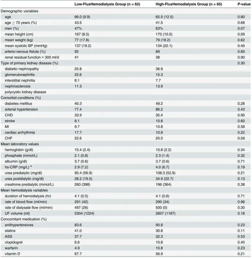
Altogether, 166 patients were included into the MINOXIS study, of which 127 patients had valid measures for all bone markers investigated at both evaluation time points (i.e., baseline and 12 months).Table 1shows the patients baseline characteristics, which were not different between the low-flux and the high-flux groups.
Primary endpoint; FGF-23
Table 1. Baseline characteristics of the study population.
Low-FluxHemodialysis Group (n = 62) High-FluxHemodialysis Group (n = 65) P-value
Demographic variables
age 66.0 (9.9) 65.5 (12.5) 0.80
age70 years (%) 43.5 41.5 0.68
men (%) 47% 63% 0.07
mean height (cm) 167 (8.5) 170 (10.0) 0.09
mean weight (kg) 77 (17.8) 79 (18.2) 0.62
mean systolic BP (mmHg) 137 (19.2) 134 (22.1) 0.40
arterio-venousfistula (%) 92 89 0.60
renal residual function>300 ml/d 41 38 0.90
Type of primary kidney disease (%) 0.30
diabetic nephropathy 25.8 36.9
glomerulonephritis 22.6 15.3
interstitial nephritis 8.1 7.7
nephrosclerosis 11.3 13.9
polycystic kidney disease Comorbid conditions (%)
diabetes mellitus 40.3 49.2 0.28
arterial hypertension 77.4 86.2 0.43
CHD 33.9 35.4 0.90
stroke 8.1 10.8 0.60
MI 9.7 10.8 0.58
cardiac arrhythmia 17.7 10.8 0.22
CHF 22.6 20.0 0.59
Mean laboratory values
hemoglobin (g/dl) 10.4 (2.4) 10.8 (2.2) 0.34
phosphate (mmol/L) 2.1 (0.8) 2.3 (1.4) 0.32
albumin (g/dl) 3.7 (0.6) 3.7 (0.6) 0.71
hs-CRP (mg/L)a 2.9 (7.2) 4.0 (6.7) 0.19
urea predialytic (mg/dl) 95.4 (56.9) 108.3 (52.9) 0.21
urea postdialytic (mg/dl) 28.2 (19.5) 34.9 (22.7) 0.13
creatinine predialytic (mmol/L) 260 (398) 196 (364) 0.38
Mean hemodialysis variables
duration of hemodialysis (hr) 4.1 (0.5) 4.1 (0.6) 0.71
rate of bloodflow (ml/min) 291 (42) 290 (34) 0.96
rate of dialysateflow (ml/min) 497 (26) 500 (0) 0.30
UF volume (ml) 2304 (1224) 2607 (1187) 0.18
Concomitant medication (%)
antihypertensives 83.6 90.8 0.23
statins 41.0 30.8 0.11
ASS 37.7 32.3 0.53
clopidogrel 6.6 10.8 0.40
warfarin 4.9 10.8 0.23
vitamin D 67.7 56.9 0.21
Values presented with numbers in parentheses are mean (SD). CHD, coronary heart disease; MI, myocardial infarction; CHF, congestive heart failure; hs-CRP, high-sensitivity C-reactive protein; UF, ultrafiltration; HMG-CoA, hydroxymethyl glutaryl coenzyme A; ASS, acetylsalicylic acid.
aMedian (interquartile range).
difference in the absolute change from baseline between the two groups was 4537 RU/ml (95% CI: 1534 to 7541, p<0.01) (Figs2and3;Table 2).
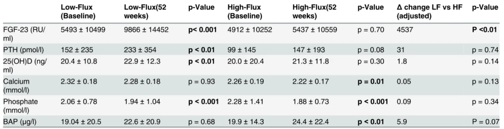
Secondary endpoints; specific markers of mineral and bone metabolism
There was a significant increase in PTH in the low-flux group during the course of the study, which was not found in the high-flux group (Table 2). However, the changes in PTH were not different between the two groups (p = 0.74). Regarding 25(OH)D levels, there was no difference in the change between the two groups (p = 0.14). We found a significant decrease in serum cal-cium levels only in the high-flux group (p = 0.01), but again, there was no difference in the change of calcium levels between the two groups (p = 0.13). Serum phosphate levels decreased in both groups during the course of the study. However, there was no difference in the change of phosphate levels between the two groups (p = 0.34). In patients who underwent high-flux di-alysis, BAP significantly increased during the study. However, the difference in absolute change between the two groups was not significant (p = 0.07).
Table 2. Outcome.
Low-Flux (Baseline)
Low-Flux(52 weeks)
p-Value High-Flux (Baseline)
High-Flux(52 weeks)
p-Value Δchange LF vs HF (adjusted)
p-Value
FGF-23 (RU/ ml)
5493±10499 9866±14452 p<0.001 4912±10252 5437±10559 p = 0.70 4537 P<0.01
PTH (pmol/l) 152±235 233±354 p<0.01 99±145 147±193 p = 0.08 31 p = 0.74
25(OH)D (ng/ ml)
20.4±10.8 22.9±12.3 p<0.01 20.0±20.4 21.3±11.8 p = 0.30 1.8 p = 0.14
Calcium (mmol/l)
2.32±0.18 2.28±0.18 p = 0.93 2.26±0.19 2.22±0.17 p = 0.01 0.05 p = 0.13
Phosphate (mmol/l)
2.06±0.78 1.94±1.04 p<0.001 2.28±1.41 1.88±0.73 p<0.001 0.09 p = 0.34
BAP (μg/l) 19.04±20.5 22.6±20.9 p = 0.68 19.9±14.3 24.4±22.4 p<0.01 5.9 P = 0.07
doi:10.1371/journal.pone.0128079.t002
Fig 2. Grouped boxplots visualizing the distributions of FGF-23 before and after the intervention stratified by randomization group.Black box: FGF-23 before the intervention, Grey box: FGF-23 after the intervention. 1: low-flux group, 2: high-flux group.
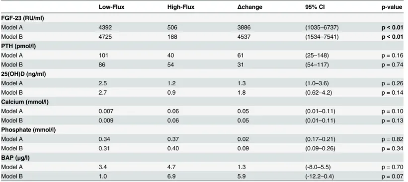
Linear regression models and multivariate mixed effect model
The results of the linear regression models are shown inTable 3. Model A, adjusting for base-line values, demonstrated a significant difference between the low-flux and the high-flux HD groups in the change of FGF-23 levels. There was no significant difference for any of the other parameters examined. Model B, additionally adjusting for age, sex, CPR, albumin, calcium, phosphate, 25-hydroxyvitamin D and PTH, was also significant for the change of FGF-23 lev-els, but not for any of the other parameters examined. Finally, we also applied a multivariate mixed effects model. Using this approach, a significant interaction between treatment group and measurement time-point was demonstrated for FGF-23 levels (Group: p = 0.78, Measure-ment: p = 0.22, Group x MeasureMeasure-ment: p = 0.05).
Fig 3. FGF-23; predicted marginal means of the absolute difference (12 months—baseline).
doi:10.1371/journal.pone.0128079.g003
Table 3. Outcome.
Low-Flux High-Flux Δchange 95% CI p-value
FGF-23 (RU/ml)
Model A 4392 506 3886 (1035–6737) p<0.01
Model B 4725 188 4537 (1534–7541) p<0.01
PTH (pmol/l)
Model A 101 40 61 (25–148) p = 0.16
Model B 86 54 31 (54–117) p = 0.74
25(OH)D (ng/ml)
Model A 2.5 1.2 1.3 (1.0–3.6) p = 0.26
Model B 2.7 0.9 1.8 (0.62–4.2) p = 0.14
Calcium (mmol/l)
Model A 0.007 0.06 0.05 (0.01–0.11) p = 0.10
Model B 0.009 0.06 0.05 (0.01–0.11) p = 0.13
Phosphate (mmol/l)
Model A 0.34 0.37 0.02 (0.17–0.21) p = 0.82
Model B 0.31 0.40 0.09 (0.09–0.26) p = 0.34
BAP (μg/l)
Model A 3.4 4.7 1.3 (-8.0–5.5) p = 0.70
Model B 1.0 6.9 5.9 (-12.2–0.4) p = 0.07
Discussion
In the present analysis of the MINOXIS trial, the effects of treatment with low- versus high-flux dialysis membranes on circulating FGF-23 concentrations were studied. To the best of our knowledge, this is the first study investigating this issue in patients undergoing HD. The main finding was that over a period of 12 months, FGF-23 levels remained stable in the high-flux di-alysis group, whereas an increase was observed in the low-flux didi-alysis group. The results were independent of potentially confounding variables including age, sex, C-reactive protein, albu-min, calcium, phosphate, 25(OH)D and PTH levels.
FGF-23 acts through activation of theklothoreceptor [11]. In the kidney, activation of the klothoreceptor has phosphaturic effectsviainhibition of proximal tubular phosphate
reab-sorption through sodium-dependent transporters [11,12]. In HD patients, levels of FGF-23 are up to 1000 times higher compared with the normal population [7]. It is thought that the early increase in FGF-23 levels in CKD patients is an adaptive mechanism for preventing phos-phate overload [4,13]. Regarding the cardiovascular system, FGF-23 has been shown to induce left ventricular hypertrophy [14,15], vascular calcification [16], arterial stiffness and endotheli-al dysfunction [17]. A recent study by Faul and colleagues demonstrated that intraventricular injection of FGF-23 directly induces pathological hypertrophy of the heart in rats [15]. These cardiovascular effects might explain why increased FGF-23 level has been found to indepen-dently predict CV mortality in CKD patients [18]. As a consequence of these detrimental ef-fects of FGF23, there is pressing need to identify potential therapeutic options to lower concentrations of FGF-23 in patients with CKD.
The present results of the MINOXIS study demonstrate that high-flux HD was associated with stable FGF-23 concentrations over 12 months. Interestingly, enhanced removal of FGF-23 has been demonstrated in patients with on-line high-efficiency hemodiafiltration compared to both low-flux and high-flux dialysis [19,20]. This finding suggests a more intense elimination by high-flux HD as a possible explanation for the differences in the FGF-23 levels observed in the MINOXIS study.
Similar to FGF-23, the secondary endpoint variable PTH has a predictive value with regard to mortality and CV events in HD patients [21,22]. In the present study, paralleling the course of FGF-23, a significant increase of the PTH concentrations in the low-flux group but not in the high-flux group was observed. This observation is in line with a prospective study in 44 children on renal replacement therapy (RRT), which demonstrated more efficient PTH remov-al of high-flux compared to flux diremov-alysis membranes [23]. PTH is a free circulating low-molecular weight protein (LMWP) with 9.500 Dalton [9] and, thus, easily permeable through high-flux membranes, but not through low-flux membranes. Nevertheless, the difference in the absolute change of the PTH levels between the two randomized groups was not significant.
The main limitation of the present study is its post hoc nature. The main strengths are the strict randomized design, the relatively large and well defined cohort, and the measurement of a large number of parameters of bone and mineral metabolism, permitting the statistical ad-justment for a large number of potential confounders of the effects of dialysis membrane char-acteristics on FGF-23 levels.
Supporting Information
S1 Fig. FGF-23 levels before and after 12 months of low-flux HD.
(TIFF)
S2 Fig. FGF-23 levels before and after 12 months of high-flux HD.
(TIFF)
Acknowledgments
We thank all patients who participated in the MINOXIS Study. We are grateful to all investiga-tors, study nurses and collaborators involved in patient recruitment, sample and data handling.
Author Contributions
Conceived and designed the experiments: AS MS CW CD. Performed the experiments: AS HS TS DK. Analyzed the data: AS MS CD. Contributed reagents/materials/analysis tools: AS MS HS CD. Wrote the paper: AS MS DK HS TS CW CD. Provided statistical support for the study: BG.
References
1. Collins AJ, Foley RN, Herzog C, Chavers BM, Gilbertson D, Ishani A, et al. Excerpts from the US Renal Data System 2009 Annual Data Report. American journal of kidney diseases: the official journal of the National Kidney Foundation. 2010; 55(1 Suppl 1):S1–420, A6-7. Epub 2010/02/10. doi:10.1053/j.ajkd.
2009.10.009PMID:20082919; PubMed Central PMCID: PMC2829836.
2. Foley RN, Parfrey PS, Sarnak MJ. Epidemiology of cardiovascular disease in chronic renal disease. Journal of the American Society of Nephrology: JASN. 1998; 9(12 Suppl):S16–23. Epub 2001/07/11.
PMID:11443763.
3. Stubbs J, Liu S, Quarles LD. Role of fibroblast growth factor 23 in phosphate homeostasis and patho-genesis of disordered mineral metabolism in chronic kidney disease. Seminars in dialysis. 2007; 20 (4):302–8. Epub 2007/07/20. doi:10.1111/j.1525-139X.2007.00308.xPMID:17635819.
4. Prie D, Urena Torres P, Friedlander G. Latest findings in phosphate homeostasis. Kidney international. 2009; 75(9):882–9. Epub 2009/02/05. doi:10.1038/ki.2008.643PMID:19190675.
5. Diniz H, Frazao JM. The role of fibroblast growth factor 23 in chronic kidney disease-mineral and bone disorder. Nefrologia: publicacion oficial de la Sociedad Espanola Nefrologia. 2013; 33(6):835–44. Epub
2013/10/26. doi:10.3265/Nefrologia.pre2013.Jul.12091PMID:24158124.
6. Gutierrez OM, Mannstadt M, Isakova T, Rauh-Hain JA, Tamez H, Shah A, et al. Fibroblast growth factor 23 and mortality among patients undergoing hemodialysis. The New England journal of medicine. 2008; 359(6):584–92. Epub 2008/08/09. doi:10.1056/NEJMoa0706130PMID:18687639; PubMed
Central PMCID: PMC2890264.
7. Kendrick J, Cheung AK, Kaufman JS, Greene T, Roberts WL, Smits G, et al. FGF-23 associates with death, cardiovascular events, and initiation of chronic dialysis. Journal of the American Society of Ne-phrology: JASN. 2011; 22(10):1913–22. Epub 2011/09/10. doi:10.1681/ASN.2010121224PMID:
21903574; PubMed Central PMCID: PMC3187186.
8. Palmer SC, Rabindranath KS, Craig JC, Roderick PJ, Locatelli F, Strippoli GF. High-flux versus low-flux membranes for end-stage kidney disease. The Cochrane database of systematic reviews. 2012; 9: CD005016. Epub 2012/09/14. doi:10.1002/14651858.CD005016.pub2PMID:22972082.
9. Neirynck N, Eloot S, Glorieux G, Barreto DV, Barreto FC, Liabeuf S, et al. Estimated glomerular filtration rate is a poor predictor of the concentration of middle molecular weight uremic solutes in chronic kidney disease. PloS one. 2012; 7(8):e44201. Epub 2012/09/07. doi:10.1371/journal.pone.0044201PMID: 22952928; PubMed Central PMCID: PMC3432070.
10. Schneider A, Drechsler C, Krane V, Krieter DH, Scharnagl H, Schneider MP, et al. The effect of high-flux hemodialysis on hemoglobin concentrations in patients with CKD: results of the MINOXIS study. Clinical journal of the American Society of Nephrology: CJASN. 2012; 7(1):52–9. Epub 2011/11/19. doi:
10.2215/CJN.02710311PMID:22096040; PubMed Central PMCID: PMC3265341.
11. Kovesdy CP, Quarles LD. The role of fibroblast growth factor-23 in cardiorenal syndrome. Nephron Clinical practice. 2013; 123(3–4):194–201. Epub 2013/08/15. doi:10.1159/000353593PMID:
12. Quarles LD. Role of FGF23 in vitamin D and phosphate metabolism: implications in chronic kidney dis-ease. Experimental cell research. 2012; 318(9):1040–8. Epub 2012/03/17. doi:10.1016/j.yexcr.2012.
02.027PMID:22421513; PubMed Central PMCID: PMC3336874.
13. Gutierrez O, Isakova T, Rhee E, Shah A, Holmes J, Collerone G, et al. Fibroblast growth factor-23 miti-gates hyperphosphatemia but accentuates calcitriol deficiency in chronic kidney disease. Journal of the American Society of Nephrology: JASN. 2005; 16(7):2205–15. Epub 2005/05/27. doi:10.1681/ASN.
2005010052PMID:15917335.
14. Gutierrez OM, Januzzi JL, Isakova T, Laliberte K, Smith K, Collerone G, et al. Fibroblast growth factor 23 and left ventricular hypertrophy in chronic kidney disease. Circulation. 2009; 119(19):2545–52.
Epub 2009/05/06. doi:10.1161/CIRCULATIONAHA.108.844506PMID:19414634; PubMed Central PMCID: PMC2740903.
15. Faul C, Amaral AP, Oskouei B, Hu MC, Sloan A, Isakova T, et al. FGF23 induces left ventricular hyper-trophy. The Journal of clinical investigation. 2011; 121(11):4393–408. Epub 2011/10/12. doi:10.1172/
JCI46122PMID:21985788; PubMed Central PMCID: PMC3204831.
16. Khan AM, Chirinos JA, Litt H, Yang W, Rosas SE. FGF-23 and the progression of coronary arterial cal-cification in patients new to dialysis. Clinical journal of the American Society of Nephrology: CJASN. 2012; 7(12):2017–22. Epub 2012/09/22. doi:10.2215/CJN.02160212PMID:22997345; PubMed
Cen-tral PMCID: PMC3513740.
17. Mirza MA, Larsson A, Lind L, Larsson TE. Circulating fibroblast growth factor-23 is associated with vas-cular dysfunction in the community. Atherosclerosis. 2009; 205(2):385–90. Epub 2009/02/03. doi:10.
1016/j.atherosclerosis.2009.01.001PMID:19181315.
18. Parker BD, Schurgers LJ, Brandenburg VM, Christenson RH, Vermeer C, Ketteler M, et al. The associ-ations of fibroblast growth factor 23 and uncarboxylated matrix Gla protein with mortality in coronary ar-tery disease: the Heart and Soul Study. Annals of internal medicine. 2010; 152(10):640–8. Epub 2010/
05/19. doi:10.7326/0003-4819-152-10-201005180-00004PMID:20479029; PubMed Central PMCID: PMC3079370.
19. Patrier L, Dupuy AM, Granger Vallee A, Chalabi L, Morena M, Canaud B, et al. FGF-23 removal is im-proved by on-line high-efficiency hemodiafiltration compared to conventional high flux hemodialysis. Journal of nephrology. 2013; 26(2):342–9. Epub 2012/05/11. doi:10.5301/jn.5000150PMID:
22573526.
20. Cornelis T, van der Sande FM, Eloot S, Cardinaels E, Bekers O, Damoiseaux J, et al. Acute Hemody-namic Response and Uremic Toxin Removal in Conventional and Extended Hemodialysis and Hemo-diafiltration: A Randomized Crossover Study. American journal of kidney diseases: the official journal of the National Kidney Foundation. 2014. Epub 2014/04/05. doi:10.1053/j.ajkd.2014.02.016PMID: 24698199.
21. Drechsler C, Krane V, Grootendorst DC, Ritz E, Winkler K, Marz W, et al. The association between parathyroid hormone and mortality in dialysis patients is modified by wasting. Nephrology, dialysis, transplantation: official publication of the European Dialysis and Transplant Association—European
Renal Association. 2009; 24(10):3151–7. Epub 2009/05/29. doi:10.1093/ndt/gfp260PMID:19474272;
PubMed Central PMCID: PMC2747498.
22. Covic A, Kothawala P, Bernal M, Robbins S, Chalian A, Goldsmith D. Systematic review of the evi-dence underlying the association between mineral metabolism disturbances and risk of all-cause mor-tality, cardiovascular mortality and cardiovascular events in chronic kidney disease. Nephrology, dialysis, transplantation: official publication of the European Dialysis and Transplant Association—
Eu-ropean Renal Association. 2009; 24(5):1506–23. Epub 2008/11/13. doi:10.1093/ndt/gfn613PMID:
19001560.
23. Makar SH, Sawires HK, Farid TM, Ali WM, Schaalan M. Effect of high-flux versus low-flux dialysis mem-branes on parathyroid hormone. Iranian journal of kidney diseases. 2010; 4(4):327–32. Epub 2010/09/