Universidade de Lisboa
Faculdade de Ciências
Departamento de Biologia Vegetal
Proof of Concept of PhageDuction: Heterologous
Transduction Mediated by Bacteriophage SPP1
Master Thesis
Nuno Filipe Brito Pais dos Santos
Mestrado em Microbiologia Aplicada
Universidade de Lisboa
Faculdade de Ciências
Departamento de Biologia Vegetal
Proof of Concept of PhageDuction: Heterologous
Transduction Mediated by Bacteriophage SPP1
Dissertação orientada por Prof. Dr. Carlos São-José (FFUL, DMI, CPM-URIA) e Prof. Dr. Mário Santos
(FCUL)
Nuno Filipe Brito Pais dos Santos
Mestrado em Microbiologia Aplicada
Proof of Concept of PhageDuction: Heterologous
Transduction Mediated by Bacteriophage SPP1
Nuno Filipe Brito Pais dos Santos
Master Thesis
2013
This thesis was fully performed at the Center of Molecular Pathogenesis-Unit of
Retroviruses and Associated Infections (CPM-URIA), Faculty of Pharmacy of the
University of Lisbon, under the direct supervision of Prof. Dr. Carlos São-José.
Prof. Dr. Mário Santos was the internal designated supervisor in the scope of
the Master in Applied Microbiology of the Faculty of Sciences of the University
of Lisbon.
Acknowledgements
This work was carried out in fulfillment of the Master in Applied Microbiology at the Faculty of Sciences of the University of Lisbon, being performed at Center of Molecular Pathogenesis-Unit of Retroviruses and Associated Infections (CPM-URIA) and Department of Microbiology and Immunology (DMI), Faculty of Pharmacy of the University of Lisbon. I would like to acknowledge the coordinator of CPM-URIA and DMI, Prof. Dr. José Moniz-Pereira, for providing the necessary support and means to perform this work.
I would like to thank Paulo Tavares and Juan Alonso for kindly providing plasmid pBT163, which was crucial for the success of this work.
I would like to thank my internal supervisor Prof. Dr. Mário Santos for his availability. I would like to acknowledge Catarina Baptista and Hugo Barreto for their precious support since the beginning of this work.
I would like to thank my mother for her immeasurable support which culminated in the opportunity of accomplishing this dissertation.
Last but definitely not least, I am incredibly grateful to my supervisor Carlos São-José and co-supervisor Sofia Fernandes for their never-ending patience and guidance.
This work has been supported through Grant EXPL/BBB-EBI/0308/2012 (to Prof. C. São-José.) from Fundação para a Ciência e a Tecnologia (FCT, MEC, Portugal).
Abstract
Technologies for gene delivery are on the basis of many biotechnological and medical applications. The currently available technologies still fail to completely fulfill the requirements of an ideal system, that is, one that allows efficient and stable gene delivery, in a cell-specific and non-toxic manner, independently of the target cell and at reasonable costs. This work is a prospect for the development of a new DNA delivering technology, here designated as PhageDuction. The conception of PhageDuction is based on bacteriophage SPP1 and its cellular receptor, the Bacillus subtilis membrane protein YueB.
There are only three essential requirements for SPP1 DNA ejection into host cells: the presence of the YueB receptor ectodomain at the B. subtilis surface, an energized cytoplasmic membrane and a calcium gradient between the extra- and intracellular milieu. Since the vast majority of cells maintain a membrane potential and a calcium gradient under physiologic conditions, it is expected that SPP1 will deliver its DNA into cells decorated with the YueB ectodomain.
To prove this concept, we have studied the capacity of SPP1 to deliver its DNAcargo to cells lacking YueB (resistant to SPP1 infection), after their decoration with the fusion protein YueB350LysM. This fusion is composed of the receptor ectodomain YueB350 and two LysM motifs, which are known to target a general component of the bacterial cell wall, the peptidoglycan. We show that SPP1 is able to deliver its DNA, plasmid DNA and heterologous DNA fragments to recipient cells decorated with YueB350LysM. The results obtained thus demonstrate PhageDuction functionality.
Resumo
As tecnologias de entrega de DNA estão na base de inúmeras aplicações biotecnológicas e médicas. As tecnologias actualmente disponíveis ainda não satisfazem completamente os requisitos de um sistema ideal, sendo este um que permita a entrega eficiente e estável de genes, de uma forma específica e não tóxica para a célula, independentemente da célula alvo e a custos reduzidos. Este trabalho é uma prospecção para o desenvolvimento de uma nova tecnologia para a entrega de DNA, aqui designada por PhageDuction. A concepção da PhageDuction é baseada no bacteriófago SPP1 e no seu receptor celular, a proteína membranar YueB de Bacillus subtilis.
Existem apenas três requisitos essenciais para a ejecção do DNA de SPP1 na célula hospedeira: a presença de um ectodomínio de YueB à superfície de B. subtilis, uma membrana citoplasmática energizada e um gradiente de cálcio entre os meios extra e intracelular. Como a larga maioria das células mantém um potencial de membrana e um gradiente de cálcio em condições fisiológicas, é expectável que SPP1 insira o seu DNA em células decoradas com o ectodomínio YueB.
Para provar este conceito, estudámos a capacidade do fago SPP1 em inserir o seu conteúdo de DNA em células desprovidas de YueB (resistentes à infecção por SPP1), após estas serem decoradas com a fusão YueB350LysM. Esta fusão é composta pelo ectodomínio receptor YueB350 e dois domínios LysM, os quais possuem afinidade para um componente geral da parede celular bacteriana, o peptidoglicano. Mostramos que, quando adicionado a células alvo decoradas com YueB350LysM, SPP1 consegue não só introduzir o seu DNA nestas, como também fazer a entrega de DNA plasmídico e de fragmentos de DNA heterólogo. Os resultados demonstram assim a funcionalidade da tecnologia PhageDuction.
Index
Acknowledgements i
Abstract ii
Resumo iii
I - Introduction 1
I.1 - Viral-based DNA delivery vectors 1
I.1.1 - Animal virus-derived vectors 1
I.1.2 - Bacteriophage-derived vectors 2
I.2 - General properties of phages 4
I.3 - Bacteriophage SPP1 of Bacillus subtilis 6
I.4 - The concept of PhageDuction: SPP1-mediated heterologous transduction 8
I.5 - Thesis goals and strategy 9
II - Results 11
II.1 - Production and purification of the receptor-ligand fusion YueB350LysM 11
II.2 - YueB350LysM triggers SPP1 DNA ejection in vitro 14
II.3 - YueB350LysM binds to target cell surface 15
II.4 - YueB350LysM-decorated cells allow productive SPP1 infection 16 II.5 - SPP1 mediates plasmid transduction to YueB350LysM-decorated cells 18 II.6 - Transduction of bacterial chromosomal genes by PhageDuction 21
III - Discussion 23
IV - Concluding Remarks 27
V - Materials and Methods 29
V.1 - Bacteria, phages, plasmids and growth conditions 29
V.2 - General techniques of molecular biology and biochemistry 30
V.2.1 - PCR amplification and cloning 30
V.2.2 - Transformation, plasmid analysis and DNA sequencing 30
V.2.3 - SDS-PAGE and Western Blot analysis 31
V.2.4 - Protein quantification 31
V.2.5 - Bioinformatics analysis 32
V.3 - SPP1 inactivation with α-SPP1 sera 32
V.4.2 - LysM350LysM production 33
V.4.3 - Monitoring of Yue350LysM solubility 34
V.4.4 - YueB350LysM purification 34
V.5 - SPP1 inactivation by YueB350LysM and DNase protection assays 35
V.6 - Binding of YueB350LysM to target cells 35
V.7 - SPP1 replication in CSJ1 cells with bound YueB350LysM 36
V.7.1 - CSJ1 decoration with YueB350LysM 36
V.7.2 - SPP1 growth in YueB350LysM-decorated cells 36 V.8 - SPP1-mediated plasmid PhageDuction to YueB350LysM-decorated cells 36
V.8.1 - Production of a SPP1 transducing lysate 37
V.8.2 - Plasmid PhageDuction 37
V.9 - PhageDuction of lacA::spec to B. licheniformis MW3 37 V.9.1 - Production of a lacA::spec SPP1 transducing lysate 37
V.9.2 - SPP1-mediated lacA::spec PhageDuction 37
VI - References 39
I – Introduction
The development of technologies to insert genetic material into target cells was preponderant in fundamental research of biological systems, and has been on the basis of many medical and biotechnological applications, like genetic manipulation, vaccination and gene therapy. The introduction of genetic material into a cell is considered the “Achilles heel” of gene therapy (1). The key for a successful gene therapy strongly depends on the discovery of the perfect delivery system, a vector that should be easy to produce, have sustained or regulated expression of DNA cargo, be immunologically inert, have tissue targeting, have variable cargo capacity, be able to replicate, segregate or integrate and infect both dividing and non-dividing cells (1).
Currently available delivery strategies can be grouped in non-viral and viral. Some remarking non-viral technologies may still involve the activity of virus-derived elements, such as the systems based on phage C31 integrase and phage P1 Cre recombinase (2, 3, 4), or transposons (5). Their main overall advantage is not to present immunity issues, but they lack transduction efficiency (6). On the other hand viral vectors, viruses or their modified forms have great transduction efficiency, but their major drawback is the host immunity response and safety (6). Nevertheless, technology improvements may outdo these issues, making viral vectors the most promising technologies for the future.
I.1 – Viral-based DNA delivery vectors
Viral vectors can be divided according to their host: animal virus-derived vectors and bacteriophage-derived vectors.
I.1.1 – Animal virus-derived vectors
The most studied and promising animal viral vectors are Retrovirus, Lentivirus, Adeno-associated vectors, all with the capacity to integrate in recipient cells, and Adenovirus that do not integrate and are maintained in cells as an episome. There are also other options, like the simplex virus and vaccinia virus.
Retroviruses are enveloped ssRNA viruses encoding an integrase that allows the correct integration of a gene in the eucromatin, leading to long and stable gene expression, even to host cell progeny (6). However, Retroviruses only infect dividing cells (6) and their large-scale production exhibit some major drawbacks, like instability, low titer supernatants and short
While Retroviruses only infect dividing cells, Lentiviruses, part of the Retroviruses family, are able to infect both dividing and non-dividing cells (6, 8). However, they also present some disadvantages. Even though Lentivirus used for gene therapy are non-replication competent, there is a safety concern that recombination may lead to replication competent Lentivirus (7, 9). Additionally, the integrase action, useful for long-term expression, also bears safety issues, as the vector-mediated integration may lead to insertional mutagenesis (9).
Adenoviruses are non-enveloped double-stranded (ds)DNA virus, with natural tropism to high respiratory tract and ocular tissues. They have high cloning capacity (until >25kb in deleted versions) and are able to infect non-dividing cells. Unlike Lentivirus, Adenovirus only lead to transient transduction, demanding repeated dosing to achieve stable gene expression (6). Unfortunately, these repeated dosing lead to strong immunity response which may result in virus and host tissue destruction (6). Natural tropism is also a disadvantage because it makes it difficult to target the vector to the intended cells or tissue (10).
Adenovirus-associated vectors (AAV) are ssDNA non-pathogenic Adenovirus derived vectors. Also able to infect both dividing and non-dividing cells, they are able to integrate in a specific site at chromosome 19 (6). AAV’s still have safety issues as they can cause chromosome fragmentation, resulting in rearrangements and deletions (11). AAV’s small capacity (only <4Kb) is also another disadvantage. Their site specific integration is still not fully understood (6). Adenovirus and AAV’s production at industrial scale is difficult due to complex virus production and the presence of empty capsid vectors, demanding increased downstream processing (7).
Herpes simplex are linear dsDNA viruses that infect the nervous system. They are used in cancer therapy for their cytopathogenicity (12). As a delivery vector, there are some safety concerns, because viral proteins are toxic even when residual and most humans had previous contact with the virus which leads to a strong immunity response (8). Vaccinia virus is a possible cytoplasmic vector that does not integrate in the nucleus (8).
Even the most promising vectors for animal applications referred here still present some disadvantages and technological challenges especially at the safety issues topic and due to high costs for their large-scale production (7).
I.1.2 – Bacteriophage-derived vectors
Bacteriophages (phages) are viruses specifically infecting bacteria. Since their discovery about one century ago they have been fundamental in molecular biology progress and in the unveiling of new possibilities in biotechnology, being used for phage therapy, phage display, vaccine delivery, gene delivery and identification and diagnostic of bacteria (13). Unlike animal
virus derived-vectors, phages lack intrinsic tropism to animal cells or tissues which makes them a safe vector. They are easy to produce at high titers, have a fast life cycle, and present genetic flexibility. Viral particles are also stable under extreme conditions like pH, DNase or proteolytic enzymes (14).
To use phages as vector, there is need of targeting the phage to the desired cell. Like the animal virus-derived vectors, this could be achieved by adding to the phage coat a ligand that has affinity to a receptor at the target cell surface (15). Larocca et al (15) elegantly showed that a modified filamentous phage, M13, could deliver selected genes to mammalian cells, serving as a vehicle to eukaryotic cells (15).
Phages are also used as vectors to deliver nucleic acid vaccines. These consist in inoculating the animal with pure DNA or RNA encoding the antigen gene regulated by a host promoter (16). The use of phage lambda (λ) virus particles as vehicle resulted in an immunity response stronger than naked DNA delivery (16). This is an easy to produce approach, where DNA is protected from the environment and it allows oral application (16). Additionally, the display of different affinity peptides at the surface of the head and tail of phage λ improved gene delivery to mammalian cells in vitro (17).
Some phages have terminal proteins (TP) that are covalently bound to the genome. Remarkably, it was observed that these TP from phages φ29, Nf, PRD1, Bam35 and Cp-1 function as nuclear localization signals (NLS) that target them to mammalian cell’s nucleus (18, 19). Different motifs or proteins can be fused to φ29 adding specific features like improving transgene expression or introducing human gene excision sites (19).
A new vector was developed from T4 DNA packaging machine, an ATP-powered efficient system (20). DNA molecules are packaged into an empty phage head that are decorated with proteins fused to outer capsid proteins for targeting (20). This system can be used for gene or enzyme delivery, nucleic acid vaccines or targeting ligands, revealing effective for single or multiple genes, PCR-amplified DNA, concatemerized DNA, short and long peptides, full length proteins and large oligomeric complexes (20).
The examples provided above illustrate that different phage properties can be used in the engineering of delivery vectors. In this work we have used a yet unexplored feature of phages to design a completely new DNA delivery strategy. To better understand what it involves, the following sections will provide relevant characteristics of phage biology, ending with a more detailed description of the phage used in our strategy, the Bacillus subtilis phage SPP1.
I.2 – General properties of phages
Phages are the most common and diverse biological entities on earth (21). There are three categories or types of phages. The most isolated type is by far that of tailed phages (Caudovirales), which possess a tail with uniform length within species members and a dsDNA monopartite genome (22, 23). Structurally, they are non-enveloped (naked) virions composed of an icosahedral capsid (head) held to the tail by and a protein disc (connector) and may present other facultative structures like spikes, baseplates or tail fibers (22). Head diameter and tail length are extremely variable within tailed phages (22). The order Caudovirales is divided in three families: Myoviridae (striated long and contractile tails), Siphoviridae (striated long and noncontractile tails) and Podoviridae (short noncontractile tails). Except for a few lipid-containing tailed phages in mycobacteria and streptococci, viral particles usually consist of DNA and protein only, having a higher DNA content than other virus, being around 50% (22). Tailed phages present specific genome features like circular permutations, terminal redundancies, packaging (pac) or cohesive (cos) sites recognized for DNA encapsidation, terminal proteins and single-stranded gaps, in addition to rare bases and sugars, and often carry methylated or hydroxylated DNA to prevent bacterial host endonuclease restriction activity (22). Phages with pac sites usually present circular permutation and terminal repeats, as in B. subtilis phage SPP1, while those with cos sites are nonredundant and nonpermuted, as λ (22).
The second category, not representing a taxonomic group, is composed of phages with medium-sized dsDNA (families Corticoviridae, Tectiviridae and Plasmaviridae) or dsRNA (family Cystoviridae), tripartite genomes inside lipid-containing capsids (23). Finally, a third category, also with no taxonomic value, comprises small single stranded (ss) DNA (families Microviridae and Inoviridae) or ssRNA (family Leviviridae) genomes (23).
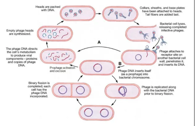
Phage infection may proceed essentially by two distinct ways, the so called lytic (virulent) and temperate cycles (Fig. 1). In both lifestyles phage infection starts with virus particle specific adsorption to host cell, followed by the phage DNA entry to the bacterial cytoplasm. The obligatory lytic phages immediately undergo expression and replication of their genomes, which culminates in the assembly of novel virions that typically escape to the media after lysis of the infected cell (Fig. 1-A) (24). In the cases where the phage DNA circularizes after entering the host cell, its replication occurs via a rolling-circle mechanism (ex. λ, P2 and SPP1), while linear DNA replication starts from internal origins of replication (ex. T4 and T7). Phages with dsDNA induce host cell lysis by the concerted action of at least two proteins, the holin and a peptidoglycan hydrolase (endolysin) (24).
Upon their DNA entry into the bacterial host, temperate phages (Fig. 1-B) have the “choice” of immediately enter the lytic cycle, as just described or follow the lysogenic pathway. In the latter case the phage genome integrates into the host bacterium’s chromosome, through the activity of integrases/recombinases. The integrated phage DNA, called prophage, can be propagated for several generations as a part of the bacterial genome. (22). Upon diverse stimuli, the prophage can excise from the bacterial chromosome and resume the typical lytic multiplication, leading to host cell lysis. Alternatively, it can persist as a plasmid in the host cytoplasm (P1) or integrate at random sites as transposons (Mu), maintaining prophages as parts of host genome or plasmids (22).
Fig. 1 – Lytic and temperate phage infection cycles. Once inside the host cell, the DNA of obligatory lytic phages immediately takes over the cell molecular machinery to drive its own replication and to produce descendent virions, which normally are released to the media through cell lysis (A); Temperate phages can “chose” between the lytic or lysogenic pathways. In the lysogenic pathway (B) phage DNA integrates in the bacterial genome, being called prophage. Prophages may be propagated through several generations as a part of the bacterial genome. Upon specific stimuli, prophages can be activated and excised from bacterial genome, undergoing the lytic cycle. Scheme adapted from (25).
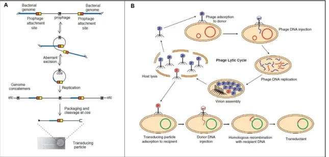
Phages can mediate generalized or specialized transduction, a process in which host bacterial DNA is transferred from one cell to another. Specialized transduction is mediated by temperate phages that followed the lysogenic cycle. It results from imprecise excisions and a limited number of bacterial genes are transferred, usually those near the prophage integration site on the bacterial chromosome (Fig 2-A). In generalized transduction virtually any bacterial DNA fragment (and in some cases bacterial plasmid DNA) can be mobilized by the infecting phage (Fig. 2-B) (26). The transfer of genetic material between bacterial cells mediated by phages is of great importance in bacterial evolution.
Fig. 2 – Specialized and generalized transduction. Specialized transduction occurs only with temperate phages, whose genomes are initially integrated in specific sites of the bacterial chromosome. Upon prophage activation, its aberrant excision may generate transducing phage particles carrying DNA fragments that lied next to attachment sites (A). In generalized transduction virtually any bacterial DNA fragment (or in some cases plasmidic DNA) can be mobilized by the infecting phage (B). Scheme adapted from (26).
I.3 – Bacteriophage SPP1 of Bacillus subtilis
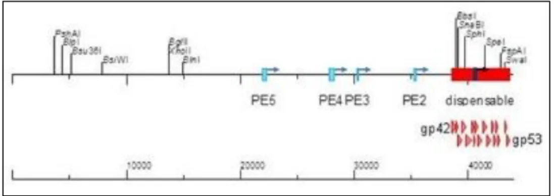
SPP1 is a tailed phage from family Siphoviridae that infects the Gram-positive model bacterium Bacillus subtilis. It was first described by Riva et al in 1968 (27), and has been broadly studied since then, being its genome extensively described (28). One potentially useful feature is that SPP1 genome has a dispensable region of 4500bp (Fig. 3).
Fig. 3 – Linear representation of the 44010bp genome of phage SPP1. The position of single restriction sites and of five early transcription promoters is indicated; note that the promoter PE1 lies within a dispensable region of the SPP1 genome (red box). Scheme adapted from (28).
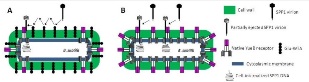
The early steps of SPP1 infection are well described. B. subtilis recognition by SPP1 virus particles is initiated by reversible interaction with poly(glycerolphosphate) wall teichoic acids (Glu-WTA’s) of the host cell wall (29). This step facilitates scanning of the cell surface for recognition of the cytoplasmic membrane receptor YueB (29). This receptor is essential for SPP1 irreversible binding and infection of B. subtilis (30) (Fig. 4-A). The extracellular domain of the YueB receptor is a dimer forming elongated fibers with an average length of 36.5nm, which are thought to be anchored to the cytoplasmic membrane (CM) via C-terminal transmembrane domains (TMDs). These fibers are long enough to cross the 30nm-thick peptidoglycan (PG) cell wall and thus to expose a receptor region for SPP1 recognition. (31). The SPP1 tail spike protein gp21 is responsible for recognition and binding to the YueB receptor (32). A result of this interaction gp21 suffers major conformational changes (33), triggering a signal that is transmitted through the tail helical structure to the phage head, causing its opening for DNA ejection (34). Remarkably, the purified receptor ectodomain YueB780 (31) or a smaller version corresponding to its central region (YueB365) (C. São-José, unpublished results) are able to bind and trigger SPP1 DNA ejection in vitro, thus leading to phage inactivation.
Bacteriophage SPP1 preferentially binds to B.subtilis cell poles (35). In mutant strains lacking Glu-WTA’s, irreversible adsorption is strongly affected (29). However, in a mutant without reversible adsorption, but over-expressing YueB, the irreversible adsorption rate is about the same as the wild-type (wt), showing that increasing YueB surface concentration bypasses the lack of reversible adsorption (Fig. 4-B) (29).
Fig. 4 – Mechanism of phage SPP1 adsorption and infection. In natural occurring infections SPP1 virions start by reversibly adsorbing to Glu-WTA. This reversible interaction allows SPP1 to “scan” the cell surface until it is irreversibly captured by receptor YueB, which preferentially accumulates at cell poles (A). In a Glu-WTA deficient cell, the irreversible adsorption rate can be about the same of wt by over-expressing YueB at cell surface, bypassing the reversible adsorption step (B).
SPP1 interaction with B.subtilis causes a very fast depolarization of the CM (1 minute post infection), depending on YueB surface concentration and SPP1 input (36). Ca2+ ions play an essential role at the first steps of infection. At submilimolar concentration it is enough to allow reversible adsorption, but it is required a milimolar concentration to DNA entry (36). Recent studies have shown that an intact proton motive force and a calcium gradient across the B. subtilis cytoplasmic membrane are absolutely required for SPP1 DNA entry into host cells (S. Fernandes et al., unpublished results)
SPP1 mediates generalized transduction (37, 38) and transduction of plasmid DNA. In the latter case SPP1 exclusively encapsidates concatemeric plasmid DNA with approximately the same length of the phage DNA (38). SPP1 DNA packaging is highly efficient, due to the presence of specific DNA segments (pac) determining packaging initiation (38), while plasmid-transducing particles are underrepresented. However, cloning SPP1 DNA fragments in plasmids leads to an increase of 100- to 1000-fold in the plasmid-transducing particles frequency (38, 39). This increment in the frequency of plasmid-transducing particles is known as facilitation of plasmid transduction (39). Following phage infection, plasmid DNA is replicated by a rolling-circle mechanism, producing concatemeric plasmid DNA large enough to be recognized for phage packaging (40).
I.4 - The concept of PhageDuction: SPP1-mediated heterologous transduction
This work aims exploring the system phage SPP1/receptor YueB in the design of a new technology for cell-targeted DNA delivery. This SPP1-based technology follows an innovative approach because the phage tropism will be artificially changed so that it can deliver its DNA
to different target cells. Since it will mimic gene transduction based on animal virus-derived vectors, we have designated this technology as PhageDuction. The major novelty of this technology is that conceptually it will be quasi universal, that is, by introducing minor modifications, PhageDuction should work with any bacterial or animal cell, because the only requirements for SPP1 infection are the presence of a YueB ectodomain at the target cell surface, an energized CM and a Ca2+ gradient between inner and outer cellular milieu. Thus, in principle the key step of this strategy will be the efficient and functional decoration of target cells with the receptor ectodomain.
At first glance one might think that PhageDuction could be developed with any phage system. However, this might not be so straightforward because SPP1 is the only currently available phage whose DNA ejection can be triggered with soluble forms of the protein receptor, in this case YueB. Another advantage of SPP1 is that its host, B. subtilis, can be easily transfected with SPP1 DNA. Thus, any recombinant SPP1 DNA, for example carrying heterologous DNA cargo, can be inexpensively amplified simply by transfecting B. subtilis.
I.5 – Thesis goals and strategy
To prove the feasibility of PhageDuction, the first checkpoint is to demonstrate that phage SPP1 will be able to deliver its DNA to yueB- B. subtilis cells, after specific decoration of their surface with a receptor ectodomain. If succeeded, the next goal will be to extend the results to other Bacillus species and other bacterial genera.
For targeting YueB to the surface of yueB- cells, it will be created a fusion protein between a YueB ectodomain and a ligand with affinity to the B. subtilis cell envelope. Since B.
subtilis is a Gram-positive bacterium exhibiting a thick PG-exposed cell wall, we have decided
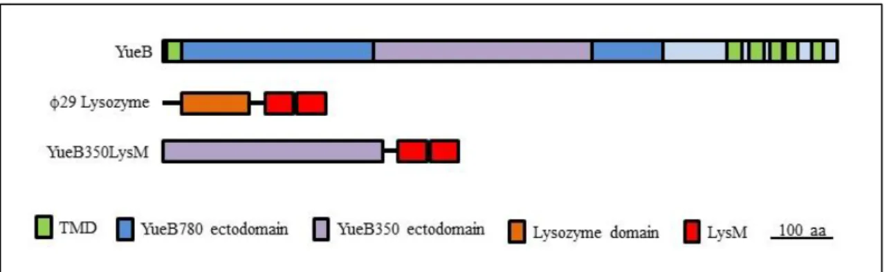
to use a ligand that targets the PG. LysM (lysin motifs) are highly conserved C-terminal repeats found in the lysozyme of B. subtilis phage φ29 (41). LysM affinity to Gram-positive bacteria cell wall has been studied (42, 43) and used previously as a ligand for targeting cell surface (44, 45, 46). Therefore, to decorate the surface of cells lacking YueB with an active receptor we will produce a fusion between an ectodomain based on YueB365 (see above) and the C-terminus of the φ29 lysozyme, which harbors two LysM motifs (45). This fusion will be named YueB350LysM. The efficiency of SPP1 DNA delivery to these YueB350LysM-decorated cells will be measured by performing transduction experiments with SPP1 transducing particles carrying plasmids conferring antibiotic resistance.
II – Results
II.1 – Production and purification of the receptor-ligand fusion YueB350LysM
In order to produce the fusion YueB350LysM, two gene fragments were amplified and fused by overlap extension-polymerase chain reaction (OE-PCR): the 5’-end segment included the coding sequence for a 350 amino acid-length central region of the YueB ectodomain; the 3’-end fragment encoded the linker region and the two LysM motifs of the B. subtilis phage
29 endolysin (Fig. 5).
Fig. 5 – YueB350LysM primary structure representation. YueB350LysM results from the fusion of the 350 amino acid-length central region of the YueB ectodomain (purple) to two LysM motifs from 29 endolysin (red), including a linker region.
The fusion gene was then cloned in the plasmid vector pIVEX2.3d, which allowed the expression of YueB350LysM C-terminally fused to a hexahistidine (His6) tag, the latter used for protein purification. The entire polypeptide has 481 amino acids (aa), with a theoretical molecular weight (MW) of 53.74 KDa and a pI of 4.64. Expression was conducted in Escherichia
coli, using a thermoinducible expression system (47).
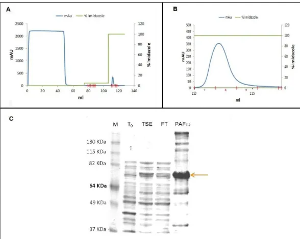
YueB350LysM was subjected to affinity chromatography purification in an ÄKTAprime™ plus system (Fig. 6). YueB350LysM presented a retarded mobility in sodium dodecyl sulfate-polyacrylamide gel electrophoresis (SDS-PAGE), migrating between the 64KDa and 82KDa bands of the marker (Fig. 6-C). An identical behavior has been observed previously for the larger ectodomain YueB780 (31) and for ectodomain YueB365, which basically corresponds to the YueB350 moiety C-terminally fused to a hexahistidine tag (see Appendix 1). This abnormal
Fractions 7 to 9 from the affinity step containing partially purified YueB350LysM were pooled and further purified by size exclusion (gel filtration) chromatography, allowing at the same time the elimination of the imidazole used in the affinity step.
Fig. 6 – YueB350LysM purification by affinity chromatography. (A) Chromatogram of the purification procedure where eluted material from the column was continuously monitored by taking absorbance measurements at 280 nm (mAU). The first broad peak corresponds to total proteins from the E. coli extract that did not bind to the affinity matrix, while the second, smaller peak corresponds to the fractions containing YueB350LysM (zoomed in (B)), after eluting matrix-bound material with 500 mM Imidazole (100% elution buffer). (C) SDS-PAGE analysis of YueB350LysM purification. M, molecular weight marker; T0, total protein extract produced before induction of protein expression; TSE, total soluble extract produced after induction and loaded in the affinity column; FT, flowthrough of the affinity column; PAF7-9, Pool of fractions 7 to 9 of the affinity peak, after a 2-fold concentration. Arrow points to YueB350LysM.
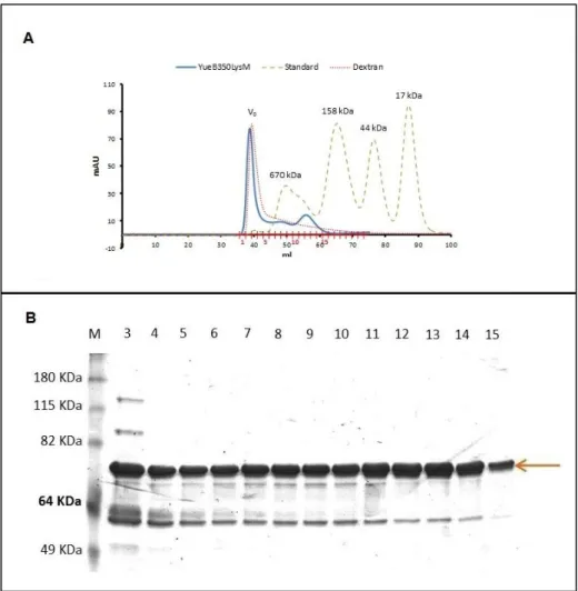
YueB350LysM eluted from the gel filtration column in one sharp peak (fractions 1 to 4, Fig. 7A) followed by two smaller, flattened and partially overlapping peaks (fractions 5 to 9 and 10 to 14, Fig. 7A). Note that YueB350LysM species eluted with apparent masses much higher than the one deduced from its primary sequence. This most probably results from
YueB350LysM adopting a fiber-like shape, as observed for YueB780 (31). SDS-PAGE analysis indicated a very similar composition for all fractions in terms of protein bands (Fig. 7B). Essentially, each fraction was composed of a major band expected for the YueB350LysM protein and a smaller band migrating between the 64 and 49kDa bands of the protein ladder. This smaller protein most probably corresponds to a major proteolysis product that remains associated to YueB350LysM during gel filtration, as observed before for YueB780 (31, see Appendix 1). The elution volume of the first peak basically corresponded to the void volume (V0) of the column, which has been determined by running in the same conditions the high
MW polymer dextran (Fig. 7-A). Thus, we have considered that fractions 1 to 4 essentially contained large aggregates of YueB350LysM.
Fig. 7 – YueB350LysM purification by size exclusion chromatography. (A) Chromatogram of the gel filtration separation of the YueB350LysM fractions resulting from the affinity step (see above). Elution of YueB350LysM was followed by absorbance measurements at 280 nm (mAU). The curves of dextran (gives the column void volume (V0)) and of a protein standard are superimposed to that of YueB350LysM. The protein standard was composed of
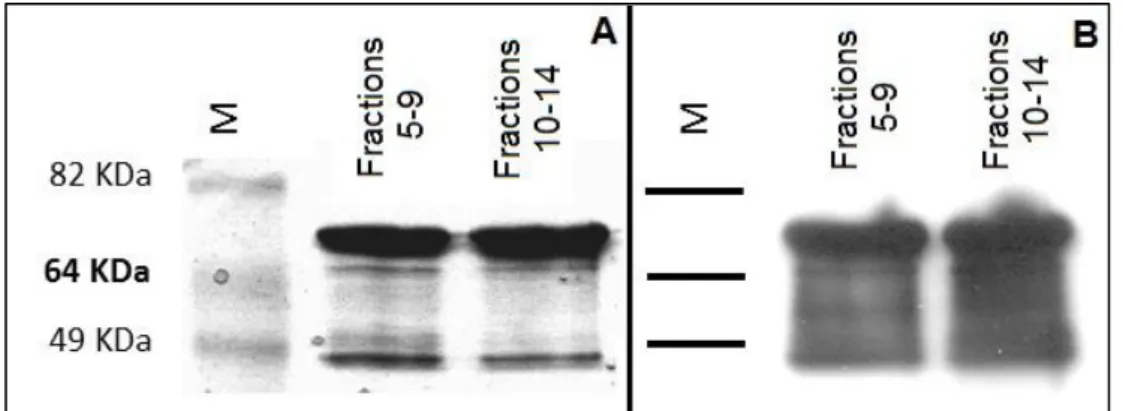
The fractions composing the other two peaks were pooled (pools 5 to 9 and 10 to14) and subjected to SDS-PAGE and western-blot analysis with α-YueB780 antibodies (31) (Fig. 8). The results confirmed that both protein pools were basically composed of purified YueB350LysM and its major truncated product, being contaminant E. coli proteins negligible. Both protein pools were concentrated 4-fold resulting in protein preparations of 1.2mg/ml (Pool5-9) and 1.4mg/ml (Pool10-14).
Fig. 8 – Analysis of the concentrated pools of fractions 5 to 9 and 10 to 14 resulting from the size exclusion chromatography step of Fig. 7. (A) Coomassie blue-strained gel after loading 5 µg of each protein preparation. An identical gel was subjected to Western blot analysis with α-YueB antibodies. M, molecular weight marker.
II.2 – YueB350LysM triggers SPP1 DNA ejection in vitro
The ability of YueB350LysM to trigger SPP1 DNA ejection was a fundamental requirement to the success of our strategy. To confirm this we have performed DNase protection assays and SPP1 inactivation experiments analogous to those reported by São-José
et al. (31) (see methods). Briefly, a CsCl-purified SPP1 lysate was incubated with increasing
concentrations of YueB350LysM from pools 5-9 or 10-14 (see above) in presence of DNase. In this type of experiments non-ejected DNA remains protected inside the phage head from DNase attack and can be directly visualized in agarose gels after deproteinization of the mixtures. In contrast, if the DNA is ejected to the medium it will be completely digested by the nuclease, leading to the disappearing of the phage DNA bands in the same agarose gels. In these assays half of the SPP1/YueB350LysM mixtures were also used to enumerate phages by plating, i.e., to measure the degree of phage inactivation after incubation with the receptor
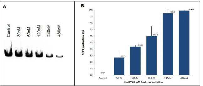
fusion. Although the protein preparation from pool10-14 was able to trigger SPP1 DNA ejection (not shown), YueB350LysM from pool5-9 revealed superior receptor activity in the DNase protection (Fig. 9-A) and in the inactivation (Fig. 9-B) assays. In vitro, 240nM of YueB350LysM were sufficient to trigger phage DNA ejection, and thus inactivate, more than 90% of the initial phage suspension (Fig. 9-B).
Fig. 9 – Trigger of SPP1 DNA ejection and phage inactivation by YueB350LysM. (A) Agarose gel electrophoresis showing the results of the DNase protection assay. SPP1 DNA ejection increases proportionally to the YueB350LysM concentrations. Only protein buffer was added to the control lane. Final concentrations of YueB350LysM (nM) are indicated above each lane. (B) SPP1 inactivation effect of the different YueB350LysM concentrations tested. Phage inactivation progressively increases with YueB350LysM concentration. Inactivation results are expressed as percentage of the phage input. Black bars indicate standard deviation errors of at least three independent experiments.
II.3 – YueB350LysM binds to target cell surface
After confirming that YueB350LysM maintains efficient receptor activity, we then tested its ability to bind to the surface of the B. subtilis strain CSJ1, a strain unable to produce the receptor YueB due to an extensive deletion of the corresponding gene (30). Most of the procedures described in literature to study the binding of LysM-carrying proteins to the bacterial cell envelope require pre-treatments that kill the target cells (42, 44, 46), something that was undesirable given our goals. Thus, we have developed a new methodology to evaluate YueB350LysM binding activity, without endangering cell viability (see methods for
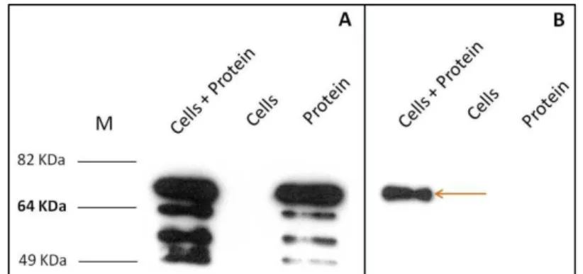
LB medium, followed by the addition of YueB350LysM at a final concentration of 200nM. After 15 min incubation at 37 ºC the mixtures were centrifuged, the cellular pellet washed and the amount of YueB350LysM present in the pellet and supernatant fractions evaluated by western blot (Fig. 10).
Fig. 10 – YueB350LysM binding to B. subtilis strain CSJ1 (yueB-). YueB350LysM polypeptides present in supernatant (A) and cell pellet (B) fractions were qualitatively estimated by western blot analysis with α-YueB780 antibodies. The equivalent of 100ng of YueB350LysM was loaded per lane. Lanes “cells” and “protein” correspond to controls with only added cells or YueB350LysM, respectively. The arrow points to the full-length YueB350LysM.
The results have indicated that in these experimental conditions a fraction of the YueB350LysM input was able to bind to CSJ1 cells in LB culture medium.
To evaluate the real contribution of the LysM motifs on the binding properties of YueB350LysM, we have performed an identical binding assay with a purified ectodomain (YueB365, see Appendix 1), which essentially corresponded to the YueB350 moiety directly fused to a hexahistidine tag. Unfortunately, the results were inconclusive as the preparation of YueB365 used seemed to precipitate in the binding assay conditions (see Appendix 2). However, the importance of the LysM motifs in the receptor fusion was evidenced in the experiments described next.
II.4 – YueB350LysM-decorated cells allow productive SPP1 infection
The next step was to study if SPP1 was able to deliver its DNA to YueB350LysM-decorated CSJ1 cells and to replicate. After targeting the receptor fusion to CSJ1 cell surface, SPP1 was added at a multiplicity of infection (moi) of 0.1. As YueB365 seemed to associate, at least not specifically, to CSJ1 cells (see above), it was also used in these experiments. B. subtilis L16601, the wild-type host strain routinely used for SPP1 propagation was used as control for
SPP1 entry and replication. After an incubation of 15 minutes that ensured phage irreversible adsorption to L16601 (29), infection mixtures were diluted 10-fold in fresh LB. A sample was immediately used for titration of free phages, to evaluate phage adsorption, showing that SPP1 was inactivated by YueB350LysM-decorated cells (Fig. 11). Since this inactivation was much greater than that observed with strain L16601, it is likely that CSJ1-decorated cells exhibited a significantly higher concentration of receptor at their surface when compared to the native levels found in the wild-type strain. The mixtures were incubated for additional 70 minutes, a period sufficient for replication and release of virion progeny from infected L16601, and free phages scored again. The results revealed that SPP1 replication occurred in CSJ1 cells decorated with YueB350LysM, although less efficiently than in the wild-type strain (Fig. 11). It is thus likely that a considerable fraction of the phages irreversibly adsorbed to receptor-decorated CSJ1 cells did not culminate in productive infection (see discussion).
Fig. 11 – SPP1 infection of CSJ1 cells decorated with YueB350LysM. After 15 and 70 minutes (min) of infection, free virus particles were collected and titrated (PFU/ml) to evaluate phage adsorption and replication, respectively. Results show that SPP1 is able to adsorb and replicate in CSJ1 decorated with YueB350LysM. Black bars indicate standard deviation errors of at least three independent experiments.
Interestingly, although YueB365 was able to associate to CSJ1 cells (see above), it did not allow either significant SPP1 adsorption or supported SPP1 replication (Fig. 11). Thus, LysM motifs seem to be critical for binding of functional receptor to the cell surface.
II.5 – SPP1 mediates plasmid transduction to YueB350LysM-decorated cells
The ability of SPP1 to infect and replicate in CSJ1 cells decorated with YueB350LysM was a very encouraging result, which gave support to the hypothesis of SPP1 being able to deliver its DNA to other cells through PhageDuction. However, we needed an expeditious and robust method to evaluate the capacity of SPP1 to deliver its DNA cargo to cells not supporting SPP1 replication. This could be particularly important in conditions of relatively low PhageDuction efficiencies, since direct measurements of intracellular SPP1 DNA could be very difficult to perform due to the high amount of contaminating DNA outside cells (DNA from intact virions and/or from virus particles that ejected their DNA to the medium).
As already mentioned, SPP1 is naturally able to mediate plasmid transduction between
B. subtilis strains susceptible to SPP1 infection. If the plasmid to be transduced presents
homology to SPP1 DNA, the transduction efficiency is 100 to 1000-fold higher (38, 39). Thus, the ability of SPP1 to mediate the transfer of a plasmid carrying a selective mark could constitute a general method to study PhageDuction efficiency. To test this idea we produced a pBT163-transducing SPP1 lysate, after infecting a B. subtilis strain carrying this plasmid (see methods). Plasmid pBT163 carried a 2.6 kb SPP1 DNA fragment containing the pac packaging site to facilitate the production of transducing particles and genes conferring resistance to chloramphenicol (Cm) and erythromycin (Ery) (48). The latter marks provided a simple method to assess entry of SPP1 DNA cargo and its expression. The transducing lysate was processed to eliminate contaminant cells and DNA (see methods).
As in the previous experiments, CSJ1 cells were decorated with YueB350LysM or YueB365. The indicator strain L16601 was used as control for pBT163 delivery and expression. After decoration, cells were infected with the pBT163-transducing lysate at a moi of 1 and incubated for 10 minutes at 37ºC with agitation. Infection mixtures were then treated with α-SPP1 serum 1834 (see Appendix 3) in order to inactivate free α-SPP1. After washing with fresh medium to remove any residual α-SPP1, cell dilutions were plated both in solid medium to enumerate colony forming units and in the same medium supplemented with Cm for transductants counting (Fig. 12). The number of transductants per surviving cell and per PFU of the lysate is shown in Table 1.
Fig. 12 – SPP1 mediated transduction of plasmid pBT163 to CSJ1 cells decorated with YueB350LysM. Infected cells were plated both in LA medium for survivors counting (blue) and LA supplemented with Cm for transductants enumeration (red). CSJ1 decorated with YueB350LysM was transduced with efficiency close to that observed in the control strain L16601. No transductants were observed with CSJ1 cells incubated with protein buffer (CSJ1 buffer) or with YueB365 (CSJ1 YueB365). Black bars indicate standard deviation errors of at least three independent experiments.
Table 1 – Efficiency of SPP1-mediated pBT163 transduction to CSJ1 cells decorated with YueB350LysM.
Transductants/ Survivor Transductants/ PFU L16601 8.8 x 10⁻³ 3.8 x 10⁻³ CSJ1 buffer 0 0 CSJ1 YueB365 0 0 CSJ1 YueB350LysM 1.6 x 10⁻³ 7.8 x 10⁻⁴
Remarkably, SPP1 could transduce CSJ1 decorated with YueB350LysM with efficiency near to that observed in the control strain L16601. As expected, SPP1 was not able to deliver pBT163 to YueB365-decorated CSJ1 cells, reinforcing that LysM motifs are essential for correct and functional cell surface targeting. CSJ1 decorated with YueB365 and non-decorated CSJ1 exhibited more survivors than L16601 and CSJ1 decorated with YueB350LysM. This is expected since the majority of SPP1 particles in the transducing lysate are “normal” infective phages leading to cell lysis.
adding the ectodomain YueB350 to the surface of the target cell. To reinforce this, Bacillus
licheniformis MW3 cells were decorated with YueB350LysM and pBT163 transduction
mediated by SPP1 studied as above (Fig. 13).
Fig. 13 - SPP1-mediated transduction of plasmid pBT163 (Cmr) to B. licheniformis strain MW3. Surviving cells (blue) and transductants (red) were enumerated as in Fig. 12. Despite with lower efficiency, SPP1 is able to mediate pBT163 transduction to strain MW3.
Even though transduction to MW3 was significantly less efficient than to L16601 (Tab. 2), SPP1 successfully delivered DNA to MW3-YueB350LysM decorated cells.
Table 2 – Efficiency of SPP1 mediated pBT163 transduction to MW3.
Transductants/ Survivor Transductants/ PFU L16601 6.0 x 10⁻³ 2.0 x 10⁻³ MW3 buffer 0 0 MW3 YueB350LysM 3.7 x 10⁻⁶ 4.4 x 10⁻⁶
For further confirmation that pBT163 was transduced, MW3 Cmr colonies were replica plated to medium supplemented with Ery. All of the 50 tested colonies were able to grow in presence of Cm and Ery, making unlikely that they corresponded to spontaneous resistant
mutants. This result strengthened the notion that SPP1 can target and deliver DNA to non-host cells, if YueB ectodomains are anchored to target cells by specific ligands.
II.6 –Transduction of bacterial chromosomal genes by PhageDuction
After successful demonstration of plasmid delivery to SPP1-resistant cells by PhageDuction, we wondered if the same strategy would be efficient enough to allow transfer of B. subtilis chromosomal marks. B. subtilis lab strains are relatively easy to manipulate, for instance to generate gene mutations. However, other SPP1-resistant B. subtilis strains, particularly undomesticated natural isolates, or related species, like B. licheniformis are very refractory to genetic manipulation by standard methods. One way to overcome this problem could be creating mutations or other genetic changes in B. subtilis lab strains and then transfer them to these strains through PhageDuction. To test this idea, an SPP1 lysate was produced in
B. subtillis 1A786 which has a spectinomycin (Spec) resistance mark in locus lacA. This locus
presents sufficient homology between B. subtilis and B. licheniformis to allow recombination (our analysis). The transducing lysate was used to infect MW3 cells decorated with YueB350LysM as described above. Since in this case we had no facilitated transduction we were expecting to observe very low transduction efficiencies, something that was confirmed by the results (Fig. 14, Tab.3).
Fig. 14 – SPP1-mediated transduction of lacA::specr to MW3. Surviving cells (blue) and transductants (red) were enumerated as in Fig. 12, except that here selection was for resistance to spectinomycin. Although with very
Table 3 – Efficiency of SPP1 mediated lacA::specr transduction to MW3. Transductants/ Survivor Transductants/ PFU L16601 1.7 x 10⁻⁶ 4.0 x 10⁻⁷ MW3 buffer 0 0 MW3 YueB350LysM 3.2 x 10⁻⁸ 5.0 x 10⁻⁸
At this low efficiency the procedure to evaluate DNA delivery is less reliable. To achieve better reliability it is necessary further testing. Nevertheless, the results obtained showed mobilization of B. subtilis chromosomal DNA to SPP1-resistant cells by PhageDuction.
III – Discussion
PhageDuction concept was built on the possibility of phage SPP1 being able to deliver its DNA cargo to phage resistant host cells, to heterologous bacterial cells or even to mammalian cells, as long as their surface is specifically decorated with a functional YueB receptor ectodomain. As a simple approach to prove this concept, we aimed to demonstrate that SPP1 could infect a yueB B. subtilis mutant, that is, a phage resistant host, after binding to its surface a YueB-LysM receptor fusion. This was fully demonstrated in this work as we have shown the effectiveness of PhageDuction in the delivery of SPP1 DNA, plasmids and even bacterial genome fragments. Although less efficiently, PhageDuction also worked when tested in another, although related, bacterial species, in this case B. licheniformis.
The receptor fusion used to make target cells sensitive to SPP1 DNA entry was YueB350LysM. The LysM motifs were responsible for the specific targeting of the receptor fusion to the cell wall of target bacteria, whereas region YueB350 provided the SPP1 receptor activity. Before this work it was already known that the active receptor ectodomain YueB780 (31, see Fig. 5) could be shortened at both N and C-terminal ends without compromising receptor activity (C. São-José, unpublished). Such truncation of YueB780 give rise to a central YueB segment, YueB365 (C. São-José, unpublished), which basically corresponds to the YueB350 moiety used in the receptor fusion. We have envisaged that an ectodomain shorter than YueB780 in the fusion would bring SPP1 to a closest proximity of the target cell surface, thus increasing the chances of good phage DNA routing to the cell cytoplasm.
Purification of YueB350LysM required an affinity step followed by size exclusion chromatography, much like what has been reported to YueB780 (31). However, YueB350LysM seemed to be more prone to aggregation as judge by the large fraction of protein that eluted close to the column void volume (Fig. 7-A). Apart from these large aggregates, YueB350LysM eluted from the size exclusion column as two broadened, partially overlapping peaks, which might reflect the presence of at least two molecular species exhibiting different oligomeric and/or conformational states. The nature of the protein species composing these two peaks was not studied in detail and we have just selected the fraction of YueB350LysM that showed better receptor activity in the in vitro tests.
It has been proposed that binding of LysM to B. subtilis cells from the external media is hindered by the cell envelope lipoteichoic acids (LTA), being required pre-treatments for example with trichloroacetic acid (TCA) to remove these anionic polymers (42, 44, 46).
spots in cell sides (42), although it binds preferentially to cell poles (44) in these conditions. As PhageDuction intends to deliver DNA to living cells and TCA treatment compromises cell viability, YueB350LysM binding procedure could not include any LTA removal step. Temperature, cell density, binding medium and pH were all important parameters considered in the optimization of YueB350LysM cell binding. Binding assays were performed at 0ºC and 37ºC, showing no relevant disparity (data not shown). Temperature was then set to 37ºC, since this is the optimal temperature for B. subtilis growth. Two cell densities were considered, OD600=0.8 (1 x 10⁸ CFU/ml) and OD600=8 (1 x 10⁹ CFU/ml). Though both densities exhibited qualitatively successful binding, OD600=8 had stronger signal (data not shown) and was better suited for the downstream manipulations. Initially, binding assays were conducted in protein buffer (Hepes, NaCl) supplemented with CaCl2 and MgCl2 and in SPP1 buffer (TBT) with the same supplements. However, the results were not always reproducible and, most importantly, SPP1 seemed to show reduced adsorption to cells after washing with these buffers, even when using the wild-type host strain L16601 (data not shown). As binding of YueB350LysM to B.
subtilis cells in LB medium (pH 6.6) seemed to be qualitatively acceptable for our purposes, we
decided to proceed to the next steps without further adjustments.
The ability of the LysM-lacking, control protein YueB365 to bind the B. subtilis cell surface was surprising. YueB350LysM and YueB365 binding results were qualitatively similar, although YueB365 binding might have been overestimated due to some protein precipitation. We should note also that the method used to evaluate binding (Western blot analysis of pellet and supernatant fractions after cell/protein contact), does not allow to ascertain binding specificity. In fact, YueB365-decorated CSJ1 cells did not allow either SPP1 productive infection or plasmid transduction, hinting for unspecific and/or misplaced binding of YueB365 on the cell surface. We believe that LysM motifs are necessary not only to confer affinity to the cell wall but also to provide the suitable localization and orientation for exposing the YueB350 ectodomain. It is worth highlighting in this regard that either exogenously added (see above) or cell endogenous LysM-containing proteins (49, 50) may preferentially locate at septa and cell poles, which is precisely the subcellular region where native YueB accumulates and the site preferred by SPP1 to enter B. subtilis cells (35).
It was observed before that repeated centrifugation and suspension of B. subtilis cells in fresh LB also affects SPP1 adsorption (S. Fernandes, unpublished results). Therefore, to minimize this effect cells were suspended in their filtrated culture supernatant and centrifugations were restricted to the strictly necessary to eliminate residual YueB350LysM. Even so, the cell decoration procedure demanded relevant cell manipulation, which may explain the observed low SPP1 irreversible adsorption to the wild-type control strain L16601,
when compared to previous studies (30, 29). In clear contrast, SPP1 adsorption to YueB350LysM-decorated CSJ1 was markedly higher than to L16601. This high irreversible adsorption resembles that observed with B. subtilis strains over-expressing YueB (30), which could suggest a high abundance of YueB350LysM at CSJ1 cells surface. However, SPP1 replication in L16601 cells is much higher than in CSJ1-decorated cells, suggesting that a significant fraction of viral particles adsorbing to CSJ with bound YueB350LysM do not proceed to productive infection.
Contrarily to SPP1 replication, the efficiency of SPP1-mediated plasmid PhageDuction to decorated CSJ1 was similar to conventional transduction to L16601, again reflecting that the high SPP1 inactivation rates produced by CSJ1-decorated cells do not translate in high, infection-productive irreversible adsorption. It is likely that a fraction of irreversible bound SPP1 cannot proceed to correct DNA routing, given the clear preference of the phage to deliver its DNA at septal and polar cell regions (35). Other hypothesis is that there was residual YueB350LysM non-associated and/or incorrectly associated to cells which inactivates SPP1 particles.
SPP1-mediated plasmid transduction was an expeditious method to follow SPP1 DNA delivery to decorated target cells. The results obtained for both L16601 and decorated CSJ1 are in agreement to those predicted for facilitated transduction (38, 39). These were very promising results, since CSJ1 became highly susceptible to SPP1-mediated transduction just by decorating its surface with YueB350 ectodomain. This was sufficient even to allow PhageDuction of a lacA::spec DNA segment between B. subtilis and B. licheniformis. In addition, CSJ1-decorated cells allowed productive SPP1 infection, strongly supporting that PhageDuction application is attainable.
Despite with lower efficiency, SPP1 could PhageDuct plasmid pBT163 to B. licheniformis MW3 decorated with YueB350LysM, reinforcing the feasibility of this new technology. Attempts to PhageDuct pBT163 to decorated B. amyloliquefaciens and B. megaterium cells were apparently unsuccessful. However, these results are not clear cut since we have confirmed neither the binding of YueB350LysM nor the plasmid capacity to replicate in these bacterial species. We have also failed in a single attempt to demonstrate SPP1-mediated PhageDuction of the known shuttle plasmid p1948 to Staphylococcus aureus strain RN4220, when using also YueB350LysM as receptor decorating fusion. Again, the ability of YueB350LysM to bind RN4220 was not studied, but these results may hint for the need of fusing to YueB350 cell species specific ligands.
MS2 (53) and T4 (20), the viral particle enters the target cell, acting like a “nanobioparticle” that just carries and protects the nucleic acid cargo (51), being the fate of this cargo completely dependent on cell post-uptake events. The PhageDuction system relies on the natural SPP1 DNA ejection mechanisms to actively deliver (by “shooting”) naked DNA into the YueB-decorated target cell, being independent of cellular events. Other difference is that in the above mentioned systems, cell targeting is based in the attachment of cell specific ligands to phage capsids, which may influence viral particle properties like hydrophobicity (52), while in PhageDuction, SPP1 is targeted to the interest cell just by decorating it with a fusion between a cell specific ligand and a YueB ectodomain.
IV – Concluding Remarks
With this work it was demonstrated that decorating the surface of SPP1-resistant cells with a YueB ectodomain fused to a cell-specific ligand is sufficient to make these cells susceptible to SPP1-mediated DNA delivery, accomplishing the proposed main goal. The results obtained with a YueB deletion mutant of a SPP1 natural host, as well as with another bacterial species, deliver tangible evidence that the concept of PhageDuction is feasible. Additionally, yet waiting for further studies, it was presented the first application of PhageDuction technology, with SPP1-mediated transduction of heterologous DNA between two Bacillus spp.
This work corresponded to the first step in the development of a new DNA delivery system. As a future perspective, to assure that PhageDuction applicability is extensible to all physiological active cells as theoretically expected, is necessary to perform a similar approach in a eukaryotic cell line, producing a fusion protein that targets the cell surface.
The main goal of proving the concept of PhageDuction was favored in detriment of developing engineered phages for general PhageDuction studies. However, in the future it cannot be neglected the engineering of SPP1 particles with genes whose expression can be monitored (for instance β-galactosidase gene bgaB, whose expression may be evaluated by measuring enzymatic activity) to provide prompt yet reliable tools to measure PhageDuction effectiveness.
Though the concept of the perfect delivery vector is merely utopian, PhageDuction system is a step towards the technology improvements required to permit suitable application in biotechnological and biomedical areas, as it bypasses several currently available vectors weaknesses, unveiling a novel and valid alternative for exploration in gene delivery.
V – Materials and Methods
V.1 – Bacteria, phages, plasmids and growth conditions
Bacterial strains, phages and plasmids used in this work are listed in Table 4. Bacterial strains were grown in LB medium (54) at 37ºC with aeration, except E. coli BL21 pGP1-2, which was grown at 28ºC. Strains carrying vectors or recombinant plasmids were selected in supplemented medium with ampicillin (Amp) and/or kanamycin (Kan), at final concentrations of 100µg/ml and 30µg/ml, respectively, Erythromycin (Ery) at 0.5µg/ml or 10µg/ml to B.
subtilis and B. licheniformis, Spectinomycin (Spec) at 200µg/ml or 300µg/ml to B. subtilis and B. licheniformis, and Chloramphenicol (Cm) at 5µg/ml or 50µg/ml to B. subtilis and B. licheniformis. Culture media were acquired from Biokar Diagnostics. Agar was added to LB
medium to a final concentration of 0.7 or 1.5% (wt/vol) in order to obtain top or bottom LB plates, respectively. The medium was supplemented with 10 mM CaCl2 for SPP1 propagation and titration (30). SPP1 suspensions were maintained and diluted in TBT buffer (100 mM Tris-HCl, 100 mM NaCl, 10 mM MgSO4; pH 7.5).
Table 4 – Bacterial strains, phages and plasmids used in this work. Bacterial strains,
phages or plasmids
Genotype and/or relevant features Reference
E. coli
MRF’ Cloning strain Stratagene
BL21 pGP1-2 Expression strain 55, 47
B. subtilis
L16601 B. subtilis 168; SPP1 indicator strain 56
CSJ1 L16601 derivative; ΔyueB; SPP1 resistant 30
YB886 B. subtilis 168 derivative free of prophages PBSX
and SPβ; SPP1 indicator strain
57
SP81 YB886 derivative containing pBT163 48
1A786 B. subtilis 168; lacA::spec BGSC1 1A786
B. licheniformis
MW3 Derivative of B. licheniformis DSM13; ΔhsdR1,
ΔhsdR2
58
Phages
SPP1 B. subtilis lytic phage 27
Plasmids
pIVEX2.3d Expression vector; T7 promoter-driven expression of C-terminally, His6-tagged fusions; Ampr
Roche Applied Science pGP1-2 Thermoinducible T7 RNA polymerase production; 55, 47
pBT163 carries a 2.6 kb SPP1 DNA fragment containing
pac; Cmr and Eryr; replicative in B. subtilis
48
pNS1 pIVEX2.3d derivative expressing YueB350LysM This work 1
BGSC – Bacillus Genetic Stock Center (http://www.bgsc.org/)
V.2 – General techniques of molecular biology and biochemistry
V.2.1 – PCR amplification and cloning
General cloning strategies used in this work were performed according to standard procedures (54). DNA amplification by PCR was conducted in thermocycler Biometra T1, with reaction conditions based on oligonucleotides (primers) melting temperatures, size of the product to be amplified and manufacturer’s indications for each DNA polymerase used.
Amplified products were purified with the kit High Pure PCR Product Purification (Roche Applied Science) and cut with endonucleases specific for primer-added restriction sites. After cutting with adequate restriction enzymes, these were heat-inactivated and cut DNA purified with dialysis discs with 0.025µm pores (Millipore). Plasmid vectors were extracted with PureLink™ Quick Plasmid Miniprep Kit (Invitrogen). Digestion products were ligated by enzyme T4 DNA ligase (New England Biolabs).
V.2.2 – Transformation, plasmid analysis and DNA sequencing
Recombinant plasmids were initially isolated in cloning strain MRF’, according to the transformation procedure described by Chung et al. (59). Transformants were in a first step screened for the presence of the desired construct by PCR using NZYTaq Green Master Mix (NZYTech) and then plasmids from positive clones prepared by the alkaline extraction method (60). After confirming the pretended plasmid structure by endonuclease restriction, recombinant plasmids used for sequencing were extracted with PureLink™ Quick Plasmid Miniprep Kit (Invitrogen). Constructions used for protein expression were previously confirmed by automatic DNA sequencing (Macrogen). For protein production appropriate plasmids were transferred to the expression strain BL21 pGP1-2.
Plasmid DNA integrity, enzymatic restrictions, DNA extractions and purifications were confirmed by agarose gel electrophoresis (0.7% agarose) with GelRed™ Nucleic Acid Gel Stain (Biotium) (0.5µg/ml). The electrophoresis buffer used was Tris-Borate-EDTA (TBE) 0.5x (45mM Tris, 45mM boric acid, 1mM EDTA, pH 8) and the molecular weight marker was the 1kb Plus
DNA Ladder (Invitrogen). Gels were photographed with a camera coupled to software (Canon Digital Camera Solution 30.2), under ultraviolet light exposition.
V.2.3 – SDS-PAGE and Western Blot analysis
Sodium Dodecil Sulfate Polyacrylamide Gel Electrophoresis (SDS-PAGE) analysis was with
resolution gels at 11%, produced from Protogel (National Diagnostic) concentrated solutions and supplemented with 5% sucrose for increased malleability after polymerization. Protein samples were supplemented with SDS sample buffer and heated 10 minutes at 100ºC before gel loading. The molecular size marker used was BenchMark™ Pre-stained Protein Ladder (Invitrogen). Electrophoresis was conducted in the Mini-Protean Cell (Bio-Rad) system. After electrophoresis, gels were immersed in a staining Bio-Safe Coomassie blue (Bio-Rad) solution for 45 minutes with gentle shaking at room temperature. Gels were then immersed several times in distilled water for washing until protein bands were visible.
For immunodetection by Western Blot, proteins were transferred from the SDS-PAGE gels to 0.45µm nitrocellulose membranes (Bio-Rad) for two hours at 100V using Mini Transblot Module (Bio-Rad) system. After the transfer, membranes were blocked overnight at 4ºC in a TBS (50mM Tris-Cl pH7.5, 150mM NaCl) blocking solution (TBS 1x, 1% Western Blocking Reagent Solution, Roche Applied Science). Membranes were washed with TBS and incubated 1h with α-His6 antibodies (Roche Applied Science) diluted 1:5000 in TBS with 1% Blocking reagent or with depleted α-YueB780 antibodies (31) diluted 1:30000 in TBS 0.5% Blocking reagent. YueB350LysM/α-His6 and YueB350LysM/α-YueB780 complexes were detected
according to the instructions of the Chemioluminescence Western Blotting Kit (Roche Applied Science) and using Amersham Hyperfilm ECL (GE Healthcare) films. Film developing was conducted in a dark room using D-19 Developer and Kodak Fixer (Carestream® Kodak ® Processing Chemicals, Sigma-Aldrich).
V.2.4 – Protein quantification
Total protein concentration in solution was quantified by Bradford method (61) following manufacturer’s instructions indicated on commercial kit Bio-Rad Protein Assay Standard II (Bio-Rad). Calibration curves were constructed using bovine gamma globulin (BGG) at final concentrations between 1 and 20µg/ml.