“E
FFECTS OF SYNTHETIC CANNABINOIDS IN
ENDOMETRIAL STROMAL CELLS
”
Renata Prata Fernandes
Master degree in Analytical, Clinical and Forensic Toxicology
Dissertation thesis for the Master Degree in Analytical, Clinical and Forensic Toxicology submitted to the Faculty of Pharmacy
Dissertação de candidatura ao Grau de Mestre em Toxicologia Analítica, Clínica e Forense apresentada à Faculdade de Farmácia
Supervisors:
Doutor Bruno Miguel Reis da Fonseca
Professora Doutora Natércia Aurora Almeida Teixeira
University of Porto August 2016
III
IS AUTHORIZED THE INTEGRAL REPRODUCTION OF THIS THESIS FOR RESEARCH PURPOSES BY MEANS OF A WRITTEN DECLARATION FOR PERMISSION OF USE.
V
Alguns agradecimentos são devidos a todos os que me ajudaram a chegar ao final desta etapa da minha vida:
Ao Doutor Bruno Fonseca, agradeço pela sua orientação e por me ter acompanhado durante quase dois anos. Obrigada pelo conhecimento transmitido, pela confiança e apoio, pela disponibilidade e paciência, e por me ter ensinado a trabalhar em Ciência. À Doutora Natércia Teixeira, por me ter recebido na família que é o laboratório de Bioquímica e pela sua orientação, quero agradecer por ter estado sempre presente e sempre com uma palavra de apoio, especialmente quando nem tudo correu bem, pois “não ter resultados já é um resultado”. À Doutora Georgina Correia-da-Silva, obrigada por todo o apoio, pelos conselhos e pelo interesse que sempre mostrou pelo meu trabalho.
À Doutora Cristina Amaral e à Doutora Marta Almada, quero agradecer por todas as dúvidas esclarecidas, pelas ideias e por todas as palavras de encorajamento que tanto me ajudaram.
À Doutora Susana Rocha e à Doutora Sandra Ribeiro, obrigada por toda a ajuda. Aos rapazes, João Maia e Tiago Augusto, agradeço por me terem ajudado mesmo quando não queriam e por me terem feito rir todos os dias durante este ano. Às minhas meninas, Fernanda Bernardo, Maria Miguel Castro, Sónia Rocha, e Mestres Lia Costa e Carina Brito, obrigada por todas as conversas de rapariga, pelos desabafos e apoio, pela ajuda que me deram, e por toda a paciência que tiveram para aturar a complicada que eu sou. Foram todos vocês que me ajudaram a manter a pouca sanidade que ainda me resta e que fizeram com que todos os dias fossem uma nova aventura.
À Ana Paula e à Dona Casimira, obrigada por toda a ajuda e por alegrarem o laboratório mesmo em dias de chuva.
Aos restantes professores e alunos do laboratório de Bioquímica, obrigada por me terem recebido e feito com que me sentisse em “casa”.
Às minhas colegas de mestrado e amigas, Maria João Martins e Mariana Soares, obrigada por estarem sempre presentes e prontas a estudar, e além disso, sempre com uma palavra amiga.
Às melhores biólogas de sempre, Sara Vieira, Francisca Diniz, Marta Teixeira e Ana João Mascarenhas, quero agradecer por existirem. Vocês são as melhores amigas e companheiras de aventuras que alguém poderia ter e conhecer-vos tornou a minha vida muito mais colorida. Ao Tiago Moreira, quero agradecer por todas as histórias e todos os risos em momentos inoportunos. Obrigada por todos os momentos partilhados nestes 5 anos e por todos os que ainda virão.
VI
interessados nos últimos capítulos da minha jornada. Obrigada por saber que sempre que precisar posso contar convosco.
Por fim, quero agradecer aos meus pais. Não há palavras para descrever o quanto estou agradecida por vocês serem como são, por me apoiarem sempre e sem hesitação, por terem sempre as palavras certas na hora certa, por me encorajarem a ser a melhor pessoa que posso ser, por me deixarem seguir os meus sonhos mesmo quando são irrealistas e, acima de tudo, por estarem ao meu lado mesmo quando não estou no meu melhor. Quero dedicar-vos este trabalho, pois é por vossa causa que cheguei aqui e é por vossa causa que não vou ficar por aqui.
VII
be divided into three categories: the phytocannabinoids, endocannabinoids and synthetic cannabinoids. Phytocannabinoids are present in the Cannabis plant, and the most studied compound is Δ9-tetrahydrocannabinol (Δ9-THC), though more than 60 natural cannabinoids have been described. Endocannabinoids were discovered in vivo and are involved in several physiological processes. Synthetic cannabinoids are molecules synthesized in laboratory with cannabimimmetic effects, primarily used for research purposes, but also as abuse drugs. Nowadays, the consumption of cannabis has increased worldwide, particularly between young adults, even though its possession and use is prohibited. These prohibitions lead to the emergence and increase in abuse of synthetic cannabinoids, known as “legal highs”, due to lack of legislation. Besides the fact that these substances have higher affinity to cannabinoid receptors than Δ9
-THC, not much is known about them. A successful pregnancy is the combination of a series of well-timed events and it was already shown that a tightly controlled endocannabinoid signalling is necessary. So, the consumption of exogenous substances may affect negatively these reproductive events. In this work, it was studied the influence of synthetic cannabinoids in endometrial stromal cells. These cells were treated for 24 and 48 hours with the synthetic cannabinoids THJ-2201, JWH-122, AB-FUBINACA, UR-144, 5F-PB-22 and WIN 55,212-2 (0.01-50 µM). Treatments with JWH-122, AB-FUBINACA, UR-144 and 5F-PB-22 caused an increase in mitochondrial activity, although no changes were seen in proliferation studies or in the ratio between mitochondrial DNA (mtDNA) and nuclear DNA (nDNA). Interestingly, WIN 55,212-2 caused a decrease in cell viability, which was accompanied by an increase in caspase -3/-7 activities, suggesting the involvement of apoptosis. Furthermore, WIN 55,212-2 induced changes in cell morphology, like chromatin condensation as observed by Giemsa and Höechst staining. The effects of these compounds in endometrial stromal cells function were also investigated by assessing the activity of matrix metalloproteinases and concentration of prostaglandin E2, although no significant alterations were observed. This work showed the impact that synthetic cannabinoids may have in the endometrium cycle and in pregnancy establishment, by influencing cell proliferation and/or death. With the increasing consumption of these substances, especially by women in fertile age, it is important to fully understand their impact in reproduction.
VIII
podem ser divididos em três categorias: os fitocanabinóides, endocanabinóides e canabinóides sintéticos. Os fitocanabinóides encontram-se na planta Cannabis, e o composto mais estudado é Δ9-tetra-hidrocanabinol (Δ9-THC), embora mais de 60 canabinóides naturais tenham sido descritos. Os endocanabinóides foram descobertos in
vivo e estão envolvidos em vários processos fisiológicos. Os canabinóides sintéticos são
moléculas sintetizadas em laboratório com efeitos canabinomiméticos, utilizados principalmente na investigação, mas também como drogas de abuso. O consumo de canábis aumentou em todo o mundo, em particular entre os jovens, embora a sua posse e consumo sejam proibidos. Estas proibições levaram ao aparecimento e aumento do abuso de canabinóides sintéticos, conhecidos como "drogas legais" devido à falta de legislação. Apesar destas substâncias terem maior afinidade para os recetores canabinóides do que Δ9-THC, pouco se sabe sobre o modo de atuação destas moléculas. Uma gravidez de sucesso é a combinação de uma série de eventos altamente regulados e já foi demonstrado que a é necessária uma sinalização endocanabinóide rigidamente controlada. Assim, o consumo de substâncias exógenas pode afetar negativamente estes eventos reprodutivos. Neste trabalho, estudou-se a influência de canabinóides sintéticos em células do estroma endometrial. Estas células foram tratadas durante 24 e 48 h com os canabinóides sintéticos THJ-2201, JWH-122, AB-FUBINACA, UR-144, 5F-PB-22 e WIN 55,212-2 (0,01-50 µM). Tratamentos com JWH-122, AB-FUBINACA, UR-144 e 5F-PB-22 induziram um aumento da atividade mitocondrial, embora não se tenham observado alterações na proliferação celular ou ainda na relação entre o DNA mitocondrial (mtDNA) e DNA nuclear (nDNA). Curiosamente, o WIN 55,212-2 causou uma diminuição na viabilidade celular, o que foi acompanhado por um aumento da atividade da caspase -3/-7. Além disso, o WIN 55,212-2 induziu condensação da cromatina como observado com a coloração de Giemsa e Höechst. Os efeitos destes compostos na função das células do estroma endometrial foram também investigados através da determinação da atividade de metaloproteinases e dos níveis de prostaglandina E2, no entanto não foram observadas alterações significativas. Este trabalho mostra o impacto que os canabinóides sintéticos podem ter no ciclo endometrial e no estabelecimento de gravidez, ao influenciar a proliferação e/ou morte das células do estroma uterino. Com o aumento do consumo destas substâncias, especialmente por mulheres em idade fértil, é crucial compreender o impacto destas drogas na reprodução.
IX
Agradecimentos ... V Abstract ... VII Resumo ... VIII Index of Figures ... XI Index of Tables ... XII List of Abbreviations ... XIII
Chapter I - Introduction ... 1 1. Cannabinoids ... 2 1.1 Historical Background ... 2 1.2 Cannabinoid Receptors ... 2 1.3 Endocannabinoids ... 3 1.4 Phytocannabinoids ... 5 1.5 Synthetic Cannabinoids ... 6
1.6 Cannabinoids and drug abuse ... 9
2. Cannabinoids and Signalling Pathways ...11
3. Cannabinoids and Pregnancy ...13
3.1 The endometrium and menstrual cycle ...13
3.2 The endometrium and pregnancy establishment ...14
3.3 Endocannabinoids and pregnancy ...15
3.4 Cannabinoids use by pregnant women ...16
Chapter II - Aims ...20
Chapter III - Materials and Methods ...22
1.1 Materials ...23
1.2 Cell cultures ...24
1.2.1 St-T1b cell line...24
1.2.2 Isolation and primary cultures of human decidual fibroblasts (HdF) ..25
1.3 Cell viability assays ...25
1.4 Cell proliferation assays ...26
1.5 Morphological studies ...27
1.6 Real-Time Polymerase Chain Reaction (RT-PCR) ...28
1.7 Caspase -3/-7 ...28
1.8 Zymography ...29
1.9 LC-MS/MS ...29
X
Chapter IV - Results ...31
1.1 Effects of synthetic cannabinoids in endometrial stromal cell viability ...32
1.2 Effects of synthetic cannabinoids in endometrial stromal cell morphology ...33
1.3 Effects of synthetic cannabinoids in endometrial stromal cell proliferation ...35
1.4 Effects of synthetic cannabinoids in endometrial stromal cell mitochondrial number ...37
1.5 Effects of synthetic cannabinoids in endometrial stromal cell cannabinoid receptors ...37
1.6 Effects of synthetic cannabinoids in endometrial stromal cell LDH release ....38
1.7 Effects of WIN 55,212-2 in endometrial stromal cell chromatin condensation 39 1.8 Effects of WIN 55,212-2 in endometrial stromal cell apoptosis ...40
1.9 Effects of WIN 55,212-2 in endometrial stromal cannabinoid receptors ...40
1.10 Effects of cannabinoids in endometrial stromal cell function ...41
1.10.1 Matrix metalloproteinase 9 activity by zymography ...41
1.10.2 Prostaglandin E2 levels by UPLC-MS/MS ...42
Chapter V - Discussion ...44
Figure 1 – The chemical structures of the major endocannabinoids ... 3
Figure 2 – The components of the endocannabinoids system ... 5
Figure 3 – The chemical structures of the main phytocannabinoids ... 6
Figure 4 – The general chemical structure of the synthetic cannabinoids………....6
Figure 5 – The chemical structures of synthetic cannabinoids ... 8
Figure 6 – Cannabis use in Europe by young adults (15-34). ... 9
Figure 7 – New psychoactive substances notified to the EU Early Warning System. ..10
Figure 8 – Cannabinoid signalling pathways after activation of the cannabinoid receptors coupled to G protein. ...11
Figure 9 – The menstrual cycle in humans ...14
Figure 10 – Illicit drug use in the United States of America by pregnant women ...17
Figure 11 – Chemical structures of the synthetic cannabinoids used in this work ...24
Figure 12 – Effects of synthetic cannabinoids in St-T1b cell viability ...32
Figure 13 – Effects of synthetic cannabinoids in HdF cell viability ...33
Figure 14 – Effects of synthetic cannabinoids in cell protein quantity. ...34
Figure 15 – Effects of synthetic cannabinoids in cell proliferation. ...34
Figure 16 – Effects of synthetic cannabinoids in St-T1b cell morphology ...35
Figure 17 – Effects of synthetic cannabinoids in HdF cell morphology ...36
Figure 18 – Effects of WIN 55,212-2 in cell morphology ...36
Figure 19 – Effects of WIN 55,212-2 in LDH release. ...37
Figure 20 – Effects of WIN 55,212-2 in chromatin condensation...38
Figure 21 – Effects of synthetic cannabinoids in St-T1b cell viability in combination with CB1 antagonists. ...38
Figure 22 – Effects of synthetic cannabinoids in HdF cell viability in combination with CBR antagonists. ...39
Figure 23 – Effects of WIN 55,212-2 in cell viability in combination with CBR antagonists……….39
Figure 24 – Effects of WIN 55,212-2 in caspase -3/-7 activity ...40
Figure 25 – Effects of synthetic cannabinoids in mitochondrial number ...41
Figure 26 – Effects of synthetic cannabinoids in MMP-9 activity.. ...42
Figure 27 – Effects of synthetic cannabinoids in PGE2 concentration in supernatant and tissue of term placenta ... 43
XII
Table 1 – Classification of synthetic cannabinoids ... 7 Table 2 – Retrospective studies on Cannabis use by pregnant women...18 Table 3 – RT-PCR conditions ...28
XIII Δ9 -THC 2-AG AA AB-AM AC AEA AM BrdU cAMP CB1 CB2 CBD CBG CBN CBR CP DAG DAGL DMEM E2 ECM EMCDDA EMT ERK1/2 EtNH2 EU FAAH FAN FBS FBS-CT GPCR GPR55 HU JNK Δ9-tetrahydrocannabinol 2-arachidonoylglycerol arachidonic acid antibiotic-antimycotic solution adenylyl cyclase arachidonoylethanolamine, anandamide Alexandros Makriyannis 5-bromo-2'-deoxyuridine
cyclic adenosine monophosphate cannabinoid receptor 1 cannabinoid receptor 2 cannabidiol cannabigerol cannabinol cannabinoid receptors Charles Pfizer 1,2-diacylglycerol diacylglycerol lipase
dulbecco’s modified eagle medium estrogen
extracellular matrix
European Monitoring Centre for Drugs and Drug Addiction endocannabinoid membrane transporter
extracellular signal-regulated kinase -1 and -2 ethanolamine
European Union
fatty acid amide hydrolase
neutral sphingomyelinase activation fetal bovine serum
fetal bovine serum-charcoal treated G-protein-coupled receptors
G protein-couple orphan receptor 55 Hebrew University
XIV LSD MAGL MAPK MDMA MMP MMP-9 mtDNA MTT NADA NAPE NAPE-PLD NAT nDNA NPS P4 PG PGE2 PI PI3K PKA PKB/Akt PLC PLC-β PPAR PPAR-γ RLU RT-PCR SM SMase SPT SRB TRPV1 UPLC-MS/MS
lysergic acid diethylamide monoacylglycerol lipase
mitogen-activated protein kinases 3,4-methylenedioxy-methamphetamine matrix metalloproteinase matrix metalloproteinase 9 mitochondrial DNA [3-(4,5-dimetylthiazol-2-yl)-2,5-dipheniltetrazolium bromide] N-arachidonoyl-dopamine N-acyl phosphatidylethanolamine N-acylphosphatidylethanolamine-specific phospholipase D N-acyltransferase nuclear DNA
new psychoactive substances progesterone prostaglandins prostaglandin E2 phosphatidylinositol phosphatidylinositol- 3-kinase protein kinase A protein kinase B phospholipase C phospholipase C-β
peroxisome proliferator-activated receptor peroxisome proliferator-activated receptor-γ relative light luminescence units
real-time polymerase chain reaction sphingomyelin
sphingomyelinase
serine palmitoyltransferase sulforhodamine B
transient receptor potential vanilloid 1
- 2 -
1. Cannabinoids
1.1 Historical Background
The cultivation of the Cannabis plant with medicinal purposes was originally described in ancient China, where it was considered effective treating pain (1, 2). However, in the 19th century, the research of the chemistry and pharmacology of
Cannabis constituents increased, culminating in 1964 with the discovery of its major
psychoactive component, the Δ9-tetrahydrocannabinol (Δ9-THC) (3, 4).
Cannabis is a flowering plant with three species: sativa, indica and ruderalis, with
the plants being assorted according to the levels of Δ9-THC they contain. It is easily cultivated outdoors in most parts of the world and anywhere indoors under proper conditions. From a Cannabis seedling, distinct products can be obtained: herbal Cannabis (marijuana) and Cannabis resin (hashish). The psychoactive effects can cause changes in memory, perception, psychomotor activity and consciousness, which are probably the reasons why Cannabis is still one of the most popular recreational drugs in Western societies, even though its use is forbidden in some countries (2, 5-7).
Cannabinoids were, until recently, characterized as terpenophenolic constituents of Cannabis sativa plant. Currently, cannabinoids can be divided in three types: the plant-derived natural products, the “phytocannabinoids”; the synthetic cannabinoids; and the animal-derived, the “endocannabinoids” (8, 9). Thus, cannabinoids include all the molecules that activate cannabinoid receptors (9, 10).
1.2 Cannabinoid Receptors
The discovery of Δ9-THC increased the study of cannabinoid pharmacology, which culminated in the discovery of cannabinoid receptors (CBR). The first cannabinoid receptor to be described was the cannabinoid receptor type 1 (CB1), which was cloned after screening of previously characterized orphan G-protein-coupled receptors (GPCRs) for their affinity for Δ9-THC, followed by a second one, the cannabinoid receptor 2 (CB
2) identified by homology cloning (11-13).
The CB1 and CB2 are transmembrane receptors coupled to protein G and present different sequences of amino acids and tissue distribution. The CB1 seems to be mainly expressed in the brain, although it can be found in peripheral tissues such as the placenta, fetal membranes, myometrium (uterus) and testes (1, 11, 14, 15). The CB2
- 3 -
receptor is primarily expressed by the immune system cells, even though it is expressed in the central nervous system and also in peripheral tissues (12, 16-18).
The cannabinoid receptor binding may cause the activation of different signalling pathways (11, 12, 19, 20). Compounds with high binding affinity for cannabinoid receptors are considered direct ligands that can act as agonist or inverse agonist. Indirect ligands can exert their effects by targeting key proteins that regulate tissue levels of endocannabinoids or allosteric sites of CB1(9).
1.3 Endocannabinoids
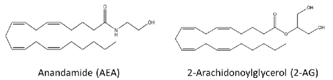
Endocannabinoids were discovered in vivo after the identification of Δ9-THC. The two endocannabinoids whose biological activity has been better characterized are N-arachidonoylethanolamine (anandamide; AEA) and 2-arachidonoylglycerol (2-AG) (21). Both chemical structures are represented in Figure 1. Like Δ9-THC, AEA and 2-AG, bind to the extracellular site of the CB1 and CB2 (11, 12, 22, 23). Despite these two well-studied endocannabinoids, there are also the 2-arachidonyl-glycerol ether (noladin-ether) (24), the N-arachidonoyl-dopamine (NADA) (25, 26) and virodhamine (27), whose metabolism and pharmacological activity are yet to be fully investigated (13). More recently, it was identified the first potent endogenous antagonist of CB1, a nonapeptide known as hemopressin, found in numerous tissues including the brain, but more studies of its metabolism and regulatory levels are needed (13).
AEA, an endogenous eicosanoid derivative, is released by the cleavage of the phospholipid precursor N-acyl phosphatidylethanolamine (NAPE). The synthesis of this precursor is catalysed by the N-acyltransferase (NAT), whereas a specific phospholipase D, the N-acylphosphatidylethanolamine-specific phospholipase D (NAPE-PLD) is responsible by the release of AEA from NAPE.
Figure 1 – The chemical structures of the major endocannabinoids. AEA and 2-AG have different
biosynthetic pathways, although both are produced on demand by cleavage of membrane phospholipds precursors.
- 4 -
2-AG is produced by the cleavage of a membrane phospholipid, by phosphatidylinositol (PI) and, sometimes, from the hydrolysis of phosphatidic acid (28) to produce 1,2-diacylglycerol (DAG). The formation of this compound is catalysed by phospholipase C (PLC), that after conversion via a membrane-bound diacylglycerol lipase (DAGL), gives rise to 2-AG.
These endocannabinoids are suppressed by enzymatic hydrolysis of their amide and ester bonds by a membrane fatty acid amide hydrolase (FAAH) and/or by a membrane-associated serine hydrolase, monoacylglycerol lipase (MAGL) (29-31). The AEA hydrolysis is mainly mediated by FAAH to generate ethanolamine (EtNH2) and arachidonic acid (AA), whereas 2-AG is degraded to AA and glycerol by MAGL or also by FAAH, though the former is considered the main degrading enzyme.
AEA binds to both cannabinoid receptors and, like Δ9-THC, it has lower efficacy and affinity for CB2 than for CB1. AEA can also activate other receptors, such as the orphan G protein-couple receptor 55 (GPR55), the transient receptor potential vanilloid 1 (TRPV1) and the peroxisome proliferator-activated receptor-γ (PPAR-γ) (2, 32-34). 2-AG affinity and efficacy to CB1 and CB2 is higher than AEA and it can also interact with PPAR-γ, but not with TRPV1 or GPR55 (21, 35).
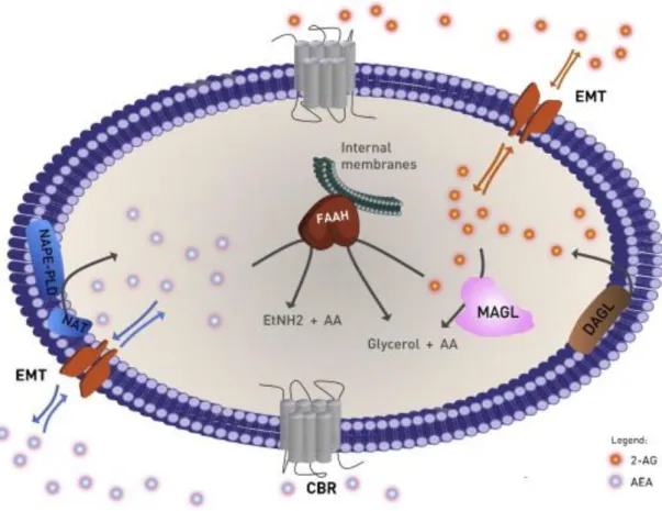
The AEA and 2-AG pharmacological effects depend on their concentration in the extracellular space, which is limited by the transport through the cell membrane, either by passive diffusion or by endocannabinoid membrane transporter (EMT) (22, 36). The endocannabinoids, together with the respective metabolic enzymes and cannabinoid receptors constitutes the endocannabinoids system, a lipid signalling network in which different proteins can control or modulate various physiological and pathophysiological processes (9, 37, 38). The endocannabinoid system elements are represented in Figure 2.
- 5 -
Figure 2 – The components of the endocannabinoids system. The biosynthesis of AEA (blue circles), is
catalysed by NAT followed by NAPE-PLD, and the biosynthesis of 2-AG (orange circles) occurs through DAGL. Endocannabinoids are transported in both directions through cell membrane by diffusion or selective transport via the EMT. In the extracellular space can interact with CBR or be internalized and degraded. AEA is hydrolyzed by FAAH into EtNH2 and AA. 2-AG is hydrolyzed through MAGL or FAAH into glycerol and AA. Adapted from (21).
1.4 Phytocannabinoids
Plant derived natural products that are able to directly interact with cannabinoid receptors or share chemical similarities with cannabinoids, or both, are known as phytocannabinoids (9).
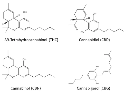
There are more than 60 naturally occurring substances found in Cannabis sativa such as Δ9-THC, cannabidiol (CBD), cannabinol (CBN) and cannabigerol (CBG) (Figure 3). Besides its psychoactive properties, Δ9
-THC also presents muscle relaxant, analgesic and antispasmodic characteristics. CBD is a nonpsychotropic constituent of Cannabis, being a versatile pharmacological agent, causing some of the effects of Δ9-THC, such as sedation, and it has the ability to act as an antagonist of CB1, evidenced by reducing Δ9 -THC psychomimetic effects. CBN, like CBD, has a low affinity for the cannabinoid receptors and causes sedation when combined with Δ9-THC (9, 39-41).
- 6 -
Figure 3 – The chemical structures of the main phytocannabinoids. The psychoactive component, Δ9 -THC, and other natural substances found in Cannabis plant, such as CBD, CBN and CBG.
1.5 Synthetic cannabinoids
Synthetic cannabinoids began being developed in the 1960s after the discovery of Δ9-THC, to help investigate the endocannabinoid system pharmacology and its potential therapeutic effects, emphasizing in the analgesic and anti-inflammatory properties and eliminating the psychoactive effects. The majority of these new synthetic cannabinoids were synthesized in four laboratories: John W. Huffman, Alexandros Makriyannis, Pfizer and Hebrew University, and now are known by their initials (JWH, AM, CP and HU) (42-45).
The structure of the major synthetic cannabinoids can be divided into four parts: the core and substituents, the link section, the ring and substituents and the tail section (46) (Figure 4).
Figure 4 – The general chemical structure of the main synthetic cannabinoids. The majority of synthetic
- 7 -
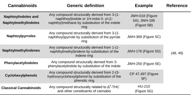
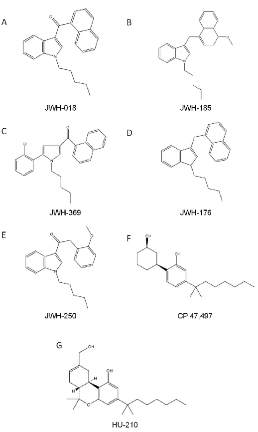
These new substances can be classified by their chemical structure, as suggested by Howlett et al. and Thakur et al. (16, 47) and the Advisory Council on the Misuse of Drugs and the European Monitoring Centre for Drugs and Drug Addiction (EMCDDA) (48, 49) divides synthetic cannabinoids into seven groups: naphtoylindoles, naphtylmethylindoles, naphtoylpyrroles, naphtylmethylindenes, phenylacetylindoles, cyclohexylphenols and classical cannabinoids (Table 1).
Table 1 – Classification of synthetic cannabinoids. The generic definition of the seven major groups of
synthetic cannabinoids and respective examples.
Cannabinoids Generic definition Example Reference
Naphtoylindoles and Naphtylmethylindoles
Any compound structurally derived from 3-(1-naphthoyl)indole or 1H-indol-3- yl-(1-naphthyl)methane by substitution of the indole
ring
JWH-018 (Figure 5A); JWH-185
(Figure 5B)
(48, 49) Naphtoylpyrroles naphthoyl)pyrrole by substitution of the pyrrole Any compound structurally derived from
3-(1-ring
JWH-369 (Figure 5C)
Naphtylmethylindenes Any compound structurally derived from 1-(1-naphthylmethyl)indene by substitution of the indene ring
JWH-176 (Figure 5D)
Phenylacetylindoles Any compound structurally derived from
3-phenylacetylindole by substitution of the indole JWH-250 (Figure 5E)
Cyclohexylphenols hydroxycyclohexyl)phenol by substitution of the Any compound structurally derived from 2-(3-phenolic ring
CP 47,497 (Figure 5F)
Classical Cannabinoids Any compound structurally related to Δ9-THC and other constituents of cannabis
HU-210 (Figure 5G)
These new substances have psychoactive effects as Δ9-THC. Also the chemical properties are similar, sharing with Δ9-THC high lipophilicity. Nonetheless, though the ability to bind to the cannabinoid receptors is higher, these are not structurally similar to classic cannabinoids (48). In fact, some of these synthetic cannabinoids have even higher affinity to the receptors than Δ9-THC due to their full agonist capability and active metabolites. In addition, the possibility that they may also interact with other receptors could explain why they are more potent than Δ9-THC (40, 50-52). This can cause a variety of symptoms that can go from mild to moderate, like nausea, emesis and agitation to more severe such as cardiac arrhythmias, psychosis, respiratory depression to coma and even death (51, 53, 54).
- 8 -
Figure 5 – The chemical structures of synthetic cannabinoids. Examples of each one of the seven major
classes of synthetic cannabinoids. A – Naphtoylindoles; B – Naphtylmethylindoles; C – Naphtoylpyrroles; D – Naphtylmethylindenes; E – Phenylacetylindoles; F – Cyclohexylphenols; G - Classical Cannabinoids.
- 9 -
1.6 Cannabinoids and drug abuse
Cannabis use, in such different forms, is prevalent in every European Union (EU),
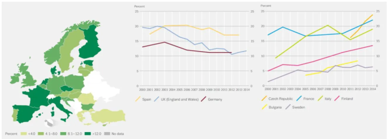
with an upward trend in young adults, as seen in Figure 6. A recent analysis made by the EMCDDA revealed that over a quarter of 15- to 64-years olds in EU is thought to have consumed the drug at least once in their lifetime. On the other hand, the use amongst young individuals (15-34 years) within the last 12 months is about 13,3 % or 16.6 million EU individuals, which makes Cannabis derivatives the most widely consumed illicit drug by both adults and youths in the world (55).
Figure 6 – Cannabis use in Europe by young adults (15-34). In the left, the countries with significant
statistically trends in Cannabis use are represented and, in the right is the most recent data (2015) about
Cannabis use in Europe. It was observed an increase in consumption in France, Italy, Finland, Czech
Republic and Sweden, but with different prevalence. However, Spain, Germany and the United Kingdom showed a decrease in Cannabis use in the last decade. Bulgaria, with less data available, has upward trends. From (55).
In the last decade the availability of new psychoactive substances (NPS) has increased in the European market (Figure 7). These new drugs, natural or synthetic, are produced to mimic effects of controlled substances and are known as “legal highs”. On the other hand, the rapid modification of chemicals, the unknown quantity of drug used and the unknown herbal components that are difficult to detect in a typical drug screen, and the lack of reference samples to make detection possible, makes these new synthetic cannabinoids potentially dangerous to human health (51, 52).
The majority of these new substances are sold as alternatives to cannabis, heroin, cocaine, amphetamines, 3,4-methylenedioxy-methamphetamine (MDMA), benzodiazepines and lysergic acid diethylamide (LSD), and can be found in either the Internet as in specialized shops – head shops – being advertised as “research chemicals”, “food supplements” or as “not intended for human consumption” (56).
- 10 -
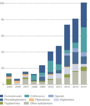
Synthetic cannabinoids is the largest group of NPS with more than 160 different synthetic cannabinoids identified till December 2015. Usually, these substances are produced in China or India and arrive in Europe where they are sold as chemical powders or herbal mixtures similar to potpourri or incense that can be smoked, and also used as infusions or even in e-cigarettes. The common street names of the new drugs are: Spice, K2, Bhang, Yucatan and Mojo. These are not marijuana, but really a combination of various synthetic compounds that can interact with the cannabinoid receptors and induce marijuana-like effects (46, 51, 52, 56).
Figure 7 – New psychoactive substances notified to the EU Early Warning System. Number and
categories of new psychoactive substance reported to the Eu Early Warning System increased 25% compared to 2013, including 30 new synthetic cannabinoids. From (57).
- 11 -
2. Cannabinoids and signalling pathways
Cannabinoids exert their effects by binding to specific cannabinoid receptors, usually activated by the endogenous ligands, the endocannabinoids. These receptors are able to regulate several central nervous system functions, such as neuronal development, neuromodulatory processes and several peripheral physiological functions, for instance cardiovascular, respiratory, digestive and reproductive systems. Additionally, they can also modulate proliferation, motility, adhesion and apoptosis of cells (58, 59).
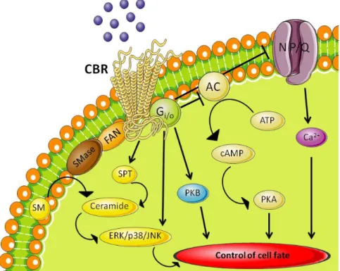
Both cannabinoid receptors belong to the GPCR superfamily, and more specific, the rhodopsin subfamily of GPCRs, and are characterized by 7-transmembrane domains, an extracellular NH2 terminus and an intracellular COOH terminus (18). They are also associated with G proteins of the Gi/o family (Gi 1, 2 and 3; Go 1 and 2) (11, 12). The activation of cannabinoid receptors leads to inhibition of the adenylyl cyclase (AC) enzyme, activation of mitogen-activated protein kinases (MAPKs) and modulation of ion channels, in this case only through activation of CB1, as represented in Figure 8.
Figure 8 – Cannabinoid signalling pathways after activation of the cannabinoid receptors coupled to G protein. This leads to inhibition of adenylyl cyclase, inducing a decrease in cAMP levels, which translates in a
decrease of PKA. The activation also activates MAPKs, such as PKB, and induces an increase in ceramide levels, through sphingomyelin hydrolysis by SMase, which regulates ERK, p38 MAPK and JNK. Additionally, CB1 can negatively couple to N- and P/Q-type voltage-operated Ca
2+
channels and induce an increase in intracellular Ca2+.
Even before cannabinoid receptors were described, it was already known that cannabinoids caused inhibition of the AC, which leads to a reduction of intracellular cyclic
- 12 -
AMP (cAMP) levels. cAMP is responsible for numerous roles inside the cell, such as the phosphorylation of proteins through protein kinase A (PKA). Essentially, decreased cAMP in cells that express CB1 and CB2 will most likely culminate in an inhibition of function of those cells (59, 60).
Both cannabinoid receptors are also able to regulate the phosphorylation and activation of the family of mitogen-activated protein kinase (MAPK). Activation of extracellular signal-regulated kinase-1 and -2 (ERK1/2), also known as p42/p44 MAPK, by CB1 receptors, requires mobilization of the phosphatidylinositol-3-kinase (PI3K) and protein kinase B (PKB/Akt) which results in the phosphorylation of Raf-1. p38 MAPK and c-Jun N-terminal kinase (JNK) can also be regulated by these receptors (58, 59, 61).
The sphingolipid-metabolising pathways also seem to be modulated by cannabinoids. Neutral sphingomyelinase activation (FAN) is the possible adaptor protein responsible for the hydrolysis of sphingomyelin (SM) via sphingomyelinase (SMase) activation. This activation can cause a peak of ceramide accumulation by enhanced synthesis de novo via serine palmitoyltransferase (SPT), which is important in the regulation of metabolic functions, such as induction of apoptosis, via a ERK/p38/JNK (59, 62).
The activation of cannabinoid receptors is also responsible for the regulation of other key proteins, including the focal adhesion kinase that plays a part in synaptic plasticity (63); activation of phospholipase A2, via MAPK, inducing the arachidonic acid cascade and production of prostaglandins (64); is also able to decrease the synthesis of growth factor receptors in some tissues, what could explain the antiproliferative effects of cannabinoids (65).
However, cannabinoid receptors agonists have different affinities to agonists and the activated signalling pathways can differ from cell to cell, which may account to the various cells responses obtained upon binding to receptors (60). CB1, additionally, can negatively couple to N- and P/Q-type voltage-operated Ca2+ channels and positively couple to A-type and inwardly rectifying K+ channels, and through G protein-dependent activation of phospholipase C-β (PLC-β) induce an increase in intracellular Ca2+ (58).
The involvement of different mitogen- and stress- activated protein kinase cascades in the control of cell fate, and the evidences that cannabinoid receptors interact with these signalling pathways, show that cannabinoids may play an important role in the cell survival/death decision (59).
- 13 -
3. Cannabinoids and Pregnancy
3.1 The endometrium and menstrual cycle
The endometrium is composed of epithelial and mesenchymal elements that suffer different morphological alterations during the menstrual cycle. The superficial epithelium is formed of epithelial cells, and the mesenchyme, or stroma, contains predominantly fibroblasts. It can also be divided into two different layers based on morphological and physiological characteristics, such as, the basalis, the deepest layer with a constant morphology during the menstrual cycle, and the functionalis, the superficial layer, where most of the alterations occur to allow the blastocyst implantation (66).
The two primary steroid hormones are estrogen (E2) and progesterone (P4), which are mainly produced in ovaries and are responsible for the cyclic changes in morphology and function during the menstrual cycle. Due to fluctuations of these hormones, menstrual cycle can be divided into three phases: in the proliferative phase, the levels of estrogen increase, inducing the thickening of the functionalis layer, by proliferation of epithelial, stromal and endothelial cells; in the secretory phase, which occurs after ovulation, the differentiation of the functional layer starts, due to the influence of progesterone, produced by the corpus luteum. It is at the end of this phase that decidualization begins. When no implantation occurs, the levels of estrogen and progesterone decrease, due to the decay of the corpus luteum, leading to shedding of the functional layer and consequent menstrual bleeding, which characterises the menstrual phase (Figure 9) (66, 67).
Studies suggest that menstrual bleeding is a secondary effect of extracellular matrix (ECM) proteolysis (68) and, although numerous proteinases and enzymes expressed in human endometrium seem to participate in menstruation, the most important enzymes for direct ECM proteolysis is the matrix metalloproteinases (MMP) family. MMP are Zn-dependent proteinases capable of degrading the ECM and regulate several signalling molecules like cytokines and chemokines. The endometrium has plenty of MMP transcripts and proteins, particularly during menstruation, and of all MMP studied only MMP-13 was not detected in the human endometrium. The major regulator of MMP expression is progesterone (66).
- 14 -
Figure 9 – The menstrual cycle in humans. The 28- to 30 days menstrual cycle begins with menstruation,
followed by the proliferative phase (red), which is under the influence of increasing estrogen levels, produce by growing ovarian follicles, which induce the proliferation of the endometrium. By midcycle, a surge of gonadotropins (LH and FSH) occur, leading to ovulation on day 14 (green). In the luteal phase, the thickening of the endometrium, formation of the corpus luteum and subsequent secretion of progesterone is the preparation for implantation of the blastocyst. Decidualization starts and the estrogen levels increasing overlapping on progesterone define the “implantation window”. In the absence of pregnancy, the window progresses to a refractory phase, leading to luteolysis, hormone withdrawal and menstruation (69).
3.2 The endometrium and pregnancy establishment
A successful pregnancy, which is the nurturing of the offspring within the womb and the production of a live birth, depends on a sequence of well-timed events since fertilization to parturition. It all starts with the fertilization of an egg by a spermatozoid, the zygote, in the Fallopian tube. Then, it travels through the oviduct, suffering multiple divisions and developing into a blastocyst, until it reaches the uterus, where it will adhere to the decidualized endometrium. This sequence of events starts with proper establishment of a receptive endometrium and a competent blastocyst (67).
Implantation is an intrusive process and occurs during the mid-secretory phase, in a 2- to 4 day period called “implantation window”, during which the endometrium is prepared by estrogen and progesterone. It can be divided into three stages: apposition, adhesion and invasion. During apposition, the first contact between blastocyst and endometrium is made and the location for implantation is found. The invasion of the
- 15 -
endometrium by the blastocyst is when the trophoblast cells infiltrate the endometrial epithelium and into the endometrial stroma until reaching blood vessels (67, 70).
The uterine endometrium is prepared every month for the possible implantation of a blastocyst and this process is known as decidualization. The rapid proliferation of epithelial and endometrial stromal cells is followed by the differentiation of the glandular epithelium into a highly secretory state. Ultimately, the fibroblast stromal cells differentiate into enlarged and glycogenic rich cells with a polygonal morphology, which characterises the decidual cells (71, 72). It also involves a tightly regulated expression of specific adhesion molecules, growth factors and cytokines (72, 73).
Decidua can be classified into three types relative to the developing conceptus: the decidua basalis, the region beneath the site of implantation; decidua capsularis, the region over the developing conceptus, and the remaining is the decidua vera or decidua parietalis (67). Decidua is mostly comprised of decidualized endometrial stromal cells, but it also has other cell types, like hematopoietic cells, macrophages, uterine natural killers and monocytes, associated with blood vessels, and uterine glands (74). To promote the adhesion of the trophoblast cells, the decidua forms a dense cellular matrix that, at the same time, helps to limit the aggressive invasion of those cells. This invasion depends upon proteolytic degradation and remodelling of the decidual extracellular matrix, which is accomplish by the MMPs secreted by the trophoblast cells, and by decidualized stromal cells, as shown by recent studies (75). Thus, decidualization results in these cells acquiring biochemical and cellular properties that facilitates them support implantation (67).
If the decidual process does not occur normally, pregnancy complications may happen, such as miscarriage, preeclampsia, foetal growth restriction and preterm labour (76). Although it was already shown that implantation and even successful pregnancies can occur without the decidua (77, 78), it is the successful decidualization that is important for implantation and success of normal pregnancies (73).
On the other hand, development and function of the placenta is the key to an adequate exchange of nutrients and gas between mother and foetus, which is intimately dependent on endometrial environment.
3.3 Endocannabinoids and pregnancy
It was already described that endocannabinoid system is implicated in several physiological and pathological processes, such as the reproductive system (79).
- 16 -
Evidences of their role in decidualization, embryo development and implantation were already shown (80).
AEA, 2-AG and respective metabolic enzymes were found in the mouse endometrium and their concentration varies during the oestrus cycle (81). In humans AEA levels increase in the menstrual cycle at the time of ovulation, the cannabinoid receptors are present in the ovaries, and NAPE-PLD is expressed by granulosa and theca cells, meaning that AEA is more likely to be produced by granulosa of growing cells than by oocytes. In addition, considering that follicular AEA concentrations are related to follicular size and that are lower in follicles from which oocytes were retrieved, it is suggested that AEA is involved in the maturation of follicles and oocytes (82-84).
Additionally, mouse embryos express cannabinoid receptors and high doses of AEA can induce a dose-dependent suspension of embryo development, and even inhibition of blastocyst hatching (85). Mouse embryos with cannabinoid receptor abnormalities also present delayed development (86). Furthermore, it was shown in mice that CB1 is important to assure embryo transport through the oviduct as CB1 knockout mice showed high levels of abortion (87).
AEA levels are higher in the endometrium than in other reproductive tissues (88, 89). Both cannabinoid receptors, NAPE-PLD and FAAH proteins were also detected in decidua from pregnant women (76). The presence of cannabinoid receptors, endocannabinoids and respective metabolic enzymes in the female reproductive system, with evidences of their presence in maternal and foetal tissues suggests that these are involved in decidualization of the endometrium, oviduct transport and implantation of the embryo (76).
3.4 Cannabinoids use by pregnant women
Cannabis is the most consumed illegal substance by pregnant women in Western
societies, with nearly 3 % of reported cases by pregnant women in the United States (Figure 10). The lipophilic characteristics of cannabinoids, allows them to cross cell barriers, including transplacental membranes (90). This explains the presence of
Cannabis metabolites in many human tissues (91), such as the placenta, amniotic fluid
and foetus (92), making the study of its effects on reproduction important (7).
Most studies show that continuous use of Cannabis is associated with reduced fertility and failed pregnancies. It is also shown that Cannabis is correlated with congenital foetal abnormalities and intrauterine growth restriction (93-95), leading to the general conclusion that maternal use of Cannabis during pregnancy affects birth outcome and
- 17 -
foetal development. However, contradictory results have also been published, as some studies show that infants exposed to Cannabis during pre-natal life did not show increased risks of birth defects (95-97).
The studies to discover the full extent of Cannabis use during pregnancy are few due to ethics problems that forbid the experiments in human tissues. In addition, the small number of studies that were published are controversial, due to the fact that women that use Cannabis frequently also consume other drugs and have adverse lifestyles (98, 99).
Figure 10 – Illicit drug use in the United States of America by pregnant women. Approximately 4% of
women admitted the use of illicit drugs while pregnant, with Cannabis as the most commonly used drug. Data from 2005. Adapted from (100).
The human studies published to date consist in following pregnant women that admitted the usage of substances that may affect the pregnancy outcome. In most of these studies the parameters that were examined were the ones related to foetal growth. In a study with 1690 children, the birth weight was lower for foetus exposed more than three times per week to Cannabis (95). In other study the increased risks of low birth weight was two times higher in foetus exposed several times a month to Cannabis (101).
One of these studies was the Generation R, that started in 2002 and consisted in assessing the foetal growth in early, mid and late pregnancy using techniques such as femur length, abdominal and head circumference and transcerebellar diameter (102, 103). In the Generation R study, 8880 pregnant women were enrolled and the results showed that foetal growth, especially birth weight, was reduced in mothers that used Cannabis in early pregnancy comparing to tobacco users and non-users. In mothers that used
Cannabis continuously during pregnancy, the birth weight was even lower comparing to
only early usage. The head circumference of foetuses was also reduced in early and continuous use during pregnancy compared to non-exposed foetuses. However, the transcerebellar diameter did not show any differences (103).
- 18 -
In Cannabis-exposed pregnant women, the use of Doppler showed an increased foetal pulsatility and resistance index of the uterine artery, which could explain the growing deficit in exposed foetuses (104, 105). Other studies also observed influence on the preterm birth rate, which increased with Cannabis use (106).
Studies have already shown that Δ9-THC can cross the placenta in animals and humans (107) and that the behaviour of Δ9-THC concentrations in the foetus are similar to the maternal. However, the concentrations in foetal blood were lower than in maternal blood in several species. The ways of intake can affect the concentrations reached in blood. In oral intake the foetal concentrations can be one-tenth of the maternal (108) comparing to the one-third if the intake occurs by intravenous or inhaled Δ9-THC (109, 110). Involving dizygotic twins, the importance of the placenta in the variability of foetal exposure to Δ9-THC was determined, in which large disparities were found regarding cannabinoid concentrations with undetectable levels in one twin and high levels in the other (40, 111).
The influence of the cannabinoid receptors in the placenta was also a subject of study and it was found that the CB1 receptor is present in all of the placental layers and that the stimulation of this receptor is responsible for the impaired foetal growth (7, 14, 112). Costa et al. studied the in vitro effects of Δ9-THC using human trophoblasts (placental epithelial cells) from term placenta (113). The results showed that Δ9-THC impaired the differentiation of cytotrophoblasts into syncytrophoblasts, and in low concentrations demonstrated to be cytoprotective, preventing cell death. Both of these findings occurred through a cannabinoid receptor-dependent mechanism. So, Δ9-THC inhibits trophoblast turnover, impairing placental development by disturbing key processes, such as proliferation, differentiation and apoptosis of cytotrophoblasts (113).
Table 2 - Retrospective studies on Cannabis use by pregnant women. Published studies analysing the
impact of Δ9
-THC in pregnancy.
Cannabinoid Effects observed Reference
Δ9
-THC
Decreased birth weight (101)
Decreased birth weight (95)
Decreased foetal growth
Decreased head circumference (103) Increased preterm birth rate (106)
The effects of synthetic cannabinoids in human reproduction are yet to be fully unveiled, but considering that Cannabis use among pregnant women seems to cause
- 19 -
adverse effects in placental development and foetal growth (113) and the fact that synthetic cannabinoids bind to cannabinoids receptors with higher affinity and potency should be of concern (2). It is also important to acknowledge that most of the users of synthetic cannabinoids use others synthetic drugs, such as synthetic cathinones, also known as bath salts (114).
- 21 -
The endocannabinoid system is a signalling network that can regulate several physiological processes, including reproduction. Cannabinoid receptors expression in the placenta, foetal membranes and endometrium, along with the endocannabinoids ability to regulate different signalling pathways involved in cell survival and their role in decidualization, embryo development and implantation shows that a tightly controlled endocannabinoid signalling is necessary for a successful pregnancy.
The emergence and increasing use of new psychoactive substances, like synthetic cannabinoids, which share similar psychoactive effects with Δ9-THC with higher affinity for the cannabinoids receptors and possible interaction with endocannabinoid system, present a new matter of concern. Furthermore, lack of the pharmacological information about the synthetic cannabinoids translated in a variety of symptoms, besides the psychoactive effects, can be quite harmful.
It was already observed that the consumption of Cannabis before and during pregnancy affect fertility, birth outcome and foetal growth. These findings together with the higher affinity to cannabinoid receptors of synthetic cannabinoids and increased abuse of these new substances during fertile age may represent a risk for fertility and for the normal progression of a future pregnancy.
Therefore, with this study we pretend to investigate the influence of different synthetic cannabinoids in endometrial stromal cells, using a cell line model, St-T1b, and primary cultures of human decidual fibroblasts, HdF, hoping to contribute to the understanding of the effects of cannabinoids abuse in endometrium development.
- 23 -
1.1 Materials
Dulbecco’s Modified Eagle Medium/F12 (DMEM/F12), foetal bovine serum (FBS), antibiotic–antimycotic solution (penicillin G sodium, streptomycin sulphate and amphotericin B) and trypsin were from Gibco/Invitrogen Corporation, Carlsbad, CA, USA. Trypan blue, activated charcoal, Triton X-100, methylthiazolyldiphenyl-tetrazolium bromide (MTT), Höechst 33342, 16S and GAPDH primers, Sulforhodamine B sodium salt, gelatin form porcine skin and BrdU antibodies were from Sigma–Aldrich Co. St. Louis, MO, USA. Ethanol and methanol were from Fisher Scientific, Loughborough, UK. DMSO was from VWR, Fontenay-sous-Bois, France. WIN 55,212-2, AM251 and AM630 were from Tocris Bioscience, Bristol, UK. AM281 and broad range molecular markers were from Santa Cruz Biotechnology, CA, USA. Δ9-THC was from Lipomed AG, Swiss. CytoTox 96 Non-Radioactive Cytotoxicity Assay Kit and Caspase-Glo 3/7 were from Promega. Giemsa was from Merck. Bradford assay reagent was from Bio-Rad, Laboratories Melville, NY, USA. TRIsure was from Bioline Reagents Ltd., UK. Prostagladin E2-d4 was from Cayman Chemical Company. BrdU was from Boenhringer Mannheim, Germany. THJ-2201, JWH-122, AB-FUBINACA, UR-144 and 5F-PB-22 (Figure 11) were provided from the Laboratory of Toxicology from the Faculty of Pharmacy of University of Porto, Portugal. Ninety-six-well white plates were from Thermo Scientific, Roskilde, Denmark.
- 24 -
Figure 11 – Chemical structures of the synthetic cannabinoids used in this work. These synthetic
cannabinoids have different affinities to CB1 and CB2. WIN 55,212-2: CB1 (1.9 nM); THJ-2201: CB1 (1.0 nM) and CB2 (2.6 nM); JWH-122: CB1 (0.69 nM) and CB2 (1.2 nM); AB-FUBINACA: CB1 (0.9 nM) and CB2 (23.2 nM); UR-144: CB1 (150 nM) and CB2 (1.8 nM); 5F-PB-22: unknown.
1.2 Cell cultures
1.2.1 St-T1b cell line
St-T1b is a telomerase-immortalized human endometrial stromal cell line, well-accepted as an endometrial cell model, kindly provided by Dr Birgit Gellersen from Endokrinologikum Hamburg (Centre for Endocrine, Metabolic and Fertility Disorders), Germany.
St-T1b cells were maintained in culture in DMEM/F12 medium supplemented with 10 % (v/v) of FBS treated with activated charcoal (FBS-CT), 1 % (v/v) of an antibiotic-antimycotic (AB-AM) solution, 1 nM 17-β-estradiol and 1 μg/ml Insulin and were incubated at 37 °C in 95% air/5% CO2 humidified atmosphere. After reaching about 80% of confluence, cells were successively sub-cultured to new culture flasks. For this, cells were treated with 0.25 % trypsin/EDTA 1 mM for 4 min at 37 ºC, washed with PBS and collected to centrifuge tubes with culture medium containing 10 % FBS-CT (v/v) to
- 25 -
inactivate trypsin. Cells were centrifuged at 180 xg for 5 min at 4 ºC. Then cells were counted in a Neubauer chamber and cultured using the cell densities: 0,5x10⁴ cells/well (final volume 200 μL) for 96-well plate, 2,5x10⁴ cells/well (final volume 500 μL) for the 24-well plate and 5x10⁴ (final volume 2000 μL) for the 6-well plate. After adherence (24 hours), the cells were washed with PBS and treated with the compounds, in cell culture medium with 2 % (v/v) FBS-CT.
1.2.2
Isolation and primary cultures of human
decidual fibroblasts (HdF)
Term placentas from caesarean section or vaginal delivery, following uncomplicated pregnancies were obtained from Centro Materno-Infantil do Norte – Centro Hospitalar do Porto. All the procedures were reviewed and approved by the Research and Ethics Board of Centro Hospitalar do Porto. After multiple placenta washes, decidua was scraped from the chorionic membrane. The decidual tissue was then dissected and enzymatically dissociated by collagenase (1 mg/ml). Red blood cells were lysed and the resulting suspension was filtered and centrifuged.
The isolated cells were maintained in culture in DMEM/F12 medium supplemented with 10 % (v/v) of FBS treated with activated charcoal (FBS-CT), 1 % (v/v) of an antibiotic-antimycotic (AB-AM) solution, 50 nM β-estradiol and 50 nM insulin and were incubated at 37 °C in 95 % air/5 % CO2 humidified atmosphere. After 24 h, the non-adherent discarded and the adherent cells were grown to confluence and sub-cultured. The purity of primary HdF was confirmed by immunocytochemical analysis for the cytoskeletal proteins vimentin and cytokeratin-7, a fibroblast and an epithelial cell marker, respectively.
Cells were cultured using the cell densities: 0,5x10⁴ cells/well (final volume 200 μL) for 96-well plate, 2,5x10⁴ cells/well (final volume 500 μL) for the 24-well plate and 5x10⁴ (final volume 2000 μL) for the 6-well plate. After adherence (24 h), the cells were washed with PBS and treated with the compounds, in cell culture medium with 2 % (v/v) FBS-CT.
1.3 Cell viability assays
To access cell viability it was used the tetrazolium salt [3-(4,5-dimetylthiazol-2-yl)-2,5-dipheniltetrazolium bromide] (MTT) assay and measured the activity of the enzyme lactate dehydrogenase (LDH) in cell culture medium. For that, St-T1b cells were plated in 96-well plates. After 24 h for adhesion, the medium was replaced with DMEM/F12 medium with 2 % (v/v) FBS-CT and 1% AB-AM solution in the presence or absence of the
- 26 -
cannabinoids (0.01-50 μM), and the cells were incubated for 24 and 48 h. Equimolar concentrations of DMSO, the solvent vehicle of the compounds, was tested did not induced any cell viability alterations. The yellow tetrazole MTT (final concentration: 0,5 mg/mL) was added, and the cells were incubated for 3 h at 37 °C. The formed purple formazan was dissolved in a solution of DMSO:isopropanol (3:1) and spectrophotometrically quantified at 540 nm by using a Multiskan Ascent microplate reader. MTT assay relies on the mitochondrial metabolism for the conversion of the yellow dye MTT on a purple dye formazan by viable cells.
Cells supernatants were collected at 24 and 48 h to perform lactate dehydrogenase (LDH) assay. LDH is a cytosolic enzyme that is only released from cells into the extracellular space when the plasma membrane is disrupted. So, the release of LDH into the culture medium was evaluated by measurement of LDH activity by the use of the CytoTox 96 Non-Radioactive Cytotoxicity Assay Kit according to the manufacturer's instructions. Released LDH activity is measured with a coupled enzymatic assay, which results in conversion of a tetrazolium salt into a red formazan product. The intensity of colour formed is proportional to the number of lysed cells. Generation of Formazan is monitored by measuring absorbance at 490 nm using BioTek Power Wave XS plate reader.
All the experiments were performed in at least three independent experiments and results were expressed as percentage of control/untreated cells.
1.4 Cell proliferation assays
To determine cell proliferation it was used the Sulforhodamine B (SRB) and BrdU assays. SRB is a fluorescent dye used for cell density determination via quantification of cellular proteins of cultured cells. The method of incubation was similar to the MTT assay till the end of incubation, and then the cells were fixed with trichloroacetic acid (final concentration: 40 %) for 1 h at 4 ºC and incubated with SRB (final concentration: 0.4 % in 1 % acetic acid) for 30 min. The bound protein stain was solubilized in Tris-base (final concentration: 10 mM) and absorbance was assessed at 492 nm spectrophotometrically.
Additionally, the thymidine analog BrdU (5-bromo-2’-deoxyuridine) proliferation assay was performed, following its incorporation by the newly synthesized genomic DNA. For that, St-T1b cells were plated in 96-well plates. After 24 h of culture, the medium was replaced with DMEM/F12 medium with 2 % FBS-CT and 1 % AB-AM solution in the presence or absence of cannabinoids (0.01-50 μM). The cells were labelled by the addition of BrdU (final concentration: 10 µM) for 28-30h. After the labelling period, the
- 27 -
cells were fixed and the anti-BrdU (final concentration: 1 µg/ml) antibody was added. The absorbance of the reaction product was measured at 450 nm using BioTek Power Wave XS plate reader.
All experiments were performed in triplicate and in at least three independent experiments. Results were expressed as percentage of control/untreated cells
.
1.5 Morphological studies
The morphological alterations caused by cannabinoids treatment were evaluated by phase-contrast microscopy, Giemsa and Höechst staining.
The cells were cultured in 24-well culture plates with coverslips and treated with the different cannabinoids (25 μM of Δ9-THC, THJ-2201, JWH-122, AB-FUBINACA, UR-144, 5F-PB-22, 2 and 5 μM of WIN) for 48 h.
Giemsa staining is a variant of the Romanowsky-type stain. This technique allows the evaluation of cell morphology. Giemsa contains a mixture of methylene blue, eosin and Azure B (methylene azure B). The eosin Y dye stains the basic components of the cells. The methylene blue and azure B dyes stain the acidic components in shades between blue and purple. Cells were washed with PBS and fixed with methanol for 30 minutes at 4 ºC. Then cells were washed with PBS and stained with Giemsa stain solution, diluted in distilled water (1:10) for 30 min. After washing with tap water, the coverslips with the stained cells were dehydrated and mounted in DPX mounting medium and observed under a bright field microscope (Eclipse E400, Nikon, Japan) equipped with image analysis software LeicaQwin.
Höechst is a dye that emits blue fluorescence when bound to DNA. This staining allows the evaluation of nuclear morphology and the identification of apoptotic nuclei, which present chromatin condensation and fragmentation. After fixation with methanol for 25 min at 4 ºC, the cells were exposed to 0.5 μg/mL Höechst 33342 (in PBS) for 20 min, washed with PBS, mounted in Fluoroshield mounting medium and observed under a fluorescence microscope equipped with an excitation filter with maximum transmission at 360/400 nm (Eclipse CI, Nikon, Japan). Images were processed by Nikon NIS Elements Image Software.
- 28 -
1.6 Real-time Polymerase Chain Reaction (RT-PCR)
To determine the amount of mitochondrial DNA (mtDNA) relative to nuclear DNA (nDNA), a quantitative real-time PCR was performed, by measuring the ratio of the mitochondrial gene, 16S rRNA, and a nuclear gene, GAPDH. For that, St-T1b cells were plated in 6-well plates. After 24 hours for adhesion, the medium was replaced with DMEM/F12 medium with 2 % FBS-CT and 1 % AB-AM solution in the presence or absence of cannabinoids (50 μM) for 48 h. Cells were collected in TRIsure reagent, and total DNA was extracted according to the manufacturer’s instructions. DNA was quantified in the NanoDrop ND-1000 spectrophotometer (NanoDrop Technologies, Inc., Wilmington, DE, USA). To determine THJ-2201, JWH-122 and AB-FUBINACA impact in the ratio of mtDNA/nDNA, DNA was amplified with specific primers: 16S rRNA and GAPDH. These reactions were carried out with KAPA SYBR FAST qPCR Master Mix 2x Kit (Kapa Biosystems, Woburn, MA, USA) in MiniOpticon Real-Time PCR Detection System (Bio-Rad Laboratories, Hercules, CA, USA), according to kit protocol. Primer sequences and RT-PCR conditions are summarized in Table 1.
Table 3 – RT-PCR conditions. Gene, primer sequences and temperatures used to obtain the ratio of
mtDNA/nDNA.
Gene Primer sequence (5’-3’) Annealing
temperature (ºC)
Melting temperature (ºC)
16S rRNA Sense: ACTTTGCAAGGAGAGCCAAA
Anti-sense: TGGACAACCAGCTATCACCA 59 ºC 83,5 ºC
GAPDH Sense: GGATGATGTTCTGGAAGAGCC
Anti-sense: AACAGCCTCAAGATCATCAGC 59 ºC 81,5 ºC
1.7 Caspase -3/-7 activity
To detect caspase -3/-7 activity, the cells were seeded in a 96-well white plate pre-incubated for 30 min with the CB1 and CB2 antagonists, AM251 (1 μM), AM281 (2 μM) and AM630 (1 μM), respectively, and treated with the cannabinoids for 24 h. At the end of the incubation time, Caspase-Glo -3/-7 reagent was added to the cells according to the manufacturer's instructions. The plate was incubated at room temperature for 1 h and the resultant luminescence was measured in relative light luminescence units (RLU) using the 96-well Microplate Luminometer (BioTek Instruments, Winooski, VT, USA). The results are expressed in percentage, comparing caspase -3/-7 activities of cannabinoid-treated cells with the untreated cells.
- 29 -
1.8 Zymography
To determine the activity of metalloproteinases (MMPs) a zymography was performed. For that, St-T1b cells were plated in 24-well plates. After 24 h of culture, the medium was replaced by DMEM/F12 medium with 2 % FBS and 1 % AB-AM solution and 12 h before experiment the medium was replaced with DMEM/F12 medium with 2 % FBS-CT and 1 % AB-AM in the presence or absence of cannabinoids (25 μM). After 48h the supernatants were collected and the Bradford assay was performed to quantify protein concentration. Then, equal amounts of total proteins (5 μg) were separated without heating and without β-mercaptoethanol on a 10 % SDS-polyacrylamide gel containing 1% gelatine. The gels were washed, incubated in developing buffer for 16 h at 37 ºC and stained with 5 % Coomassie blue R250. Bands representing gelatinase activity were photographed and analysed by densitometry.
1.9 UPLC-MS/MS
To determine the concentration of prostaglandin E2 in term placenta supernatant and tissue, an ultra-performance liquid chromatography-mass spectrometry (UPLC-MS/MS) analysis was performed. Explants of decidua from term placentas were collected within two hours after delivery and plated in 6-well plates with DMEM/F12 medium with 2 % FBS-CT and 1 % AB-AM. After two hours, the medium was replaced in the presence and absence of cannabinoids (25 μM). After 24 h, supernatant and tissue were collected and frozen at -80 ºC.
1.9.1 Sample preparation
In the day of the experiment, prostaglandin extraction was performed. In supernatant, 500 μl of sample were added to 0.4 g of MgSO4, 0.1 g of NaCl, 500 μl of ethyl acetate and 20 μl of PGE2-d4, and centrifuge for 15 min at 3500 xg. At the end of centrifugation, supernatant was collected and evaporated to dryness under nitrogen. The samples were reconstituted with 100 μl of methanol. Tissue extraction was performed by adding 500 μl of methanol, 20 μl of PGE2-d4 and C18 to a small fragment of tissue, and centrifuge for 30 min at 3500 xg. At the end of centrifugation, supernatant was collected and evaporated to dryness under nitrogen. Samples were reconstituted with 100 μl of acetonitrile and injected in the UPLC-MS/MS (10 μl injection volume).