Origin of frontal lobe spikes in the early onset benign occipital
lobe epilepsy (Panayiotopoulos syndrome)
Alberto J.R. Leal
a,*, Jose´ C. Ferreira
b, Ana I. Dias
c, Eula´lia Calado
caDepartment of Neurophysiology, Hospital Ju´lio de Matos, Avado Brasil nr 53, 1749-002 Lisbon, Portugal bDepartment of Neurology, Hospital Egas Moniz, Lisbon, Portugal
c
Department of Paediatric Neurology, Hospital Dona Estefaˆnia, Lisbon, Portugal Accepted 30 April 2008
Available online 11 July 2008
Abstract
Objective: Early onset benign occipital lobe epilepsy (Panayiotopoulos syndrome [PS]) is a common and easily recognizable epilepsy. Interictal EEG spike activity is often multifocal but most frequently localized in the occipital lobes. The origin and clinical significance of the extra-occipital spikes remain poorly understood.
Methods: Three patients with the PS and interictal EEG spikes with frontal lobe topography were studied using high-resolution EEG. Independent component analysis (ICA) was used to decompose the spikes in components with distinct temporal dynamics. The compo-nents were mapped in the scalp with a spline-laplacian algorithm.
Results: The change in scalp potential topography from spike onset to peak, suggests the contribution of several intracranial generators, with different kinetics of activation and significant overlap. ICA was able to separate the major contributors to frontal spikes and con-sistently revealed an early activating group of components over the occipital areas in all the patients. The local origin of these early potentials was established by the spline-laplacian montage.
Conclusions: Frontal spikes in PS are consistently associated with early and unilateral occipital lobe activation, suggesting a postero-anterior spike propagation.
Significance: Frontal spikes in the PS represent a secondary activation triggered by occipital interictal discharges and do not represent an independent focus.
Ó 2008 International Federation of Clinical Neurophysiology. Published by Elsevier Ireland Ltd. All rights reserved.
Keywords: Epilepsy; Occipital lobe; ICA; Panayiotopoulos syndrome
1. Introduction
The early onset benign idiopathic occipital lobe epilepsy, presently reported as Panayiotopoulos syndrome (PS), has generated a lot of interest in recent years (Lada et al., 2003; Carabalo et al., 2007; Koutroumanidis, 2007). Despite the relatively easy recognition of clinical symptoms (Ferrie et al., 2006), the highly variable characteristics of the interictal EEG activity have prevented the determination of a consistent localization for the epileptic focus
(Panayiotopoulos, 2002). Such variability has even sug-gested to some researchers that a generalized hyper-excit-ability might be present (reviewed in Koutroumanidis, 2007). This theory fails to explain why there are consistent clinical manifestations of the seizures along time in the same patient and among different subjects.
An alternative view suggests that the variable EEG topographies of the spikes are due to fast spread of epilep-tic activity between distant corepilep-tical areas (Leal et al., 2007a,b) with the epileptic activity originating in the pos-terior parts of the brain. In order to test the latter hypoth-esis we decided to study patients with the typical clinical picture but with predominant frontal lobe spikes in the interictal EEG. The demonstration of an occipital origin
1388-2457/$34.00Ó 2008 International Federation of Clinical Neurophysiology. Published by Elsevier Ireland Ltd. All rights reserved. doi:10.1016/j.clinph.2008.04.299
*
Corresponding author. Tel.: +351 969851734; fax: +351 217819809. E-mail address:a.leal@aleeg.com(A.J.R. Leal).
for this extreme topography might be a good test of the theory postulating a common posterior onset of the epilep-tic discharges.
2. Patients and methods
Three patients with the diagnosis of Panayiotopoulos syndrome and predominant or exclusive frontal lobe spikes in the EEG were selected from the pool of patients evalu-ated at our department with this syndrome. All the patients had a history of seizures with prominent autonomic mani-festations (Table 1) and expressed the major features of the syndrome as set forth in a recent consensus view (Ferrie et al., 2006).
We performed a 2-h long EEG recording, including a sleep period, using a 64-channel montage in an electrode cap (EasyCap, Herrsching-Breitbrunn Germany) with the 10–10 positions (Fig. 2). The electrodes were made of sin-tered AgCl and were connected to an EEG amplifier (Micr-omed, Italy). The sampling rate was 512 Hz, and we used high- and low-pass filters at 0.5 and 70 Hz, respectively. The EEG was imported to a software environment which allowed offline high-pass filtering at 1 Hz, detection of frontal spikes, segmentation in epochs of 1 s around the spike peaks and visual inspection to remove the ones with artifacts (Scan 4.3, Neuroscan El Paso, USA).
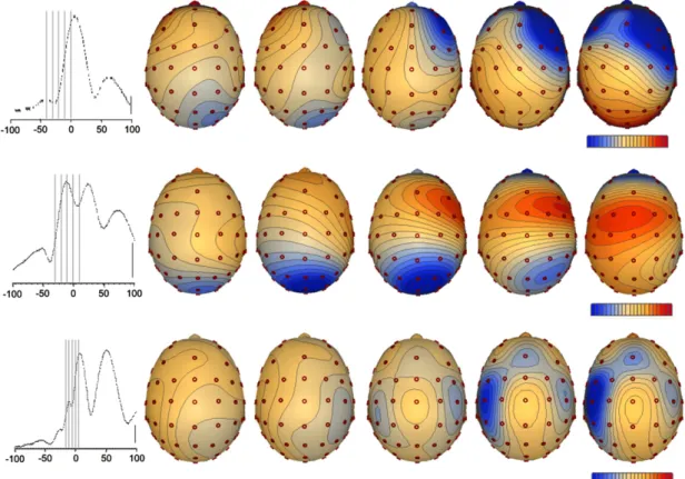
The evaluation of potential maps of the average frontal spikes at fixed intervals before the spike peak revealed a pattern of early negativity over occipital areas which moved to the frontal lobes when approaching the spike maximum (Fig. 1). The instant in time when this change took place was determined for each patient and later used to separate early onset activity from the late onset one. The processed individual spikes were imported into the EEG-LAB software (Delorme and Makeig, 2004) where
decom-position in independent components was performed using the Infomax algorithm (Bell and Sejnowski, 1995). We selected the independent components (ICs) with a clear sin-gle maximum around the raw spike peak, classifying them in either early components if they had a maximum before the instant in time where the shift of topography from occipital to frontal lobes occurred, or late components with a maximum after that instant. The EEG recovered from these two groups of components consistently separated the frontal and occipital contribution to the recorded spikes (Figs. 2a, 3a and 4a).
To better establish the localization of origin of the two components of the average spike we increased the spatial resolution of our recordings using a spline-laplacian mon-tage (Babiloni et al., 1996) as implemented in the software ASA 2.2 (ANT, Enschede, The Netherlands). The scalp surface was obtained from an age-matched normal subject MRI and the standard electrode coordinates of the cap were fitted to the reconstructed scalp in order to match the configuration used in our patients.
The ICA decomposition was repeated in a sub-sample of 19 EEG channels, corresponding to the electrodes of the 10–20 system, to determine whether this analysis method is able to reproduce the same results of the full dataset. 3. Results
The preliminary analysis of the average frontal spikes revealed a significant change in scalp potential topography from spike onset to spike peak (Fig. 1). A consistent pat-tern of early negativity at posterior electrodes was followed by a progressive shift towards the frontal areas. Such behaviour suggested to us the existence of multiple intra-cranial generators with different temporal dynamics.
The decomposition of the spikes using ICA, consistently separated the contribution of frontal and non-frontal ICs (Figs. 2b, 3b and 4b). The temporal activation of the two groups of ICs demonstrated a pattern of early activation over the posterior areas of the scalp, and a later frontal involvement (Figs. 2a, 3a and 4a) in the three patients studied.
The spline-laplacian improved the spatial resolution of the scalp EEG and demonstrated a complex pattern of cur-rent sources and sinks over one occipital lobe in three patients (Figs. 2b, 3b and 4b, above) and also over the pos-terior temporal lobe in one (Fig. 2b, above), for the early activity. This component could be clearly identified in most single spikes for all patients (Figs. 2d, 3d and 4d), which suggests that the large majority, if not all, frontal dis-charges originate in the posterior brain areas. The late activity produced an even more complex, bilateral and widespread, configuration of current sources and sinks over frontal and anterior temporal scalp areas (Figs. 2b, 3b and 4b, below).
The repetition of the ICA analysis with the 19 electrodes of the 10–20 system produced similar early-onset compo-nents over the posterior scalp areas, with a similar scalp
Table 1
Clinical and neurophysiological data
Patient 1 Patient 2 Patient 3
Age 6 y 8 y 6 y
Sex M M M
Cog. develop. Normal Normal Normal Seizures
Age of onset 3 y 6 y 5 y
Nausea Yes No Yes
Vomiting Yes No Yes Urinary
incontinence
Yes No No
Eye deviation Yes Yes Yes Reduced
responsiveness
Yes Yes Yes
Pallor No Yes Yes
Loss of muscle tone
Yes Yes No
Duration >15 min >10 min >15 min Seizure freq. Single event Single event Single event MRI Normal Normal
EEG Frontal lobe spikes only
Frontal lobe spikes
Frontal lobe spikes
topography (Figs. 2c, 3c and 4c, left). The comparison of these components with the average spikes (Figs. 2c, 3c and 4c, right) demonstrates that they explain well the early negative component seen in the average spikes of patients 2 and 3, but they are very small and hard to recognize in patient 1 (Fig. 2c, right).
A dynamic model with onset of spike activity over one occipital lobe followed by widespread and bilateral activity over frontal and anterior temporal areas explains well the temporal changes in scalp potential for the three patients and suggests a posterior origin for the interictal activity. 4. Discussion
The main conclusion from this study is that in 3 patients with the PS the epileptic interictal spikes over the frontal lobes are in fact propagated events from an earlier epileptic discharge at posterior localizations within the occipital lobes.
The absence of a consistent EEG topography for the high amplitude spikes seen in PS has led to speculation that this form of epilepsy might not be related to a particular onset zone, but to a general brain hyper-excitability (reviewed in Koutroumanidis, 2007). Despite this sugges-tion, very few detailed EEG studies have been made in these patients, and most authors used only the 10–20
map of electrodes to sample the scalp potential spikes (Ohtsu et al., 2003; Lada et al., 2003). Studies using a higher electrode density (Leal et al., 2007a) and a more careful evaluation of the temporal spike dynamics (Leal et al., 2007b) suggest that significant spatial spread of the potential over the scalp takes place from spike onset to spike peak, with the posterior brain areas consistently being the earliest to activate. These studies imply the possi-bility that the multifocal spike activity described in the scalp might be due to fast propagation of the epileptic activity between different brain areas, from a common area of onset. The severe spatial under-sampling of the 19 elec-trodes of the standard 10–20 system, associated with simple methods of analysis, did not allow a clear distinction between the two possibilities.
The significant variation in scalp topography of the scalp potential at different moments from spike onset till spike peak in our data (Fig. 1) supports a differential tem-poral involvement of distinct brain areas. The concomitant activation of several generators is expected to be picked up by the surface electrodes, producing volume conductor mixing, which makes the visual analysis of the raw EEG a difficult one.
The techniques of blind source separation based on ICA are powerful tools in the temporal dynamic analysis of such type of signals, where there is a linear mix of the potentials
Fig. 1. Maps of scalp potential at several moments in time, from spike onset till spike peak. The maps show significant changes in this temporal interval, with a pattern of the maximum negativity shifting from the posterior areas to the frontal lobes. Each row represents a patient (1 to 3 from top down). The mean global field power is in the left and the time point of each map is represented in doted lines. Temporal scale of the graph is in milliseconds; potential scale is 6.3, 10.0 and 25.2 lV/step for patients 1, 2 and 3; vertical scale is 10 lV.
generated by several generators (Makeig et al., 2004). In this study, the ICA algorithm was very effective in separat-ing the dominant frontal potential from an early activatseparat-ing one over the occipital brain areas (Figs. 2a, 3a and 4a),
sug-gesting that these two components explain most of the scalp potential complexity of the spike. While this propaga-tion could be suspected from the raw visual analysis of the spikes in two patients (Figs. 3 and 4), in the remaining
Fig. 2. (a) Average spike for patient 1 (left), with a clear maximum over the frontal lobes. The two components produced by the ICA procedure demonstrate a early posterior component (middle) and a late frontal one (right). Vertical scale is 12 lV for the middle trace and 250 lV for the other traces. (b) Potential maps and laplacians of the early (above) and late (below) components. For each component the potential map on the plane and the spline-laplacian map on the scalp surface, at spike peak, are represented. A localized configuration of sources and sinks is apparent over the right posterior area in association with the early component, while a more widespread and bilateral configuration is associated with the late component. (c) Early component recovered with the ICA procedure from a sub-sample of 19-channels of the original record. The configuration is similar to the one recovered from the original 64-channel record (b, above). Superposition of the original potential and the one associated with the early component (right). The early component contributes very little to the overall potential seen at the posterior scalp electrodes. Vertical scale 30 lV, horizontal scale 20 ms. (d) Color representation of the potential of the early component for every spike included in the average. A consistent brief negative potential is apparent 30 ms before spike peak in almost every spike (blue vertical strip). Left scale is the number of the spike, right scale is lV.
patient the small amplitude of the early component pre-vents such recognition.
The inference of which brain areas are involved in the generation of particular potential components in the scalp is a difficult one due to the uncertainties of the inverse
problem in EEG (Plummer et al., 2008). Because our aim was to establish the dynamic relation of the occipital versus frontal activity, we decided to use a standard method to improve the resolution of the EEG using a spline-laplacian montage. This method does not recover the intracranial
Fig. 3. (a) The average spike for patient 2 (left) reveals a negative spike over the posterior scalp areas just before the frontal spikes, with a partial overlap. The ICA decomposition separates the two components (middle and right). Vertical scale 100 lV. (b) Potential and spline-laplacian maps of the early (above) and late (below) components, demonstrating a focal posterior activation preceding the more widespread frontal one. (c) The ICA procedure in a sample of 19-channels recovers the early component (left). The recovered early component explains most of the early component of the raw potential. Vertical scale 30 lV, horizontal scale 20 ms. (d) The negative potential of the early component is present in most spikes. Scales as forFig. 2.
generators of the scalp potential, but its high selectivity to sources near the recording electrodes (Nunez and Pilgreen, 1991) suggests a contribution of the underlying cortex when focal configurations of sources and sinks are found in the scalp. The spline-laplacian maps of the ICA sepa-rated components implicate the occipital and frontal cortex in the production of the epileptic spikes in the three
patients studied, with the occipital activity leading consis-tently the frontal one. This consistent relative timing of the discharges strongly suggests that the unknown genera-tors of the epileptic spikes are localized over posterior brain areas, justifying the early occipital activation.
The previous effect could also be obtained with the 19-electrode sampling, suggesting that better methods of
anal-Fig. 4. (a) Average spike for patient 3 (left), with the ICA decomposed early component (middle) and the late component (right). Vertical scale is 250 lV for the left and right traces, while for the middle trace it is 20 lV. (b) Potential and spline-laplacian maps at the spike peak for the early (above) and late components (below). An early and posterior area of onset is associated with a late widespread frontal activation. (c) Early component recovered from the 19-channel EEG sample. The early component explains most of the negativity seen in the scalp posterior electrodes. Vertical scale 30 lV, horizontal scale 20 ms. (d) A brief negative potential can be seen in most spikes preceding the spike peak. Scales as inFig. 2.
ysis of conventional EEG recordings can also support the main conclusions of our study.
Our conclusion regarding the consistent secondary con-tribution of frontal lobes to spike generation is in contrast with the study ofSaitoh et al. (2006)in which they found an independent MEG frontal lobe dipole in an atypical case of PS. The reportedly low goodness of fit (0.65–0.70) suggests that the choice of a single dipole solution was inappropriate and most likely a more complex magnetic field was present than can be explained by such a simple model. The search for additional generators that could pro-vide a better fit to the recorded data was not done.
Overall our data demonstrate that the EEG frontal spikes in patients with PS are consistently preceded by occipital lobe activation, suggesting that they are propa-gated events from earlier discharges originated in the pos-terior part of the brain, and supporting the dynamical model postulating an occipital onset of epileptic activity in PS.
Acknowledgements
The authors are grateful to Daniel Carvalho, Rita Pinto and Elisabete Lage for technical assistance and to Dr. Jan de Munck for reviewing the manuscript and helpful suggestions.
References
Babiloni F, Babiloni C, Carducci F, Fattorini L, Onorati P, Urbano A. Spline laplacian estimate of EEG potentials over a realistic magnetic resonance-constructed scalp surface model. Electroencephal Clin Neurophysiol 1996;98:363–73.
Bell A, Sejnowski T. An information-maximization approach to blind source separation and blind deconvolution. Neural Comput 1995;7:1004–34.
Carabalo R, Cerso´simo R, Fejerman N. Panayiotopoulos syndrome: a prospective study of 192 patients. Epilepsia 2007;48(6):1054–61. Delorme A, Makeig S. EEGLAB: an open source toolbox for analysis of
single-trial EEG dynamics including independent component analysis. J Neurosci Methods 2004;134:9–21.
Ferrie C, Caraballo R, Covanis A, Demirbilek V, Dervent A, Kivity S, et al. Panayiotopoulos syndrome: a consensus view. Dev Med Child Neurol 2006;48:236–40.
Koutroumanidis M. Panayiotopoulos syndrome: an important electro clinical example of benign childhood system epilepsy. Epilepsia 2007;48(6):1044–53.
Lada C, Skiadas K, Theodorou V, Loli N, Covanis A. A study of 43 patients with Panayiotopoulos syndrome, a common and benign childhood seizure susceptibility. Epilepsia 2003;44(1):81–8.
Leal A, Nunes S, Martins A, Secca MF, Jorda˜o C. Brain mapping of epileptic activity in a case of idiopathic occipital lobe epilepsy (Panayiotopoulos syndrome). Epilepsia 2007a;48(6):1179–83. Leal A, Nunes S, Dias AI, Vieira JP, Moreira A, Calado E. Analysis of the
generators of epileptic activity in early-onset childhood benign occipital lobe epilepsy. Clin Neurophysiol 2007b;118:1341–7. Makeig S, Debener S, Onton J, Delorme A. Mining event-related brain
dynamics. Trends Cogn Sci 2004;8:204–10.
Nunez P, Pilgreen KL. The spline-laplacian in clinical neurophysiology: a method to improve EEG spatial resolution. J Clin Neurophysiol 1991;8(4):391–413.
Ohtsu M, Oguni H, Hayashi K, Funatsuka M, Imai K, Osawa M. EEG in children with early-onset benign occipital lobe seizure susceptibility syndrome: Panayiotopoulos syndrome. Epilepsia 2003;44(3):435–42. Panayiotopoulos CP. Panayotopoulos syndrome – a common and
benign childhood epileptic syndrome. London: John Libbey & Company; 2002.
Plummer C, Harvey A, Cook M. EEG source localization in focal epilepsy: where are we now? Epilepsia 2008;49(2):201–18.
Saitoh M, Kubota M, Kimura I, Mizuguchi M, Igarashi T. A case of Panayiotopoulos syndrome showing an atypical course. Seizure 2006;15:643–8.