895
https://doi.org/10.1590/0004-282X20170154
IMAGES IN NEUROLOGY
Multiple sporadic cerebral cavernous malformations
Múltiplos cavernomas cerebrais esporádicos
Rodrigo Alencar e Silva
1, Thadeu Alexandre Paulino de Souza
2, Thiago Cardoso Vale
3A 32-year-old previously healthy man presented with
head-ache that progressively worsened during the day. He denied
any previous history of headache and trauma. here was no
family history of neurological diseases. Examination revealed
nuchal rigidity. Cranial computed tomography disclosed a
left frontal hemorrhage. Brain magnetic resonance imaging
revealed multiple cerebral cavernous malformations (CCM).
he patient received conservative treatment. Cerebral cav
-ernous malformations are commonly described in the
famil-ial form and are frequently asymptomatic. When symptoms
do occur, seizures are the most common followed by focal
deicits and headache
1. Several mutations in CCM genes have
already been identiied in patients with sporadic disease
2.
1Hospital Monsenhor Walfredo Gurgel, Divisão de Neurologia, Natal RN, Brasil; 2Hospital Monsenhor Walfredo Gurgel, Divisão de Radiologia, Natal RN, Brasil;
3Universidade Federal de Juiz de Fora, Faculdade de Medicina, Hospital Universitário, Unidade de Distúrbios do Movimento, Serviço de Neurologia, Juiz de Fora MG, Brasil.
Correspondence: Rodrigo Alencar e Silva; Avenida Campos Sales, 682 / Ap. 901B; 59020-300 Natal RN, Brasil; Email: [email protected]
Conflict of interest: There is no conflict of interest to declare.
Received 25 January 2017; Received in final form 28 August 2017; Accepted 05 September 2017.
Figure 1.
Axial cranial CT scan: an oval-shaped hyperdense
lesion in the left frontal lobe with perilesional vasogenic edema
and a small focus of blood in the contralateral frontal lobe.
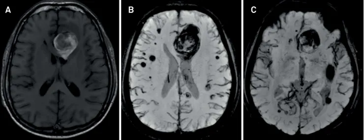
Figure 2.
Brain MRI in axial T1 (A), Axial-susceptibility-weighted (B) and multiplanar reconstruction (C) showing an heterogeneous
lesion with a hyperintense signal associated with perilesional vasogenic edema suggestive of acute hematoma. Additionally, there
are multiple nodules with hypointense signal throughout the parenchyma and a subarachnoid hemorrhage in the left Sylvian fissure.
A
B
C
References
1. Chahine LM, Berg MJ. Clinical reasoning: cerebral cavernous malformations. Neurology. 2009;73(9):e44-9.
https://doi.org/10.1212/WNL.0b013e3181b59a5b