Universidade de Lisboa
Faculdade de Ciências
Departamento de Química e Bioquímica
The role of mRNA translation on nonsense-mediated
decay inhibition of transcripts carrying a short open
reading frame
Isabel Cristina Ramos Peixeiro
Tese orientada por:
Doutora Luísa Romão Loison (Instituto Nacional de Saúde Dr. Ricardo Jorge)
Professora Doutora Margarida D. Amaral (Faculdade de Ciências da Universidade de Lisboa)
Doutoramento em Bioquímica
(Genética Molecular)
“I like nonsense, it wakes up the brain cells.”
Theodor Seuss Geiseil
Prefácio
O trabalho de investigação descrito na presente tese de Doutoramento foi realizado na Unidade de Investigação e Desenvolvimento do Departamento de Genética do Instituto Nacional de Saúde Dr. Ricardo Jorge, sob a orientação da Doutora Luísa Romão Loison e co-orientação da Professora Doutora Margarida Amaral, membro da Faculdade de Ciências da Universidade de Lisboa.
Este estudo teve como objectivo principal identificar o mecanismo molecular subjacente à inibição do mecanismo de nonsense-mediated mRNA decay (NMD) previamente descrita para transcritos portadores de um codão de terminação prematuro localizado na proximidade do codão de iniciação. Este trabalho contribuiu para a identificação de novos determinantes implicados na indução do mecanismo de NMD e na tradução. Em conformidade com o disposto no nº 5 do artigo 41º do Regulamento dos Estudos Pós-Graduados da Universidade de Lisboa, deliberação nº 93/2006, publicado em Diário da República, 2ª série – Nº 209 – 30 de Outubro de 2006, esta tese apresenta-se em língua inglesa e inclui um resumo em português com mais de 1200 palavras (ver Resumo).
De acordo com o nº 1 do mesmo artigo do referido Regulamento, na elaboração desta tese foi aproveitado o resultado do trabalho de colaboração já publicado numa revista de circulação internacional, estando a minha contribuição pessoal devidamente indicada:
Peixeiro I, Inácio A, Barbosa C, Silva AL, Liebhaber SA and Romão L. (2011) Interaction of
PABPC1 with the translation initiation complex is critical to the NMD resistance of AUG-proximal nonsense mutations. Nucleic Acids Research doi:10.1093/nar/gkr820.
No âmbito do trabalho realizado para a obtenção desta dissertação foi ainda publicado um artigo de revisão numa revista de circulação internacional:
Peixeiro I, Silva AL, Romão L. (2011) Control of human beta-globin mRNA stability and its
Este trabalho foi financiado pela Fundação para a Ciência e Tecnologia (FCT) na forma de uma Bolsa de Doutoramento com a Referência SFRH/BD/35962/2007 e através do Programa de Financiamento Plurianual do Center for Biodiversity, Functional and
Integrative Genomics (BioFIG) e do Centro de Investigação em Genética Molecular
Humana (CIGMH), e pela Fundação Luso-Americana para o Desenvolvimento (FLAD). O desenvolvimento deste trabalho não teria sido possível sem a colaboração de várias pessoas, às quais quero expressar os meus agradecimentos.
Gostaria de começar por agradecer à minha orientadora, Dra. Luísa Romão Loison, pela oportunidade de trabalhar no seu grupo. Foram a sua paciência, entusiasmo e orientação que constituíram os pilares desta tese. Durante o desenvolvimento deste trabalho, partilhou comigo as suas ideias brilhantes e palavras de encorajamento. Agradeço-lhe ainda pelo seu apoio, pelos seus conselhos e amizade.
Gostaria de agradecer à Prof. Margarida Amaral pela co-orientação deste trabalho de Doutoramento, pelo seu apoio e pelo interesse demonstrado no desenvolvimento do mesmo.
Gostaria de expressar os meus agradecimentos ao Prof. Steve Liebhaber pelas discussões produtivas e ideias que tanto contribuíram para o desenvolvimento do trabalho aqui apresentado.
Gostaria ainda de expressar o meu reconhecimento ao Dr. João Lavinha por me acolher no Departamento de Genética do Instituto Nacional de Saúde Dr. Ricardo Jorge e pelas suas sugestões e comentários nos manuscritos produzidos durante o desenvolvimento desta tese.
Agradeço também ao Dr. José Manuel Furtado pelo seu apoio na cultura de células e pelas suas palavras encorajadoras.
Aos meus colegas no instituto gostaria de agradecer por partilharem comigo os altos e baixos que o trabalho científico acarreta. Agradeço também por ter tido a oportunidade de conhecer excelentes pessoas, como os meus colegas de laboratório, Cristina Barbosa, Ana Luísa Silva, Ana Morgado, Francisco Pereira, Rute Martins, Alexandre Teixeira, Ana Ramos, Rafaela Lacerda, David Cruz e Cláudia Onofre; com quem pude partilhar as experiências e dificuldades não só laboratoriais, mas também de vida. Agradeço principalmente à Cristina, que é agora uma das minhas melhores e mais queridas
amigas, pela sua compreensão, apoio e incentivo durante todo o processo, e acima de tudo, pela sua amizade e constante presença nos melhores e nos piores momentos. Não poderia deixar de agradecer à Cristina e à Rute pela paciência e ajuda preciosa nas correcções desta dissertação.
Agradeço a todos os meus amigos, alguns dos quais já mencionados. Agradeço à Inês, à Sylvie, à Tânia, ao Hugo, ao Rui, à Sónia, à Andreia e ao resto do grupo da Faculdade de Ciências pelo seu apoio e amizade, e por conseguirem estar sempre presentes, mesmo quando uma grande distância nos separa. Agradeço ainda à Cláudia, a minha amiga de sempre, pelo seu constante apoio; à Bisca pela sua alegria e boa disposição; à Maria, à Daniela e a tantos outros por todo o apoio, pelo carinho e amizade.
Gostaria de agradecer também à minha família pelo apoio e amor com que, sempre, me envolveram. Agradeço aos meus pais, irmão e irmã por sempre me incentivarem a seguir o meu coração e os meus sonhos. Foram os sábios conselhos e o apoio incessante dos meus pais que me forneceram as ferramentas necessárias para enfrentar a vida, e para tal, um simples obrigada não é suficiente.
Acima de tudo, agradeço ao meu marido André pela sua paciência e apoio constante. Sem o seu encorajamento e compreensão teria sido impossível concluir este trabalho. Obrigada pelo teu amor total e incondicional que me faz ultrapassar tudo. Por fim, gostaria de agradecer ao meu filho. O Tiago ilumina a minha vida e, por isso, lhe dedico esta tese.
Acknowledgements
This work would have not been completed without the support of all the kind people around me. Though it will not be enough to express my gratitude in words, I would still like to thank to those who gave me the possibility to complete this thesis.
First of all, I would like to give my sincere thanks to my supervisor Dr. Luísa Romão Loison, for accepting me in her research group. Her patience, enthusiasm and guidance have provided a good basis for the present thesis. Throughout my PhD, she provided encouragement, excellent teaching and lots of good ideas. For her support, good advice and friendship I am extremely grateful.
I would like to thank Prof. Margarida Amaral for the co-supervision of my PhD, for her support and interest in the development of this work.
I am most grateful to Prof. Steve Liebhaber for the productive discussions and valuable ideas related to my work, which have been very helpful.
I also acknowledge Dr. João Lavinha for allowing me to carry out this study at
Departamento de Genética from Instituto Nacional de Saúde Dr. Ricardo Jorge and for
his helpful suggestions.
I am also grateful to Dr. José M. Furtado for his support in cell culture, for his generosity and encouragement.
To my colleagues in the institute I would like to thank for sharing the glory and sadness of day-to-day research. It was a pleasure to share doctoral studies and life with wonderful people like Cristina Barbosa, Ana Luísa Silva, Ana Morgado, Francisco Pereira, Rute Martins, Alexandre Teixeira, Ana Ramos, Rafaela Lacerda, David Cruz and Cláudia Onofre. I am especially grateful to Cristina, who had become a very close friend, for her understanding, support and encouragement throughout, but above all, for her friendship and help in difficult times. I also thank Cristina and Rute for their precious help in proof reading this document.
To all of my friends, some of whom have already been mentioned, I express my gratitude. I thank Inês, Sylvie, Tânia, Hugo, Rui, Sónia, Andreia and the rest of the group from Faculdade de Ciências for their support and devoted friendship, and for always being around. I also thank Cláudia, Maria, Bisca, Daniela and so many others for their support and caring.
I would also like to thank my family for the support and love they provided me through my entire life. I thank my parents, brother and sister who had always encouraged me to follow my heart. My parents provided me a good foundation with which to meet life, for which my mere expression of thanks likewise does not suffice.
Above all I would like to thank my husband André for his great patience and support, without any complain or regret. Without his encouragement and understanding, it would have been impossible for me to finish my PhD. His complete and unconditional love carries me through always. In addition, these acknowledgements would not be complete if I did not mention my son, Tiago. He has been a bright light in my life. To him I dedicate this thesis.
Resumo
O mecanismo de decaimento do mRNA mediado por mutações nonsense, ou
nonsense-mediated mRNA decay (NMD), é um processo de controlo de qualidade da expressão
génica que permite a eliminação de transcritos portadores de codões de terminação da tradução prematura (CTPs). A eliminação dos transcritos anómalos permite reduzir a produção de proteínas truncadas, protegendo a célula dos efeitos dominantes negativos resultantes da acumulação de péptidos potencialmente tóxicos (Behm-Ansmant & Izaurralde, 2006; Chang et al, 2007). Este mecanismo desempenha não só uma função como modulador de doenças genéticas, mas também uma importante função na regulação de transcritos fisiológicos envolvidos em vários processos celulares (McGlincy et al, 2010; Mendell et al, 2004; Rehwinkel et al, 2006).
Nos mamíferos, durante o processo de splicing, é depositado um complexo proteico, cerca de 20-24 nucleótidos (nt) a montante de cada junção exão-exão, designado complexo junção exão-exão (exon junction complex – EJC) (Le Hir et al, 2001). Estes complexos permanecem associados ao mRNA durante o seu transporte para o citoplasma e são removidos pelo ribossoma durante a primeira ronda de tradução (Ishigaki et al, 2001; Lejeune et al, 2002; Maquat, 2004). Assim, admite-se que, no caso de existir um CTP situado a 50-55 nt a montante da última junção exão-exão, o transcrito permanecerá associado a pelo menos um EJC, que poderá induzir a activação do mecanismo de NMD. A relação entre os EJCs e o NMD é mediada por três factores UPF1, UPF2 e UPF3, considerados os principais efetores do processo de NMD. A interacção das proteínas UPF2 e UPF3, componentes do EJC, com a proteína UPF1, que é recrutada para o complexo de terminação pelos factores de terminação da tradução eRF1 e eRF3, resulta na activação do mecanismo de NMD (Kashima et al, 2006; Le Hir et al, 2001). Por outro lado, quando o codão de terminação se localiza depois da última junção exão-exão, o ribossoma atinge o codão de terminação depois de remover todos os EJCs, que não poderão, assim, desencadear a degradação do transcrito (Maquat, 2004).
No decorrer do últimos anos, vários estudos têm vindo a evidenciar o envolvimento da proteína citoplasmática 1 de ligação à cauda poli(A), PABPC1, no processo de discriminação entre codões de terminação normais e prematuros. A proteína PABPC1
compete com a UPF1 pela interacção com o complexo de terminação e estimula a eficiência da terminação da tradução. De facto, existem evidências experimentais de que a proximidade da PABPC1 ao codão de terminação é suficiente para inibir o mecanismo de NMD, mesmo na presença de um EJC a jusante (Eberle et al, 2008; Ivanov et al, 2008; Silva et al, 2008; Singh et al, 2008). Desta forma, quanto maior a distância entre o codão de terminação prematuro e a cauda poli(A), maior será a probabilidade de desencadear o NMD, e a presença de um EJC a jusante poderá funcionar como um potenciador da degradação do transcrito (Stalder & Muhlemann, 2008).
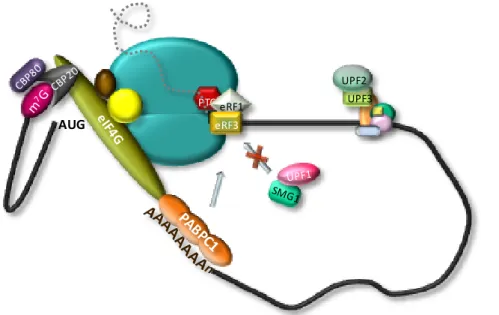
De acordo com este modelo, a localização de um codão de terminação prematuro na proximidade do codão de iniciação (AUG) será suficiente para activar o NMD. No entanto, verificou-se que os transcritos da β-globina humana portadores de CTPs no início do primeiro exão constituem uma excepção a esta regra, uma vez que apresentam uma estabilidade semelhante à do transcrito normal (Romao et al, 2000). A análise funcional destes transcritos permitiu concluir que a resistência ao mecanismo de NMD não é uma consequência da ocorrência de re-iniciação da tradução e é independente quer da identidade do transcrito quer do contexto da sequência (Inacio et al, 2004; Silva et al, 2006). De facto, estudos anteriores indicam que a proximidade do codão de terminação ao AUG é o principal determinante na inibição do NMD (Silva et al, 2006; Silva et al, 2008). Com o objectivo de compreender o mecanismo molecular subjacente ao efeito da proximidade do CTP ao AUG foi proposto um modelo que postula que a PABPC1, por se encontrar na vizinhança do AUG iniciador durante o processo de tradução de uma grelha de leitura curta, poderá estar implicada na resistência ao NMD dos transcritos portadores de um CTP próximo do codão de iniciação (Silva et al, 2008; Silva & Romao, 2009). Este modelo baseia-se na capacidade da PABPC1 interagir com a cauda poli(A), na extremidade 3’ do mRNA, e em simultâneo com o factor de iniciação eIF4G, associado à extremidade 5’ do transcrito. Esta interacção entre as duas extremidades dos mRNAs durante a tradução resulta na aquisição de uma conformação circular (Wells et al, 1998). As evidências experimentais que indicam que alguns dos factores de iniciação da tradução envolvidos no processo de scanning permanecem associados ao ribossoma durante os primeiros passos do alongamento da tradução (Poyry et al, 2004) levaram-nos a sugerir que a PABPC1 poderia ser trazida para a vizinhança do AUG em associação com o complexo de iniciação. Assim, no caso de um
evento de terminação demasiadamente prematuro, a PABPC1 manter-se-ia numa posição favorável para interagir com o complexo de terminação e estimular a eficiência da terminação da tradução, com a consequente supressão do NMD (Silva & Romao, 2009).
O trabalho apresentado nesta dissertação focou-se na identificação dos determinantes da resistência ao NMD de transcritos portadores de um CTP na proximidade do AUG, contribuindo, assim, para a compreensão do mecanismo de discriminação de um codão prematuro e do mecanismo geral da tradução.
Com o objectivo de confirmar o papel da PABPC1 na inibição do NMD de transcritos portadores de um CTP localizado na proximidade do codão de iniciação, analisámos o efeito da ausência da interacção PABPC1-eRF3 na estabilidade destes transcritos. Mediante a inibição específica da expressão da proteína PABPC1 endógena por interferência de RNA e expressão de uma proteína mutante PABPC1 com uma deleção na região C-terminal (domínio responsável pela interacção com eRF3), observámos uma desestabilização dos mRNAs portadores de CTP na proximidade do AUG. Este resultado foi corroborado pela análise da estabilidade destes transcritos em condições de inibição da expressão da eRF3 endógena e expressão de uma proteína mutante eRF3 sem a região de interacção com PABPC1 (domínio N-terminal). Estes resultados sugerem que, à semelhança do que acontece nos casos de codões de terminação na parte 3’ do último exão, quando o CTP se encontra posicionado na vizinhança do AUG, a PABPC1 interage com o complexo de terminação, impedindo a interacção deste complexo com a UPF1, com a consequente inibição do NMD.
Adicionalmente, demonstrámos que a resistência ao NMD dos transcritos portadores de um CTP próximo ao AUG pode ser reduzida pela inibição da interacção entre a PABPC1 e o factor do complexo de iniciação eIF4G resultante da sobreexpressão da proteína PAIP2. De facto, foi possível reverter o efeito destabilizador da sobreexpressão desta proteína mediante a inibição da expressão da UPF1, o principal mediador do mecanismo de NMD.
O factor de iniciação da tradução eIF3 é um complexo multiproteico que interage com o ribossoma e a proteína eIF4G, funcionando como um elo de ligação entre os dois durante o processo de scanning (Hinnebusch, 2006; LeFebvre et al, 2006). O facto deste complexo ter a capacidade de se manter associado ao ribossoma durante os primeiros
passos de elongação (Pestova, 2007) levou-nos a questionar se algumas subunidades do eIF3 poderiam estar envolvidas no posicionamento da PABPC1 na vizinhança do CTP próximo do AUG. Os nossos resultados demonstraram não só que as subunidades eIF3F e eIF3h são essências para a interacção do complexo eIF3 com o factor eIF4G e o ribossoma, respetivamente, mas também para a supressão do NMD de transcritos portadores de um CTP na proximidade do codão de iniciação.
Por outro lado, uma vez que existem evidências experimentais em plantas que demonstram que a subunidade eIF3h é essencial para a re-iniciação da tradução num codão AUG a jusante do codão de terminação de pequenas grelhas de leitura a jusante (upstream open reading frame – uORF) da ORF principal; a destabilização dos transcritos em estudo em células com uma expressão reduzida da eIF3h poderia ser um resultado da incapacidade do ribossoma re-iniciar a tradução num AUG a jusante do CTP. No entanto, o estudo do efeito da eIF3h na re-iniciação da tradução após a tradução da uORF da eritropoietina humana demonstrou que a eficiência da re-iniciação da tradução não é afectada em condições de inibição da expressão da eIF3h.
Em resumo, o trabalho desenvolvido nesta dissertação contribuiu para a elucidação da base molecular subjacente ao efeito da proximidade do AUG na inibição do NMD de transcritos portadores de um CTP próximo ao codão de iniciação. Os resultados obtidos demonstram que esta resistência é dependente da interacção da PABPC1 com o complexo de iniciação da tradução, através do eIF4G e das subunidades eIF3f e eIF3h, que, no caso de um CTP localizado na proximidade do AUG iniciador, posiciona a PABPC1 numa situação favorável para interagir com o complexo de terminação, através do factor de terminação eRF3, com a consequente supressão do NMD. Deste modo, o trabalho aqui apresentado, que inclui resultados já publicados num artigo científico e outros que serão submetidos em breve para publicação, contribui para a caracterização dos determinantes da discriminação de um codão prematuro e activação/inibição do mecanismo de NMD. A elucidação da função de factores de iniciação da tradução no efeito supressor de NMD resultante da proximidade do AUG ao CTP contribui também para a compreensão de conceitos da tradução, nomeadamente para o caso de pequenas grelhas de leitura.
Palavras-chave
mRNA; β-globina humana; codão de terminação prematuro; decaimento do mRNA mediado por codões nonsense; controlo de qualidade do mRNA; tradução
Abstract
Nonsense-mediated mRNA decay (NMD) is a surveillance pathway that recognizes and rapidly degrades mRNAs containing premature termination codons (PTC). The unified model for NMD, proposes that the decision of NMD triggering is the outcome of the competition between the cytoplasmatic poly(A)-binding protein 1 (PABPC1) and the NMD effector UPF1 for the termination complex. Consequently, PTCs located far, in a linear sense, from the poly(A) tail and associated PABPC1 in mRNAs containing residual downstream exon junction complexes (EJCs) are expected to elicit NMD. Nevertheless, it has been reported that mRNAs containing PTCs in close proximity to the translation initiation codon (AUG-proximal PTCs) can substantially evade NMD. The work described in this thesis was focused on the mechanistic basis for this NMD resistance, exemplified by human β-globin transcripts carrying an AUG-proximal PTC. We demonstrated that translation termination at an AUG-proximal PTC lacks the ribosome stalling that is evident in an NMD-sensitive PTC. In fact, we have shown that the establishment of an efficient translation termination reaction at the AUG-proximal PTC is dependent on PABPC1 interaction with the initiation factor eIF4G and with the release factor eRF3 at the terminating ribosome. These interactions underlie critical 3’-5’ linkage of translation initiation with efficient termination at the AUG-proximal PTC and contribute to an NMD-resistant PTC definition at an early phase of translation elongation. Furthermore, we provide strong evidence that the eukaryotic initiation factor 3 is involved in delivering eIF4G-associated PABPC1 into the vicinity of the AUG-proximal PTC through eIF3h and eIF3f subunits. These data support the “AUG-proximity effect” as a major inhibitor of the NMD pathway.
The work presented here corroborates a role for PABPC1 on NMD evasion of AUG-proximal transcripts and provides further insights into the mechanistic details of PTC definition and translation initiation.
Keywords
mRNA; human β-globin; premature termination codon; nonsense-mediated mRNA decay; mRNA surveillance; translation
Abbreviations
4E-BP1 eukaryotic translation initiation factor 4E-binding protein 1
A adenosine
Aa aminoacid
Ab antibody
ADP adenosine diphosphate
A-site aminoacyl-site
ATP adenosine triphosphate
bp base pairs
BRCA1 breast cancer protein 1
BTZ Barentz
C cytidine
CBC cap binding complex
CBP cap binding protein
cDNA mRNA-complementary DNA
CH cysteine- and histidine-rich domains
CHX cyclohexamide
C-terminal carboxyl-terminal
DCP decapping-protein
DEAD Asp-Glu-Ala-Asp motif
DECID decay-inducing complex
DHX34 DEAH box polypeptide 34
DMD Duchenne muscular dystrophy
DMEM Dulbecco’s modified Eagle medium
DMSO dimethyl sulfoxide
DNA deoxyrybonucleic acid
DNase deoxyribonuclease
dNTP deoxynucleoside triphosphate
DSE downstream sequence element
DTT dithiothreitol
EDTA ethylenediaminotetraacetic acid
eEF eukaryotic translation elongation factor
eIF eukaryotic translation initiation factor
EJC exon junction complex
EPO erythropoietin
eRF eukaryotic translation release factor
E-site exit-site
G guanosine
GAPDH glyceraldehyde-3-phosphate dehydrogenase
GCN2 general control non-derepressible-2 protein
GDP guanosine diphosphate
GFP green-fluorescent protein
GTP guanosine triphosphate
HDAC histone deacetylase 1
Ig immunoglobulin
IP immunoprecipitation
IRES internal ribosome entry site
LUC luciferase
m7G 7-methylguanosine
MAGOH mago-nashi homolog
Met methionine
Met-tRNAi methionine-loaded initiator tRNA
MIF4G middle of eIF4G-like domain
mRNA messenger ribonucleic acid
mRNP messenger ribonucleoprotein particle
mTOR mammalian target of rapamycin
NLS nuclear localization signals
NMD nonsense-mediated mRNA decay
NP40 nonidet-P40
nt nucleotide
N-terminal amino-terminus
ORF open reading frame
p53 tumour protein 53
PABP poly(A)-binding protein
PABPC1 cytoplasmic poly(A)-binding protein 1
PABPN nuclear poly(A)-binding protein
PAGE polyacrilamide gel electrophoresis
PAIP2 poly(A)-binding protein-interacting protein 2
PAN poly(A) nuclease
PARN poly(A) ribonuclease
P-bodies processing bodies
PBS phosphate buffer saline
PCR polymerase chain reaction
PERK PKR-like endoplasmic reticulum kinase
Pi inorganic phosphate
PIC pre-initiation complex
PIKK phosphatidylinositol 3-kinase-related protein
PIN PilT N terminus
PKR protein kinase activated by double-stranded RNAs
Poly(A) poly-adenilate
PP2A protein phosphatase 2a
Pre-mRNA messenger ribonucleic acid precursor
P-site peptidyl-site
PTC premature translation termination codon
Puror puromycin resistance
PVDF polyvinylidene difluoride
REF RNA and export factor binding protein
RNA ribonucleic acid
RNAi RNA interference
RNase ribonuclease
RPA ribonuclease protection assay
rpS6 ribosomal protein small subunit 6
RRL rabbit reticulocyte lysate
RRM RNA recognition motif
RT reverse-transcription
RT-qPCR reverse transcription quantitative polymerase chain reaction
SDS sodium dodecyl sulphate
Ser serine
siRNA short interfering RNA
SKI superkiller
SMG suppressor of morphological defects on genitalia
sORF short open reading frame
Srm160 SR-related nuclear matrix protein of 160 kDa
STAU1 staufen-1
SURF SMG1-UPF1-eRFs complex
T thimidine
TCR T‐cell receptor
TERRA telomeric repeat-containing RNA
Tet tetracycline
TPI triosephosphate isomerase
Tris tris(hydroxymethyl)aminomethane
tRNA transfer ribonucleic acid
U uridine
uORF upstream open reading frame
UPF up-frameshift protein
UTR untranslated region
WT wild type
WT1 Wilms tumour protein 1
XRN1 5’-3’ exoribonuclease 1
β15 human β‐globin transcript with PTC at codon 15
β39 human β‐globin transcript with PTC at codon 39
Table of Contents
Prefácio vii Acknowledgements xi Resumo xiii Palavras-chave xvii Abstract xix Keywords xix Abbreviations xxi Table of Contents xxvChapter I – General Introduction 29
I.1. mRNA metabolism and quality control 31
I.1.1. mRNA translation 32
I.1.2. Translation regulation 35
I.2. Nonsense-mediated mRNA decay as a surveillance mechanism 37
I.2.1. Aberrant and natural NMD targets 37
I.2.2. Nonsense-mediated mRNA decay implications in disease 39
I.2.3. Molecular basis of the NMD pathway 42
I.2.3.1. UPFs: the core of the NMD machinery 44
I.2.3.2. SMGs – determining the phosphorylation status of UPF1 46
I.2.3.3. Splicing and the EJC 47
I.2.4. PTC definition 49
I.2.4.1. Pioneer round of translation 50
I.2.4.2. Proper versus aberrant termination 52
I.2.4.3. Alternative branches 53
I.2.4.4. An evolutionary model with different second signals 54
I.2.4.5. AUG-proximal PTCs unexpectedly evade NMD 57
I.2.5. Degradation of NMD targets 59
I.2.6. Feedback regulatory mechanisms 61
I.2.7. NMD effectors: other functions & new players 63
I.2.7.1. NMD factors and genome stability 63
I.2.7.2. Functions in other mRNA turnover pathways 64
I.2.7.3. NMD factors and translation 64
I.2.8. Human β-globin as a model system for studying NMD 65
Chapter II – Interaction of PABPC1 with the translation initiation complex is critical to the NMD-resistance of AUG-proximal nonsense mutations
69
Author’s note 71
II.1. Abstract 73
II.2. Introduction 73
II.3. Materials and methods 75
II.3.1. Plasmid constructs 75
II.3.2. Cell culture, plasmid and siRNA transfection 77
II.3.3. RNA isolation 77
II.3.4. Reverse transcription-coupled quantitative PCR 77
II.3.5. Semi-quantitative RT-PCR 78
II.3.6. SDS-PAGE and Western blotting 78
II.3.7. In vitro transcription and capping 79
II.3.8. Toeprinting assay 79
II.4. Results 80
II.4.1. Translation termination at an AUG-proximal PTC obviates ribosome stalling characteristic of NMD-sensitive PTCs.
80
II.4.2. PABPC1 plays an essential role in establishing NMD-resistance of an AUG-proximal nonsense-mutated mRNA
83
II.4.3. Interaction of PABPC1 with the ribosome release factor, eRF3, is critical to NMD-evasion by an AUG-proximal nonsense-mutated mRNA
86
II.4.4. Inhibition of PABPC1 function by PAIP2 sensitizes an AUG-proximal nonsense-mutated mRNA to NMD
87
II.4.5. The eIF3 subunits that tether the 40S ribosomal subunit to eIF4G are required for NMD-resistance of an AUG-proximal nonsense-mutated mRNA
89
II.5. Discussion 92
II.6. Funding 96
II.6. Acknowledgements 96
Chapter III – Further insights into the role of eIF3f and eIF3h non-conserved subunits on translational regulation
97
Author’s note 99
III.1. Abstract 101
III.2. Introduction 101
III.3. Materials and methods 104
III.3.1. Plasmid constructs 104
III.3.2. Cell culture, plasmid and siRNA transfection 105
III.3.4. Semi-quantitative RT-PCR 106
III.3.5. Dual luciferase assay 107
III.3.6. In vitro transcription and capping 107
III.3.7. Immunoprecipitation 107
III.3.8. SDS-PAGE and Western blotting 108
III.3.9. GST-Pull down assays 108
III.4. Results 109
III.4.1. mRNA acquires a closed-loop conformation during in vitro translation in rabbit reticulocyte lysate
109
III.4.2. eIF3f silencing affects eIF3h expression 110
III.4.3. eIF3f depletion decreases eIF4G-eIF3 interaction and increases CBP80-mRNA association
112
III.4.4. eIF3h is involved in eIF3 interaction with the 40S ribosomal subunit 113
III.4.5. Efficiency of translation reinitiation downstream the erythropoietin uORF is not affected by the knockdown of eIF3 non-conserved subunits
114
III.5. Discussion 115
III.6. Funding 118
III.6. Acknowledgements 119
Chapter IV – General Discussion 121
Conclusions and future directions 127
Chapter V – References 129
Figures
I.1. The canonical pathway of eukaryotic translation 33
I.2. Nonsense-mediated mRNA decay modulates the disease phenotype 40
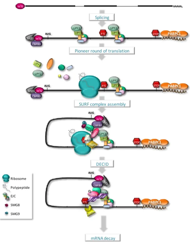
I.3. Nonsense-mediated mRNA decay occurs during the pioneer round of translation
51
I.4. The unified model for nonsense-mediated mRNA decay 56
I.5. Model for the AUG-proximity effect on NMD-evasion of transcripts harboring an AUG-proximal PTC
59
I.6. Decay pathways of human NMD 61
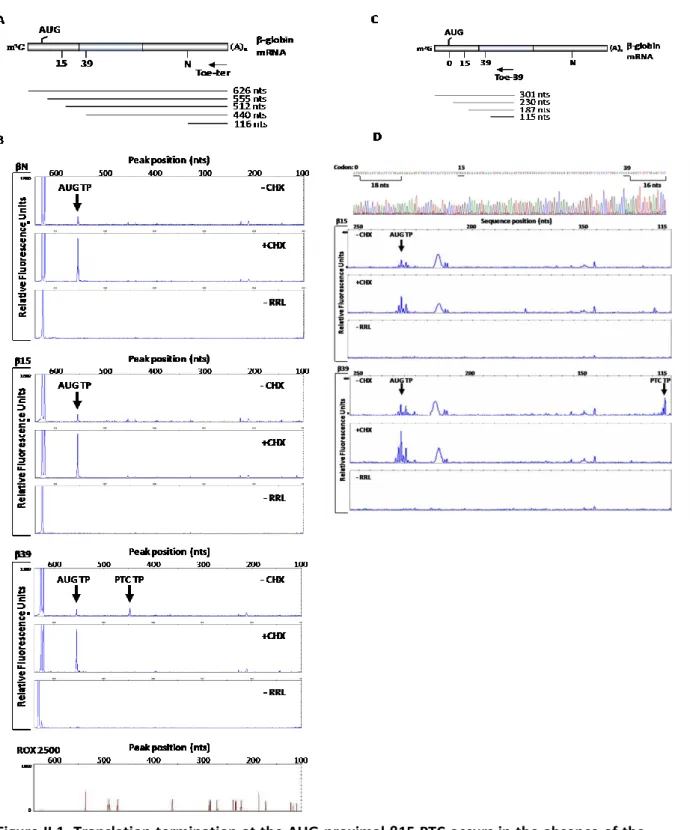
II.1. Translation termination at the AUG-proximal β15 PTC occurs in the absence of the ribosome stalling evident at the more distal β39 PTC
81
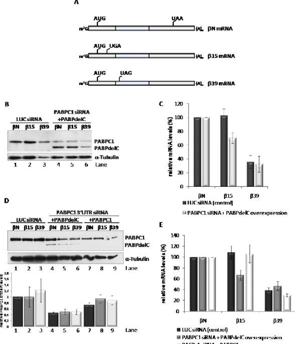
II.2. PABPC1 plays an essential role in NMD-resistance of an AUG-proximal PTC 84
II.3. The N-terminal domain of eRF3 is critical to NMD-evasion by an AUG-proximal nonsense-mutated mRNA
II.4. Over-expression of PAIP2 induces NMD sensitivity in the β15 nonsense-mutated mRNA
88
II.5. The translation initiation factors eIF3h and eIF3f are required for the NMD-resistant phenotype of the β15 nonsense-mutated mRNA
90
II.6. A model for NMD-resistance of AUG-proximal nonsense-mutated mRNAs 92
III.1. 5’-3’ mRNA ends interaction during translation in rabbit reticulocyte lysates 110
III.2. Knocking down eIF3f subunit destabilizes eIF3h expression 111
III.3. eIF3f depletion decreases eIF4G-eIF3 interaction and increases CBP80-mRNA association
113
III.4. eIF3h depletion affects eIF3 interaction with the 40S ribosomal subunit 114
III.5. Efficiency of translation reinitiation downstream the erythropoietin uORF is not affected by eIF3h, eIF3f or eIF3e depletion
116
Tables
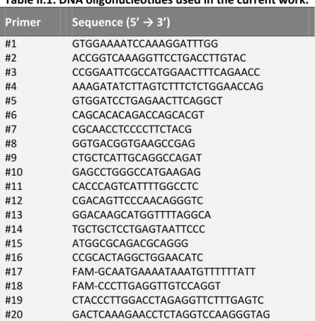
II.1. DNA oligonucleotides used in the current work 76
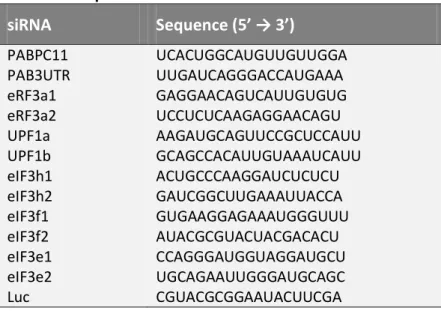
II.2. Sequences of the siRNAs used in the current work 78
III.1. DNA oligonucleotides used in the current work 105
C
C
h
h
a
a
p
p
t
t
e
e
r
r
I
I
General Introduction
I.1. mRNA metabolism and quality control
Eukaryotic gene expression involves a sequence of biochemical processes that initiates with transcription of the genetic material into messenger RNAs (mRNAs) followed by protein synthesis and that, ultimately, culminates in mRNA and protein degradation. The key intermediate, mRNA precursor, is produced by DNA transcription in the nucleus. The pre-mRNA is then subjected to removal of introns (splicing) and addition of the cap structure and the poly(A) tail, and this mature mRNA is transported into the cytoplasm where translation occurs. Throughout this process, mRNA molecules are in dynamic association with RNA-binding proteins constituting the messenger ribonucleoprotein particle (mRNP) (Dreyfuss et al, 2002; Isken & Maquat, 2007; Moore, 2005). The mRNP content influences the mRNA fate by mediating its cellular localization, translation and decay, responding to cellular signal networks (Moore, 2005). This post-transcriptional regulatory mechanism allows the cells to rapidly adapt to changes in their environment by altering the patterns of gene expression (Silva & Romao, 2009). A tight control of such a mechanism is therefore essential to guarantee cell survival. Thus, to ensure the quality and fidelity of RNA metabolism and function, cells have evolved quality control mechanisms that preferentially degrade aberrant transcripts or nonfunctional RNAs (Doma & Parker, 2007). These quality control mechanisms, also called mRNA surveillance pathways, operate in the nucleus, where improperly processed mRNAs are degraded before export; and in the cytoplasm, where the quality control mechanisms assess the translatability of the mRNA and degrade those that lack translation termination codons or that carry premature translation termination codons (Behm-Ansmant et al, 2007b). In fact, the quality-control mechanisms do not need to recognize specific defective features of an RNA or RNP but, instead, can function on a broad range of defects affecting the rate of a given step in metabolism or function (Doma & Parker, 2007). Moreover, the function of these mRNA surveillance pathways extends to the post-transcriptional regulation of gene expression, since they act not only in aberrant mRNAs, but also regulate the expression of naturally occurring transcripts that present features recognized by the surveillance machinery (Behm-Ansmant et al, 2007b).
I.1.1. mRNA Translation
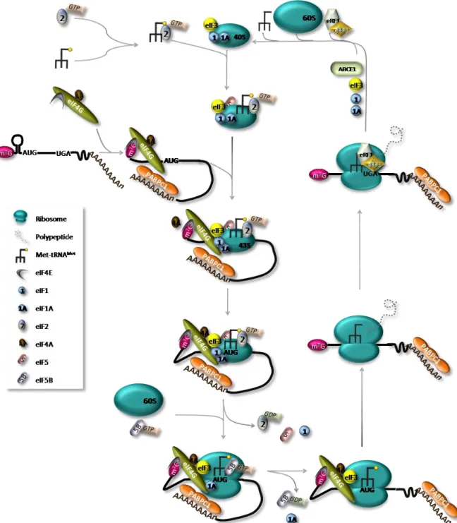
Translation regulation of existing mRNAs allows for a spatial and temporal fine-tuning of levels of the encoded proteins, allowing the maintenance of cellular homeostasis (Gebauer & Hentze, 2004; Sonenberg & Hinnebusch, 2009). The translation process can be divided into four stages - initiation, elongation, termination and ribosome recycling – each of which requires a particular set of conditions and factors (Figure I.1) (Sonenberg & Hinnebusch, 2009). Translation initiation is the rate-limiting step and requires the function of several eukaryotic translation initiation factors (eIFs) (Livingstone et al, 2010). This complex event involves the assembly of elongation-competent 80S ribosomes at the initiation codon (AUG). On most mRNAs, the start codon is identified by a scanning mechanism, where the 43S pre-initiation complex binds to the mRNA near the 5’ end and scans the 5’ untranslated region (5’UTR) for an AUG codon. The 43S pre-initiation complex comprises the small (40S) ribosomal subunit, the eIFs 3, 1, 1A and 5, and a ternary complex, which consists of the methionine-loaded initiator tRNA (Met-tRNAiMet) and eIF2 coupled to GTP (Gebauer & Hentze, 2004). Binding of this complex to the mRNA requires the cooperative action of eIF4F and eIF4B or eIF4H, which unwind the 5’UTR of the mRNA allowing ribosomal attachment. eIF4F is composed by eIF4E protein, that binds the m7G cap structure, the DEAD-box RNA helicase eIF4A and by eIF4G, which functions as a scaffold protein by interacting with eIF4E, eIF4A, eIF3 and with the cytoplasmic poly(A)-binding protein 1 (PABPC1) (Jackson et al, 2010). The simultaneous interaction of eIF4E and PABP with eIF4G is believed to circularize the mRNA, bringing the 3’UTR in close proximity to the 5’end of the mRNA (Wells et al, 1998). The functional connection between the mRNA ends during translation is highlighted by the fact that most known regulatory sequences are found within the 3’UTR (Gebauer & Hentze, 2004).
After loading to the mRNA, 43S complex scans downstream to the initiation codon. The scanning process is assisted by eIF4A, eIF4G, eIF4B and possibly by eIF3 (Jackson et al, 2010; Pestova & Kolupaeva, 2002). eIF3 plays a key role in recruiting the 43S pre-initiation complex to the mRNA and, at least in mammals, also forms a bridge to the mRNA by interacting with eIF4G (Hinnebusch, 2006; Pestova, 2007).
Figure I.1. The canonical pathway of eukaryotic translation. The ternary complex, comprising eIF2-GTP-Met-tRNAiMet, binds the 40S ribosomal subunit together with eIFs 1, 1A, 3 and 5, creating the 43S pre-initiation complex. eIF4G functions as a scaffold protein by interacting with other components of the eIF4F complex, eIF4E and eIF4A, and with the poly(A)-binding protein PABPC1. This interaction is presumed to circularize the mRNA. The eIF4F and eIF4B or eIF4H, will then unwind the 5’ UTR of the mRNA allowing 43S complex attachment and 5’ to 3’ scanning by the 43S complex. After recognition of the AUG initiation codon and 48S initiation complex formation, eIF1 is displaced and eIF5 mediates the hydrolysis of eIF2-bound GTP. Joining of the 60S ribosomal subunit will cause the displacement eIF2-GDP and other eIFs mediated by eIF5B and the assembly of 80S elongation-competent ribosomes induces the release of eIF1A and eIF5B. After translation termination, recycling generates separated ribosomal subunits. Although not represented, the potential interaction of PABPC1 with the release factor eRF3 may also result in formation of a closed-loop structure during translation termination.
eIF1 has a crucial role in the selection of initiation codons, allowing scanning 43S complexes to discriminate against codon-anticodon mismatches and preventing premature eIF5-induced hydrolysis of eIF2-GTP and Pi release (Holcik & Pestova, 2007). eIF1A also regulates start codon selection promoting continued scanning at non-AUG codons or by arresting scanning and promoting eIF1 release at AUG codons (Sonenberg & Hinnebusch, 2009). The Met-tRNAiMet is anchored to the 43S complex by the GTP-bound form of eIF2, and a perfect match with an AUG start codon [usually the first AUG triplet in an optimum context – GCC(A/G)CCAUGG, with a purine at the -3 and a G at the +4 positions (relative to the A of the AUG codon, which is designated +1) (Kozak, 1991)]
triggers the arrest of scanning and irreversible hydrolysis of GTP in the eIF2-GTP-Met-tRNAi ternary complex. This step, during which the ribosome becomes committed to initiation at that codon, is catalyzed by eIF5, an eIF2-specific GTPase-activating protein (Gebauer & Hentze, 2004; Jackson et al, 2010). With the release of eIF2-GDP and other eIFs, mediated by eIF5B, the large (60S) subunit joins to form an 80S initiation complex ready to accept the appropriate aminoacyl-tRNA into the A- (aminoacyl) site and synthesize the first peptide bond (Pestova, 2007). Because eIF3 binds mainly to the solvent side of the 40S subunit, the dissociation of this factor is not essential for subunit joining and may thus be delayed (Szamecz et al, 2008; Valasek et al, 2002).
During the elongation stage, amino acids are added sequentially to the growing polypeptide chain (Abbott & Proud, 2004). The ribosome has three tRNA-binding sites: the A-site, which accepts the incoming aminoacyl-tRNA, the P- (peptidyl) site, which holds the tRNA with the nascent peptide chain and the E- (exit) site that holds the deacylated tRNA before it leaves the ribosome (Proud, 1994). The key factor in this process is the elongation factor (eEF) 1A which delivers cognate tRNA to the A-site, upon hydrolysis of GTP. The nascent peptide chain is then transferred to the amino acid of the A-site aminoacyl-tRNA. The translocation of peptidyl-tRNA from A- to P- and of deacylated tRNA from P- to E- sites is then promoted by eEF2; this process is also dependent on GTP hydrolysis (Abbott & Proud, 2004; Pisarev et al, 2007).
Termination of translation is triggered by peptide release factors eRF1 and eRF3. When a stop codon enters the A-site, eRF1 induces the hydrolysis of the ester bond of the P-site peptidyl-tRNA (Pestova, 2007). The function of eRF3 is to ensure the rapid and efficient hydrolysis of the peptidyl-tRNA by eRF1 (Alkalaeva et al, 2006). It is suggested that eRF1,
eRF3 and GTP bind the pre-termination complexes as a ternary complex. However, the mechanism of post-termination dissociation of the release factors is unknown (Pisarev et al, 2007).
After translation termination, the mRNA and P-site deacylated tRNA remain associated with ribosomes in post-termination complexes. eIFs 3, 1, 1A and 3j cooperatively dissociate such complexes into free 60S subunits and mRNA- and tRNA-bound 40S subunits (Pisarev et al, 2007). According to the model proposed by Pisarev et al. (2007), eIF1 promotes dissociation of P-site deacylated and eIF3j mediates release of mRNA. Ribosomal recycling might influence post-termination events such as reinitiation and possibly mRNA decay pathways.
Reinitiation may occur after translation of an upstream open reading frame (uORF), if the 40S ribosomal subunit remains bound to the mRNA molecule upon translation termination. Following translation-termination and the consequent release of the 60S ribosomal subunit, the 40S subunit may resume scanning downstream from the uORF, exhibiting a conditional loss of reinitiation competence. The de novo recruitment of the eIF2-GTP-Met-tRNAiMet ternary complex, that will allow recognizing the AUG of the main ORF, determines the acquirement of full reinitiation competence (Kozak, 1987; Kozak, 2001). The reinitiation efficiency depends on i) cis-acting mRNA features; ii) the time required for the uORF translation, which is determined by the relative length of the uORF and the translation elongation rate; and iii) the translation initiation factors involved in the translation initiation event (Kozak, 2001; Poyry et al, 2004). It has been hypothesized that the eIFs involved in resuming scanning remain at least transiently associated with the elongating ribosome, and increasing the uORF length or the ribosome transit time increases the odds of these factors dissociate (Kozak, 2001). Corroborating this hypothesis, in yeast, eIF3 remains 80S-bound for several rounds of elongation and enhances the reinitiation competence of post-termination 40S ribosomes (Szamecz et al, 2008).
I.1.2. Translation regulation
Global control of protein synthesis is mostly exerted at the stage of translation initiation. The mechanisms regulating initiation may impact on the eIFs or ribosomes, affecting
scanning-dependent mechanisms; or may impact directly on the mRNA, through sequence-specific RNA-binding proteins or microRNAs.
The best well-known examples of regulation mechanisms impacting on the eIFs are the control of the availability of active eIF2 and eIF4F by reversible protein phosphorylation. Phosphorylation of the α subunit of eIF2 at Ser51 blocks the GTP-exchange reaction by reducing the dissociation rate of eIF2 from eIF2B (Gebauer & Hentze, 2004). There are four mammalian protein kinases that phosphorylate eIF2α at Ser51 decreasing global translation: haem-regulated kinase, which is stimulated by haem depletion; PKR (protein kinase activated by double-stranded RNAs), which is activated by viral infection; GCN2 (general control non-derepressible-2), which is activated by amino-acid starvation; and PERK (PKR-like endoplasmic reticulum kinase), which is activated under circumstances of endoplasmic reticulum stress (Jackson et al, 2010). The availability of the cap-binding protein eIF4E is also used to regulate general translation rates. When hypophosphorylated, a eIF4E-binding protein (4E-BP) binds eIF4E competitively displacing eIF4G; but phosphorylation of the 4E-BP at multiple sites (mainly by mTOR) releases eIF4E for assimilation into eIF4F, thus promoting translation initiation (Gebauer & Hentze, 2004). The phosphorylation of several other eIFs (eIF1, eIF2β, several eIF3 subunits, eIF4G, eIF4B, eIF4H, eIF5 and eIF5B) and of ribosomal protein 6 (RPS6) increasing under conditions in which translation is activated has also been described. However, there is no concrete evidence that these phosphorylation events are the cause of such activation (Jackson et al, 2010).
Regulation of translation initiation through the mRNA itself, by a sequence-specific RNA-binding protein, is exclusive of the mRNAs that contain the relevant RNA sequence motif and is generally inhibitory, with the activation of such mRNAs requiring the inactivation or the degradation of the inhibitory protein. The regulation by specific 5’UTR-protein interactions is surprisingly rare, being the ferritin mRNAs the only well-studied example. In contrast, there are several cases, most of them important in development, of control by 3’UTR-protein interactions (Jackson et al, 2010).
While the mentioned protein-RNA interactions usually result in decreased translation, the binding of PABPC1 to the poly(A) tail stimulates translation (Borman et al, 2000). This stimulatory effect appears to result mainly from the ability of PABPC1 to interact with eIF4G, mediating mRNA circularization by linking the cap and the poly(A) tail
(Imataka et al, 1998; Wells et al, 1998). By interacting with eIF4G, PABPC1 enhances eIF4F binding to the cap of the mRNA (Kahvejian et al, 2005) and, therefore, stimulates mRNA binding do the 43S pre-initiation complex (Pestova, 2007). In fact, an in vitro study showed that the formation of the closed-loop occurs before or during the 48S complex assembly, independently of a prior termination event (Amrani et al, 2008). The formation of a second closed-loop mRNP, comprising PABPC1, eIF4G, eIF4E, eIF3 and the termination factors eRF1 and eRF3, was also described during the subunit joining to form the 80S ribosome (Amrani et al, 2008). The formation of such a circular conformation could facilitate reinitiation by post-termination ribosomes. Moreover, PABP also stimulates 60S subunit joining (Kahvejian et al, 2005) and is required to block the inhibitory effect of RNA helicases on subunit joining factor eIF5B (Searfoss et al, 2001).
I.2. Nonsense-mediated mRNA decay as a surveillance
mechanism
The best characterized mRNA quality control system in eukaryotes is nonsense-mediated mRNA decay (NMD). This surveillance mechanism detects and ensures the rapid degradation of faulty mRNAs harboring premature translation termination codons (PTCs) that would otherwise result in the synthesis of C-terminally truncated proteins. Hence, the quality control function of NMD relies in protecting the cell from the deleterious dominant-negative or gain-of-function effects of these truncated proteins (Behm-Ansmant & Izaurralde, 2006; Chang et al, 2007).
I.2.1. Aberrant and natural NMD targets
Cells produce a large number of faulty mRNAs harboring PTCs that are recognized and rapidly eliminated by NMD. PTC-harboring transcripts can arise at the DNA level as the result of germline or somatic alterations (direct nonsense-mutations; frame-shifting deletions and insertions that can lead to PTCs downstream of the shift; mutations within either an intron or an exon that results in inaccurate intron removal from the pre-mRNA and create an intron-derived PTC or a frameshift) or at the RNA level due to errors in transcription or pre-mRNA processing (Kuzmiak & Maquat, 2006; Stalder & Muhlemann,
2008). Other types of faulty mRNAs are transcripts from non-functional pseudogenes, endogenous retroviral and transposon RNAs or mRNA-like non-protein coding RNAs from intergenic regions (Mendell et al, 2004; Nicholson et al, 2010; Rehwinkel et al, 2006). By targeting these transcripts for decay, NMD exerts a role in reducing genomic noise.
In addition, PTCs can also be generated by non-faulty regulated processes of the mRNA metabolism, such as somatic rearrangements in the DNA, alternative splicing or utilization of alternative AUG initiation sites that lead to a shift in the main ORF (Holbrook et al, 2004; Kuzmiak & Maquat, 2006). In fact, unproductive alternative splicing is believed to constitute the major source of PTC-containing mRNAs in mammals, since bioinformatic approaches studies suggested that about one-third of the alternatively spliced human mRNAs contain a PTC that is targeted for NMD (Lewis et al, 2003); and, at least for same cases, it constitutes a form of autoregulation of the expression of the canonical protein-encoding isoforms, as the protein products of these genes are responsible for NMD triggering (Neu-Yilik & Kulozik, 2008). Thus, besides ensuring the degradation of faulty transcripts, NMD also has a crucial role in regulating gene expression. Indeed, genome-wide screens in NMD-deficient budding yeast, fruitfly and human cells have revealed that NMD controls the abundance of 3% - 10% of the transcriptome (He et al, 2003; Mendell et al, 2004; Rehwinkel et al, 2005; Rehwinkel et al, 2006). Examples of natural NMD targets include transcripts containing a regulatory ORF that resides upstream of the primary downstream ORF (uORFs); introns in the 3’ UTR; programmed frameshifts; and long 3’ UTRs (Kuzmiak & Maquat, 2006; Nicholson et al, 2010). Notably, mRNAs containing UGA triplets that direct selenocysteine incorporation can also elicit NMD, since the UGA codon can be interpreted as a PTC when the selenium concentration in the cell is low [reviwed in(Bhuvanagiri et al, 2010; Nicholson et al, 2010)]. The process of maturation of the immune system is other example of the physiological role of NMD. This programmed somatic rearrangements and hypermutations generate PTCs at a high frequency (Kuzmiak & Maquat, 2006; Nicholson et al, 2010).
Remarkably, many of the identified natural NMD targets encode proteins that could be grouped in clusters acting in related pathways (Neu-Yilik et al, 2004; Weischenfeldt et al, 2005). Such mRNAs are involved in a variety of cellular processes such as stress
responses, hematopoietic stem cell development, regulation of alternative splice forms, genomic stability, cell-cycle, telomere length maintenance and embryonic development (Bhuvanagiri et al, 2010; Stalder & Muhlemann, 2008).
I.2.2. Nonsense-mediated mRNA decay implications in disease
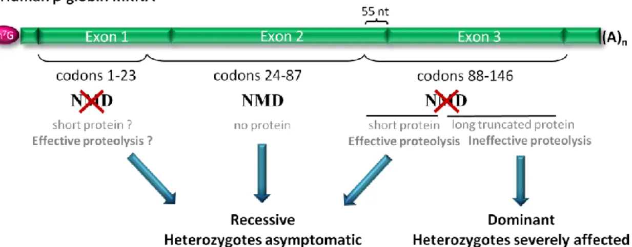
The biological and medical significance of the NMD pathway is evidenced by the fact that approximately 30% of all inherited genetic disorders are due to PTCs, and in many of these cases, NMD influences the severity of the clinical phenotype (Holbrook et al, 2004; Stalder & Muhlemann, 2008). In most cases, NMD has a beneficial effect by eliminating transcripts harboring PTCs that would otherwise give rise to truncated proteins either with a complete loss of function or with a dominant-negative function leading to toxicity (Bhuvanagiri et al, 2010). The benefits that can be achieved by the elimination of mRNAs encoding these faulty proteins are clearly illustrated in β-thalassemia (Frischmeyer & Dietz, 1999). β-Thalassemia is characterized by the absence or reduction in the synthesis of β-globin polypeptide chains, one of the hemoglobin subunits (Weatherall, 2000). This disorder exemplifies the phenotypic impact of the polar effect of PTCs at different positions between the same β-globin gene (Holbrook et al, 2004). If a PTC is located at a position that activates NMD, the disease results in a recessive mode of inheritance with the heterozygous being asymptomatic (Figure I.2). In this case the defective β-globin transcript is degraded by NMD resulting in a limited synthesis of the truncated product. The excess of free α-globin, as well as the limited amount of truncated β-globin protein (if translated) are proteolytically degraded. Indeed, it has been shown that these defects result in very low amounts of β-globin chain production from the affected allele causing a reduction about 50% of total β-globin chain synthesis in the heterozygote. This produces the clinically asymptomatic phenotype of β-thalassemia trait (also known as thalassemia minor), as heterozygotes usually synthesize enough β-globin from the remaining normal allele to support near-normal haemoglobin levels (Holbrook et al, 2004; Peixeiro et al, 2011b). If the PTC is located at a position that does not induce NMD, at less than 55 nucleotides (nts) upstream the last exon-exon junction or at the 5’ part of exon 3, the amount of truncated protein that is synthesized might be small enough to be efficiently degraded
by the proteolytic system of the red blood cell, together with the excess of free α-globin chains. In this case, the heterozygotes are also asymptomatic and the disease still results in a recessive mode of inheritance (Figure I.2). In contrast, if the PTC is located further downstream at exon 3 of the β-globin mRNA, NMD is not triggered; instead, substantial amounts of mutant β-globin mRNA are present in the patients, encoding for translation products that are long enough to overburden the cellular proteolytic system (Figure I.2). Under these conditions, accumulation of the truncated products, as well as free α-globin chains in excess, act in a dominant negative manner, leading to deleterious effects on the cell, as the nonfunctional globin chains cause toxic precipitation of insoluble globins (Holbrook et al, 2004; Neu-Yilik & Kulozik, 2008; Peixeiro et al, 2011b). This condition is related to a symptomatic form of the disease in heterozygotes named “thalassemia intermedia” with a dominant mode of inheritance (Hall & Thein, 1994).
Figure I.2. Nonsense-mediated mRNA decay modulates the disease phenotype. The example
here presented shows the human β-globin mRNA. AUG-proximal PTCs do not trigger NMD but heterozygotes are asymptomatic, as the translated short β-globin peptides, along with the β-globin chains in excess, are effectively degraded. If the PTC is located downstream of codon 23 and more than 55 nucleotides (nt) upstream of the last exon-exon junction, the corresponding transcript is targeted for NMD and heterozygotes are also protected from thalassemia. In contrast, transcripts bearing PTCs located less than 55 nt upstream of the last exon-exon junction, or in the 5’-part of exon 3, escape NMD, resulting in the production of truncated proteins that are small enough to be efficiently degraded, along with the α-globin chains in excess, and heterozygotes are also asymptomatic for β-thalassemia. However, if the PTC is located further downstream, the encoded truncated nonfunctional β-globin proteins overwhelm the cellular proteolytic system and cause toxic precipitation of insoluble globin chains. Heterozygotes with these mutations are affected with dominantly inherited β-thalassemia intermedia.
Other examples of the protective impact of NMD and of its disease-modulator function are retinal degeneration, von Willebrand disease, myotonia congenital and factor X
deficiency (Behm-Ansmant et al, 2007b; Khajavi et al, 2006). A potential influence of NMD in cancer has also been suggested. In fact, transcripts of several mutant forms of the tumor suppressor proteins breast cancer 1 (BRCA1), p53 and Wilms tumor (WT1) have been shown to be eliminated by NMD. The targeting for degradation of these nonsense-transcripts that would convert the tumor suppressors into dominant-negative oncoproteins, protects the heterozygous carriers from developing cancer (Behm-Ansmant et al, 2007b; Bhuvanagiri et al, 2010).
However, it is important to note that NMD can also aggravate the disease phenotype by eliminating mRNAs that would otherwise support the synthesis of partially functional proteins, leading to haploinsufficiency (Khajavi et al, 2006). Examples of the detrimental effect of NMD are cystic fibrosis, Hurler syndrome and X-linked nephrogenic diabetes insipidus (Neu-Yilik & Kulozik, 2008). The clinical picture of protein deficiency induced by NMD is also clearly illustrated in Duchenne muscular dystrophy (DMD), where the rare truncating mutations that occur near the 3’ end of dystrophin gene result in variable mild phenotypes while the 5’ PTCs are associated with a severe form of DMD and fail to rescue the phenotype as a consequence of NMD triggering (Khajavi et al, 2006). In these cases, where NMD has such a detrimental effect, therapeutics that specifically suppresses premature translation termination by promoting translational readthrough would be clinically useful (Holbrook et al, 2004; Kuzmiak & Maquat, 2006; Neu-Yilik & Kulozik, 2008). The use of aminoglycosides as a therapeutic approach has been tested in the treatment of the above mentioned genetic disorders. The aminoglycoside antibiotics bind to the decoding center of the ribosome and reduce the accuracy requirements for the codon-anticodon pairing, promoting the incorporation of an amino-acid at the stop codon (Eustice & Wilhelm, 1984), generating functionally active, although missense-mutated, proteins. This therapeutic strategy was tested by using gentamicin in clinical trials performed in patients with DMD, Becker muscular dystrophy (Wagner et al, 2001) and cystic fibrosis (Clancy et al, 2001), showing that these antibiotics can promote in
vivo readthrough of nonsense mutations and lead to the expression of full-length
proteins. However, the variability in the response to the aminoglycoside treatment, the side-effects (such as kidney damage), and the fact that these suppressing agents may lead to a general loss in the fidelity of translation termination by promoting nonspecific
readthrough of normal termination codons, limited the use of its application (Bhuvanagiri et al, 2010; Frischmeyer & Dietz, 1999).
Recently, PTC124, a new drug similar to gentamicin, has been shown to effectively promote the selective readthrough of nonsense codons without interfering with translation termination at the normal stop codon. Unlike aminoglycosides, PTC124 is efficient at very low dosages and its side effects appear to be rare and mild (Welch et al, 2007), which makes this new drug a promising clinical application for the treatment of PTC-associated genetic disorders.
The elimination of the PTC-carrying portion of a nonsense-mutated transcript constitutes another viable approach for the treatment of NMD-associated diseases. One example of these repairing strategies is the use of antisense oligoribonucleotides to redirect splicing avoiding the creation of PTCs and it was first implemented to correct aberrant splicing of the β-globin gene (Dominski & Kole, 1993). The major drawback of this strategy relates to the lack a proper delivery system, issues of transfection efficiency and undesired side effects (Bhuvanagiri et al, 2010).
Biological evidence suggests that individuals with similar genetic mutations may exhibit different phenotypic severities as a result of variability in NMD efficiency (Kerr et al, 2001). This points out the modulation of NMD itself as another potential therapeutic strategy, by stimulation or inhibition of this decay pathway, rather than modulating recognition of PTCs. Even though this could benefit cases where the truncated proteins retain a certain level of function, the variety of functions of NMD and its cofactors in RNA surveillance and gene expression makes very challenging to identify a strategy to attack NMD systemically without undesired side-effects (Bhuvanagiri et al, 2010; Holbrook et al, 2004). Therefore, a balance between the potential side-effects and efficacy of these drugs will have to be found for therapeutic strategies targeting the physiological NMD function.
I.2.3. Molecular basis of the NMD pathway
NMD was first described in 1979 through the observation that in Saccharomyces
cerevisiae (S. cerevisiae) nonsense mutations in the Ura3 resulted in reduced amount of
β-globin expression in β0-thalassemic patients was due to the presence of PTCs within the coding sequence of the transcripts (Chang & Kan, 1979). Subsequent studies revealed NMD as a well phylogenetic conserved surveillance mechanism by documenting that the fast degradation of nonsense-mutated organisms is common to other organisms such as
Caenorhabditis elegans (C. elegans) (Pulak & Anderson, 1993), Drosophila melanogaster
(D. melanogaster) (Brogna, 1999) and plants (Jofuku et al, 1989; Voelker et al, 1990). Three key steps constitute the NMD pathway: i) the identification of the PTC; ii) the assembly of the surveillance complex on the mRNA and iii) the degradation of the NMD target. The mechanism by which premature stop codons are recognized and discriminated from natural stop codons is translation-dependent and leads to the recruitment of NMD trans-acting factors. Once assembled, the surveillance complex interacts with eukaryotic translation termination factors, eRF1 and eRF3, and triggers the fast degradation of the aberrant mRNA (Behm-Ansmant et al, 2007b; Conti & Izaurralde, 2005).
The NMD machinery comprises three trans-acting factors designated up-frameshift (UPF) proteins and seven proteins designated SMGs (for suppressor of morphological defects on genitalia). These effectors were first identified through genetic screens in S.
cerevisiae and C. elegans and were later identified in other organisms by homology
searches (Amrani et al, 2006b; Cali et al, 1999; Conti & Izaurralde, 2005; Hodgkin et al, 1989; Leeds et al, 1991; Leeds et al, 1992; Pulak & Anderson, 1993). The UPF1, UPF2 and UPF3 yeast factors, homologous of C. elegans SMG2, SMG3 and SMG4, respectively, are essential for NMD and orthologues are present in all eukaryotes. The SMG1, SMG5, SMG6 and SMG7 are conserved in almost all high eukaryotes, with the exception of D.
melanogaster, which contains no clear orthologue for SMG7. Homologues of SMG1,
SMG5 and SMG6 as regulators of NMD are not present in S. cerevisiae (Behm-Ansmant et al, 2007b; Muhlemann, 2008; Neu-Yilik & Kulozik, 2008; Nicholson et al, 2010). In human cells, the NMD pathway comprises the factors UPF1, UPF2 and UPF3A and UPF3B and the factors SMG1, SMG5, SMG6 and SMG7 (Culbertson & Leeds, 2003). However, additional NMD effectors are likely to exist. Indeed, recently, new NMD factors have been described: NAG and DHX34, the human homologous of C. elegans SMGL-1 and SMGL-2 (Longman et al, 2007); SMG8 and SMG9, which were shown to regulate the
SMG1 kinase activity (Yamashita et al, 2009) and RUVBL1 and RUVBL2, which also regulate SMG1 (Izumi et al, 2010).
I.2.3.1. UPFs: the core of the NMD machinery
UPF1 is the most conserved NMD factor and appears to be a key factor of this surveillance pathway in all organisms (Culbertson & Leeds, 2003). UPF1 is an RNA helicase and nucleic-acid-dependent ATPase. The conserved central region of this complex phosphoprotein harbors two cysteine-rich zinc-fingers and seven group I helicase motifs, two of which are responsible for the ATPase activity. The ATPase activity is essential for NMD in yeast and humans and is linked to the ATP-dependent 5’ to 3’ helicase activity (Bhattacharya et al, 2000; Chang et al, 2007). In spite of being primarily a cytoplasmic protein, UPF1 has been shown to shuttle between the nucleus and the cytoplasm (Mendell et al, 2002). UPF1 interacts with UPF2 through its N-terminal cysteine- and histidine-rich domains (CH), which forms three zinc-binding motifs arranged in two tandem modules (Kadlec et al, 2006). UPF1 also interacts with the termination factors eRF1 and eRF3 (Czaplinski et al, 1998; Ivanov et al, 2008) and immunoprecipitation studies revealed an association of UPF1 with SMG1, eRF1 and eRF3 that constitutes the SURF complex (Kashima et al, 2006). In higher eukaryotes, UPF1 activity is regulated by phosphorylation/dephosphorylation cycles (Ohnishi et al, 2003). Phosphorylation of its serine-glutamine C-terminal motifs is catalyzed by SMG1 and requires UPF2 and UPF3 (Grimson et al, 2004; Kashima et al, 2006; Page et al, 1999; Yamashita et al, 2001). Nevertheless, recent studies have provided evidence for the existence of UPF2-independent and UPF3-independent NMD pathways (Chan et al, 2007; Gehring et al, 2005; Ivanov et al, 2008). SMG5, SMG6 and SMG7 mediate the dephosphorylation of phospho-UPF1 by the protein phosphatase 2A (PP2A) (Anders et al, 2003; Chiu et al, 2003; Ohnishi et al, 2003). In coimmunoprecipitation studies, UFP1 has been found to interact with a variety of other NMD factors, such as exon-junction complex (EJC) components, but also with other proteins with no obvious role in this decay mechanism, such as the initiation factor eIF3, through eIF3e subunit (Isken et al, 2008; Morris et al, 2007). Moreover, a recent study showed that the cap binding protein CBP80 at the 5’ mRNA end chaperones SMG1 and UPF1 to an EJC, thus promoting NMD