FACULDADE DE
ENGENHARIA DA
UNIVERSIDADE DO
PORTO
Computer-aided Detection of Malaria
Parasites
Luís Filipe Caeiro Margalho Guerra Rosado
Programa Doutoral em Engenharia Biomédica Supervisor: Jaime S. Cardoso
Co-Supervisor: Dirk Elias
Computer-aided Detection of Malaria Parasites
Luís Filipe Caeiro Margalho Guerra Rosado
Programa Doutoral em Engenharia Biomédica
Abstract
Malaria is a leading cause of death and disease in many developing countries, where young children and pregnant women are the most affected groups. In 2016, there were an estimated 216 million cases of malaria in 91 countries, which caused approximately 445 000 deaths. Around 90% of those cases occurred in Africa, where the lack of access to malaria diagnosis is largely due to shortage of expertise and equipment.
Microscopy examination has been the pillar of malaria diagnosis, being the recommended procedure when its quality can be maintained. However, the need for trained personnel and adequate equipment limits its availability and accessibility in malaria-endemic areas. These drawbacks are closely related with the increasing interest in the development of computer-aided diagnosis systems, particularly distributed solutions that provide access to complex diagnosis in rural areas. Promising advances have been reported in the area of computer-aided detection of malaria parasites during the past few years. However, the majority of the proposed approaches in the literature are based on two main requirements unsuitable for most malaria-endemic areas: images acquired under well-controlled conditions and the need of proper microscopic equipment. Both criteria are difficult to accomplish in those areas, where this type of equipment and the know-how to maneuver it are scarce or nonexistent.
The search for suitable alternatives for these scenarios has been the main driving force behind this thesis. Consequently, the objectives of this thesis revolved around making contributions in the field of computer-aided detection of malaria parasites using mobile devices. These contributions were envisioned to be the main building blocks for the development of a mobile-based framework supporting the pre-diagnosis of malaria in medically-underserved areas, being simultaneously low cost and easy to use, even for non-experts in microscopy. To achieve that goal, the development of both hardware and software components were explored.
We started by developing a fully automated 3D-printed smartphone microscope with a motorized stage, termed µSmartScope. The developed prototype allows autonomous acquisition of a pre-defined number of images at 1000x magnification, by using a motorized automated stage fully powered and controlled by a smartphone, without the need of manual focus. In order to validate the prototype as a reliable alternative to conventional microscopy, we evaluated the µSmartScope performance in terms of: resolution; field of view; illumination; motorized stage performance (more specifically the mechanical movement precision/resolution and power consumption); and the proposed automated focus procedure.
Furthermore, we also explored the development of computational approaches for the automated detection of malaria parasites in microscopic blood smear images acquired with the µSmartScope. Particularly, we proposed two different image processing methodologies using supervised classifi-cation: i) an approach to detect the presence and count the number of malaria parasites in thick blood smears; and ii) an approach to determine the species and life cycle stage of malaria parasites in thin blood smears. The promising results achieved by both methodologies attested the potential of using these approaches as a valid alternative to conventional microscopy examination.
The work presented throughout this thesis clearly demonstrates the potential of using a mobile-based framework to support the pre-diagnosis of malaria, specially in areas with limited access to healthcare services. In addition, new research topics emerged from the conducted work, namely the development of similar frameworks that support the diagnosis of other neglected tropical diseases, such as lymphatic filariasis or Chagas’ disease.
Keywords: Malaria, Computer-aided Diagnosis, Image Processing and Analysis, Machine Learn-ing, Microscopy, Mobile Devices.
Resumo
A malária é uma das principais causas de morte e doença em muitos países em desenvolvimento, onde os grupos mais afetados são crianças pequenas e mulheres grávidas. Em 2016 foram estimados um total de 216 milhões de casos de malária em 91 países, o que provocou aproximadamente 445 000 mortes. Cerca de 90% desses casos ocorreram em África, onde a falta de acesso ao diagnóstico de malária deve-se em grande parte à falta de pessoal especializado e equipamentos.
O exame microscópico tem sido o pilar do diagnóstico de malária, sendo o procedimento re-comendado quando sua qualidade pode ser mantida. No entanto, a necessidade de pessoal treinado e equipamento adequado limita a sua disponibilidade e acessibilidade nas áreas endémicas de malária. Essas desvantagens estão intimamente relacionadas com o crescente interesse pelo desenvolvimento de sistemas de diagnóstico assistidos por computador, particularmente soluções distribuídas que fornecem acesso a diagnósticos complexos em áreas rurais. Avanços promissores foram reportados na área de detecção assistida por computador de parasitas da malária nos últimos anos. No entanto, a maioria das abordagens propostas na literatura baseiam-se em dois requisitos que se mostram inadequados para a maioria das áreas endémicas de malária: imagens adquiridas em condições bem controladas e a necessidade de equipamento microscópico apropriado. Assegurar estes critérios na maioria destas regiões torna-se de facto extremamente complicado, dado os condicionalismos respeitantes ao acesso a equipamento necessário e/ou recursos humanos diferenciados.
A busca de alternativas adequadas para esses cenários foi a principal força motriz por detrás desta tese. Consequentemente, os objetivos desta tese concentraram-se em contribuições no campo da detecção assistida por computador de parasitas da malária usando dispositivos móveis. Essas contribuições constituem os componentes principais para o desenvolvimento de uma framework móvel de suporte ao pré-diagnóstico da malária em áreas carenciadas, sendo simultaneamente de baixo custo e fácil de usar, mesmo para utilizadores não especialistas em microscopia. Para alcançar esse objetivo, foi explorado o desenvolvimento tanto de componentes de hardware como de software.
Começámos por desenvolver um microscópio adaptável a smartphones produzido através de impressão 3D (apelidado de µSmartScope), o qual é automatizado através de uma plataforma motorizada. O protótipo desenvolvido permite a aquisição autónoma de um número pré-definido de imagens com ampliação de 1000x, usando uma plataforma automática motorizada que é totalmente alimentada e controlada por um smartphone, sem a necessidade de foco manual. Para validar o protótipo como uma alternativa viável ao microscópio convencional, avaliámos o desempenho do µSmartScope em termos de: resolução; campo de visão; iluminação; desempenho da plataforma motorizada (mais especificamente a precisão/resolução do movimento mecânico e o consumo de energia); e a abordagem de focagem automática proposta.
Além disso, também explorámos o desenvolvimento de abordagens computacionais para a detecção automatizada de parasitas de malária em imagens microscópicas de amostras de sangue adquiridas através do µSmartScope. Em particular, propusémos duas metodologias de processa-mento de imagem diferentes usando classificação supervisionada: i) uma abordagem para detectar
a presença e contar o número de parasitas de malária em lâminas de gota espessa de sangue; e ii) uma abordagem para determinar as espécies e os estágio de desenvolvimento de parasitas da malária em lâminas de esfregaço de sangue. Os resultados promissores alcançados em ambas as metodologias atestam o potencial de usar essas abordagens como uma alternativa válida ao exame de microscopia convencional.
O trabalho apresentado ao longo desta tese evidencia claramente o potencial de usar uma frame-work baseada em dispositivos móveis que visa apoiar o pré-diagnóstico da malária, especialmente em áreas com acesso limitado aos serviços de saúde. Além disso, novos tópicos de pesquisa emer-giram a partir do trabalho realizado, nomeadamente o desenvolvimento de frameworks semelhantes que apoiem o diagnóstico de outras doenças tropicais neglicenciadas, tais como a filariose linfática ou a doença de Chagas.
Palavras-chave: Malária, Diagnóstico assistido por computador, Processamento e análise de imagem, Aprendizagem computacional, Microscopia, Dispositivos móveis.
Acknowledgements
Undertaking this PhD over the last five years has been a truly challenging but rewarding experience. Looking back, it’s curious to realize how my current sense of fulfillment comes not only from the process of tackling the raised research questions, but also from achievements that I not even considered at the begin of this journey. And for that I would like to start by expressing my deepest gratitude to my supervisor Professor Jaime S. Cardoso and co-supervisor Professor Dirk Elias. Their guidance, teaching and advices impacted me in ways that went far beyond my scientific and technical growth. It shaped the way I reason and teached me the importance of keep constantly checking if the right questions are being asked. For that I will be always grateful to them.
Similar, profound gratitude goes to Professor José Manuel Costa. I am particularly indebted to José for his constant availability and enthusiasm on this work since day one, which was essential for the birth of the MalariaScope project, and consequently to my decision of pursuing a PhD on this topic.
This journey would also not have been possible without the unconditional support and encourage-ment received from Fraunhofer Portugal AICOS. I embraced the FhP family seven years ago, and since then I had the opportunity to work with so many amazing people that contributed directly or indirectly for the success of this thesis. So here it goes a massive thank you to the entire FhP team. A very special word of gratitude has to be done to the current and former members of the MalariaS-cope team: Maria Vasconcelos, João Oliveira, José Faria, Paulo Silva, Luís Moreira, Fábio Pinho and Fernando Correira. Having the opportunity to work with such a gifted multidisciplinary team was a true privilege and essential for the outcomes of this thesis.
An acknowledgment is also due to the FhP colleagues that followed more closely my PhD -Liliana Ferreira, Rui Castro, Filipe Sousa, David Ribeiro, Vânia Guimarães, João Gonçalves, Nino Rocha, Juarez Souza, Susana Hotz, Liliana Flores, Maria Costa, António Antunes, Filipe Soares, Inês Sousa and Joana Silva - thank you for your support and friendship during the last years. At the personal level, I have to start by sending a big thank you to my group of childhood friends from Alentejo. Despite the increasing distances and absences, our friendship is one of the oldest and irreplaceable certainties in my life, untouchable as if we were still little kids that never left Vila Viçosa.
Many thanks to the "Porcelain Dogs", Pedro Costa, Miguel Heleno, Ricardo Rodrigues, Rui Oliveira and Paulo Pereira for being the most legendary host family I could ask for when I first arrived in Porto.
Finally, I dedicate this thesis to my parents, Joaquim and Margarida, to my sister Célia, and to the love of my life Dora. You are the most important persons in my life and I owe you everything, so I thank you from the bottom of my heart for all your support and patience during this journey.
“ Scientific knowledge is hard to take, because it removes the reassuring crutches of opinion, ideology, and leaves only what is demonstrably true about the world. And the reason why so many people may be thinking about throwing away those crutches is because, thanks to science and technology, they have begun to know that they don’t know so much.”
James Burke
Contents
List of Figures xiii
List of Tables xvii
List of Abbreviations xix
I Introduction and Theoretical Background 1
1 Introduction 3 1.1 Motivation . . . 4 1.2 Objectives . . . 5 1.3 Contributions . . . 5 1.4 List of Publications . . . 6 1.5 Document Structure . . . 7
2 Fundamentals of Computer-aided Malaria Parasites Detection 9 2.1 Introduction . . . 13
2.2 Malaria Disease Characterization . . . 14
2.2.1 Malaria Parasites Stages . . . 14
2.2.2 Malaria Parasites Species . . . 14
2.3 Malaria Diagnosis Characterization . . . 15
2.3.1 Image Characteristics . . . 15 2.3.2 Performance Metrics . . . 18 2.4 Literature Review . . . 18 2.4.1 Segmentation . . . 18 2.4.2 Feature Extraction . . . 23 2.4.3 Feature Selection . . . 25 2.4.4 Classification . . . 26
2.5 Summation and Critical Appreciation . . . 28
2.6 Conclusion . . . 31
2.7 *AI-powered Microscopes for Malaria Parasites Detection . . . 32
II Methodologies and Results 35 3 µSmartScope: Towards a Fully Automated 3D-printed Smartphone Microscope with Motorized Stage 37 3.1 Introduction . . . 39
3.2 Related Work . . . 40 3.3 µSmartScope Overview . . . 41 3.3.1 Optics Module . . . 43 3.3.2 Illumination Module . . . 43 3.4 µStage Module . . . 44 3.4.1 XY-plane Submodule . . . 45 3.4.2 Z-axis Submodule . . . 46 3.4.3 Electronics . . . 47 3.5 Automated Focus . . . 48
3.5.1 Focus Region Selection . . . 49
3.5.2 Focus Measurement . . . 49
3.5.3 Focus Point Search Logic . . . 50
3.6 Results and Discussion . . . 53
3.6.1 Resolution . . . 53 3.6.2 Field of View . . . 54 3.6.3 Illumination . . . 55 3.6.4 µStage Performance . . . 55 3.6.5 Automated Focus . . . 57 3.6.6 Applicability Examples . . . 59
3.7 Conclusions and Future Work . . . 61
4 Automated Detection of Malaria Parasites on Thick Blood Smears via Mobile Devices 63 4.1 Introduction . . . 65
4.2 Related Work . . . 66
4.3 Mobile-based Framework for Malaria Parasites Detection: An Overview . . . 67
4.4 Methodology . . . 67
4.4.1 WBCs Detection . . . 68
4.4.2 Trophozoites Detection . . . 71
4.5 Results . . . 71
4.6 Conclusions and Future Work . . . 72
5 Mobile-Based Analysis of Malaria-Infected Thin Blood Smears: Automated Species and Life Cycle Stage Determination 75 5.1 Introduction . . . 77
5.2 Malaria Disease Characterization . . . 78
5.3 Related Work . . . 79
5.4 Mobile-Based Framework for Malaria Parasites Detection: An Overview . . . 81
5.5 Methodology . . . 82
5.5.1 mThinMPs Database . . . 82
5.5.2 Pre-Processing . . . 84
5.5.3 Segmentation and Filtering . . . 86
5.5.4 Feature Extraction . . . 91
5.5.5 Classification . . . 93
5.6 Results and Discussion . . . 95
CONTENTS xi
III Conclusion 101
6 Conclusions and Future Work 103
6.1 Conclusions . . . 103 6.2 Future Work . . . 105
List of Figures
1.1 Conventional flow for thick and thin blood smear analysis using manual microscopy
examination. . . 4
2.1 Illustrative example of a thick smear full view image (from [17]). . . 16
2.2 Illustrative example of a thin smear full view image (from [60]). . . 16
2.3 Illustrative examples of thick smear cropped sub-images (from [46]). . . 17
2.4 Illustrative examples of thin smear cropped sub-images (from [39]). . . 17
2.5 Illustrative examples of thin smear cropped sub-images acquired with mobile devices, with positive cases at the top and negative cases at the bottom (from [47]). 18 2.6 EasyScan GO solution: (Left) A researcher inserts a cassette with a blood smear into the EasyScan GO; (Right) Transport case (from [41]). . . 32
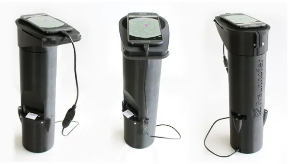
2.7 SightDx Parasight Platform: (A) Desktop scanning device; (B) Loading cartridge, which holds five patient samples; (C) Image of the monolayer at x20 (from [18]). 33 3.1 The µSmartScope prototype, with smartphone attached and microscopic slide inserted. . . 42
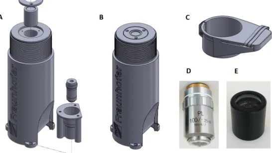
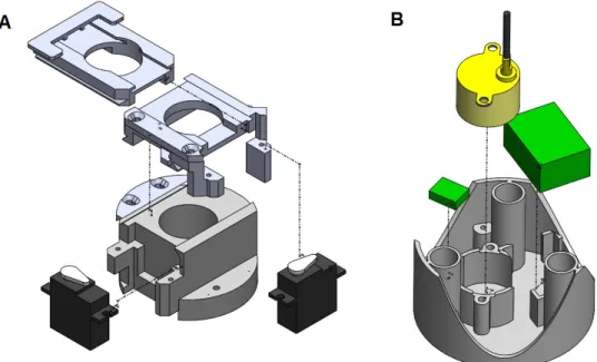
3.2 µSmartScope render models: (A) External view with microscope slide inserted (at yellow); (B) Cut view with optical and electrical components highlighted; (C) Detail of the cut view. . . 42
3.3 Optics module: (A) Exploded view; (B) Assembled view; (C) Smartphone holder; (D) Planachromat 100x oil-immersion objective lens; (E) Wide angle 10x eyepiece lens. . . 43
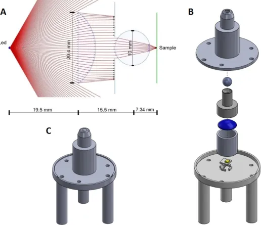
3.4 Illumination module: (A) Schematic of the developed light condenser generated with OpticalRayTracer optics design software; (B) Exploded view of the lightR condenser with lenses (at blue) and LED light (at yellow); (C) Assembled view of the light condenser. . . 44
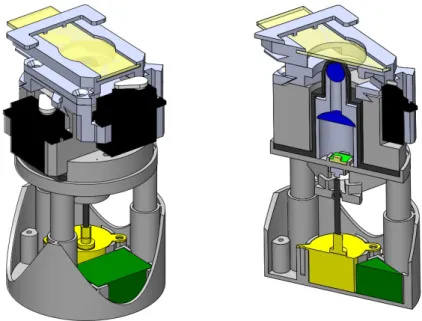
3.5 µStage module: External view (on the left) and cut view (on the right), with detail of servo motors (at black), step motor (at yellow), electronics (at green) and optics (at blue). . . 45
3.6 µStage functional submodules: (A) Exploded view of the XY-plane submodule, with servo motors (at black); (B) Exploded view of the Z-axis submodule, with step motor (at yellow) and electronics (at green). . . 46
3.7 Prototype PCB to control the µStage. [52] . . . 47 3.8 Focused image obtained using the µSmartScope, and respective central square
resulting from focus region selection: (A) Thin blood smear; (B) Thick blood smear. 49
3.9 Variation of the Tenenbaum focus metrics while the µStage is ascending in the vertical axis. Illustrative examples of preview images obtained by the smartphone camera at different Z positions are presented: (A) During the Rough phase; (B) Entering the Precise phase; (C) Focus point; (D) Stopping condition after focus point detected. . . 52 3.10 Images of READY OPTICS USAF 1951 microscope resolution target: (A)
Ac-quired using Bresser Microscope-5102000-Erudit DL; (B) Detail of Group 10 using the Microscope; (C) Acquired using the µSmartScope; (D) Detail of Group 10 using the µSmartScope. [52] . . . 53 3.11 Minimum Michelson contrast for USAF Resolution Target Elements of Group
10. [52] . . . 54 3.12 µSmartScope illumination uniformity analysis: (A) Original image; (B) Mean
pixel intensity of the 10x10 pixel boxes on the diagonal direction; (C) Standard deviation of the 10x10 pixel boxes on the diagonal direction. [52] . . . 55 3.13 Measured step size values after 100 repetitions: (A) XY plane; (B) Z axis. . . 56 3.14 Illustrative examples of focused images autonomously acquired with the µSmartScope
and different smartphone models for 5 different malaria-infected thin blood smears: (A) Asus Zenfone 2; (B) HTC One M8; (C) Motorola Moto G5; (D) LG Nexus 5; (E) Samsung Galaxy 6. . . 59 3.15 Illustrative examples of focused images autonomously acquired with the µSmartScope
and different smartphone models for 4 different malaria-infected thick blood smears: (A) Asus Zenfone 2; (B) HTC One M8; (C) Motorola Moto G5; (D) LG Nexus 5; (E) Samsung Galaxy 6. . . 60 3.16 Images of different smears acquired with the µSmartScope: (A) Thick blood
smear infected with malaria parasites (P.falciparum species); (B) Thin blood smear infected with malaria parasites (P.ovale and P.malariae species); (C) Thin blood smear infected with Chagas parasites (Trypanosome cruzi species); (D) Liquid-based Pap smear with high grade squamous lesions; (E) Thick blood smear infected with Lymphatic Filariasis parasites (Brugia malayi species); (F) Thick blood smear infected with Lymphatic Filariasis parasites (Wuchereria bancrofti species). Images (A), (B) and (C) were acquires with a LG Nexus 5, while images (D), (E) and (F) with a Samsung Galaxy S5. All images were acquired with magnification of 1000x, except image d) which has magnification of 400x. [52] . . . 61 4.1 Cropped microscopic sub-images of P.falciparum trophozoites and WBCs on thick
blood smear acquired with: (A) proper microscopic equipment [9]; (B) smartphone coupled to a Optical Magnification Prototype (see Section 3). . . 66 4.2 Mobile-based Framework for Malaria Parasites Detection: (A) Smartphone
Appli-cation; (B) Optical Magnification Prototype. . . 67 4.3 Microscopic Image Dataset: (A) Trophozoites manual annotation; (B) White Blood
Cells manual annotation. . . 68 4.4 Segmentation Results: (A) Original image with region of interest (ROI) at green;
(B) Optical Circle segmentation mask; (C) WBCs candidates segmentation mask of ROI; (D) Trophozoites candidates segmentation mask of ROI; (E) Chromatin dots candidates segmentation mask of ROI. . . 69 5.1 Blood smear analysis flow for both quantification and species/life cycle stage
LIST OF FIGURES xv
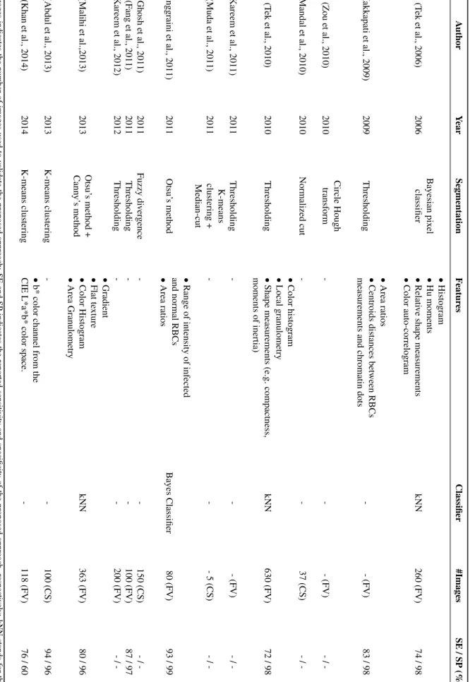
5.2 Mobile-based framework for malaria parasites’s detection: (A) µSmartScope with smartphone attached and blood smear inserted; (B) smartphone application screen-shots; (C) exemplificative usage of the solution (from left to right): (i) blood smear insertion; (ii) start image acquisition through the smartphone app; and (iii) visual feedback of the automated detection. . . 82 5.3 Diagram of the proposed methodology for the automatic analysis of thin smear
images. . . 83 5.4 Illustrative examples of different MPs species and life cycle stages from the Mobile
Thin Smear Malaria Parasites (mThinMPs) database. . . 84 5.5 Effect of brightness and contrast adjustment, with cumulative histograms: (A)
original image; (B) processed image after α and β correction, followed by mean-shift filtering. . . 84 5.6 Pre-processing: (A) original image; (B) brightness and contrast adjustment; (C)
sharpening applied over green channel of adjusted image; (D) RBCs segmentation applied over the sharpened image; (E) blue channel of the original image; (F) optical circle segmentation applied over the blue channel of the original image. . 85 5.7 Examples of trophozoites ring stage candidates: (A) original image (cropped ROI);
(B) brightness and contrast enhancement; (C) cytoplasm grayscale sharpening; (D) cytoplasm segmentation and filtering; (E) chromatin grayscale sharpening; (F) chromatin segmentation and filtering; (G) final candidates (cytoplasm in red; chromatin in yellow; RBC with candidate inside in green). . . 88 5.8 Examples of mature trophozoite stage candidates: (A) original image (cropped
ROI); (B) brightness and contrast enhancement; (C) cytoplasm grayscale sharpen-ing; (D) cytoplasm segmentation and filtersharpen-ing; (E) chromatin grayscale sharpensharpen-ing; (F) chromatin segmentation and filtering; (G) final candidates (cytoplasm in red; chromatin in yellow; RBC with candidate inside in green). . . 89 5.9 Examples of schizonts candidates: (A) original image (cropped ROI); (B) brightness
and contrast enhancement; (C) cytoplasm grayscale sharpening; (D) cytoplasm segmentation and filtering; (E) merozoites’ chromatin grayscale sharpening; (F) Merozoites’ chromatin segmentation and filtering; (G) final schizonts candidates (cytoplasm in green; chromatin in yellow). . . 90 5.10 Gametocytes candidates: (A) original image (cropped ROI); (B) brightness and
contrast enhancement; (C) grayscale sharpening; (D) segmentation and filtering; (E) final candidates (at green). . . 91 5.11 Illustrative examples of the data augmentation procedure. . . 94 5.12 Performance metrics criteria. . . 96 5.13 Examples of false negatives’ candidates for different species and life stages after
segmentation and filtering. . . 97 5.14 Heat maps of the SVM parameters’ selection process for each species-stage
combi-nation. . . 98 5.15 Classification models workflow. (a) Diagram of the classifier models workflow for
the detection of multiple species-stage combinations in a single image. (b) Illustra-tive examples with detection of: (I) P. falciparum trophozoites and gametocyte; (II)
List of Tables
2.1 Proposed approaches for the detection and/or segmentation of malaria parasites in thick blood films. . . 29 2.2 Proposed approaches for the detection and/or segmentation of malaria parasites in
thin blood films. . . 30 3.1 Power consumption test results. [52] . . . 57 3.2 Average seconds per image for different smartphone models and thin blood smears,
calculated over 100 automated focus attempts for each combination. . . 58 3.3 Average seconds per image for different smartphone models and thick blood smears,
calculated over 100 automated focus attempts for each combination. . . 58 4.1 Summary of the extracted image features. . . 70 4.2 Results for WBCs and P.falciparum trophozoites candidates after segmentation and
filtering. . . 72 4.3 Results for WBCs and P.falciparum trophozoites detection after machine-learning
classification. . . 72 5.1 MPs’ manual annotations by species and life cycle stage in the mThinMPs database. 83 5.2 Maximum length and respective Dcirclerelative ratios of RBCs’ and MPs’
struc-tures [13]. . . 86 5.3 Summary of the extracted image features. . . 92 5.4 Results after the segmentation step for each MP stage. . . 96 5.5 Results after machine learning classification for each species-stage combination. . 97
Abbreviations
AC Accuracy
AGNES Absence of Gradients and Nernstian Equilibrium Stripping AI Artificial Intelligence
ARR Annular Ring Ratio AUC Area under the curve BFL Back-Focal Length CAD Computer-aided diagnosis CHT Circle Hough Transform
CIE Commission International de l’Élairage FFF Fused Filament Fabrication
FOV Field-of-view F1 F1score
fps frames per second
GLMSR General Linear Model Simple Regression HSI Hue, saturation, intensity
INF Informedness
IQFT Inverse Quaternion Fourier transform kNN K-nearest neighbors
L*a*b* Lightness, green/red coordinate, blue/yellow coordinate L*C*h◦ Lightness, chroma, hue
mHealth Mobile health MPs Malaria parasites NN Neural Networks
NTDs Neglected tropical diseases PCA Principal Component Analysis QFT Quaternion Fourier Transform RBCs Red blood cells
RDTs Rapid diagnostic tests RGB Red, green, blue
ROC Receiver operating characteristic ROI Region of interest
SE Sensitivity SP Specificity
SVM Support vector machines WBCs White blood cells
WHO World Health Organization
Part I
Introduction and Theoretical
Background
Chapter 1
Introduction
Between 2010 and 2015, malaria mortality rates dropped by an estimated 29% globally and by 31% in the African region due to the scale-up of malaria interventions. Despite this remarkable progress, the global tally of malaria in 2015 was still 212 million new cases and 429,000 deaths, with an estimation that malaria surveillance systems only detected 19% of the cases occurred globally. Most of these deaths occurred in the African region (92%), but it is estimated that nearly half of the world’s population is at risk of malaria. This disease is considered endemic in 91 countries [74], and across Africa millions of people still lack access to the tools they need to properly prevent and treat it. Prompt diagnosis can not only prevent the development of severe malaria, but also reduce the length of time that patients carry malaria parasites (MPs) in their blood, which in turn reduces the risk of onward transmission [74].
Since 2010, WHO has recommended that all persons with suspected malaria should undergo malaria diagnostic testing, by either microscopy or rapid diagnostic tests (RDTs). Microscopy examination consists of preparing a blood smear, staining it (most often with Giemsa stain) and examining it through a microscope. This process remains the mainstay of malaria diagnosis in most large health clinics and hospitals. However, microscopy-based diagnosis in rural areas is frequently non-existent or inadequate in terms of quality due to scarce resources (equipment and trained staff). When stained MPs are spotted by the microscopist, the diagnosis of malaria is confirmed by identifying the respective stage, species and infection density [71]. Microscopic examination can be made through the usage of thin and thick blood smears. While the thin smear consists of a single layer of red blood cells (RBCs), the thick smear is 6 to 20 times thicker, allowing for a greater volume of blood to be examined. Thus, thick smears are firstly used to check the presence of MPs, while thin smears are subsequently analyzed for the identification of MP species. The conventional flow for thick and thin blood smear analysis using manual microscopy examination is presented in Figure1.1.
Malaria diagnosis through RDTs is accomplished by detecting specific malaria antigens in a per-son’s blood. However, the use of the RDT does not eliminate the need for malaria microscopy for two main factors: (i) the RDT may not be able to detect some infections with low parasite density; (ii) the currently approved RDT in the U.S. only detects two different malaria antigens
Figure 1.1: Conventional flow for thick and thin blood smear analysis using manual microscopy examination.
(one is specific for P. falciparum, and the other is found in all four human species of malaria). Thus, microscopy is further needed to determine the parasite species and to quantify the proportion of RBCs that are infected, which is an important prognostic indicator [10].
1.1
Motivation
A recent report [73] considers that current funding distribution on malaria control commodities (US $1.6 billion in 2014) is not addressing the fundamental weaknesses in health systems of developing countries, suggesting that innovative ways may be required to rapidly expand access to malaria interventions. It is worth underlining that the mobile phone is currently Africa’s most important digital technology [6], and just as African telecommunications largely skipped over landline infrastructure and went straight to mobile phones, some experts say African medicine can skip over centralized labs [16]. Moreover, the combination of mobile devices with image processing and artificial intelligence for malaria diagnosis can bring several advantages, like potentially reducing the dependence of manual microscopic examination, which is an exhaustive and time-consuming activity that requires considerable expertise of the laboratory technician.
The development of new microscopic devices, ideally portable and low cost, is also an area that can greatly improve the chances of the successful deployment of computer-aided diagnosis solutions for malaria diagnosis in the field. Considering the high customs taxes and import duties currently in practice in most of the African countries, the easy replicability of these microscopy devices in developing countries is a topic that should also be taken into account. Other requirements
1.2 Objectives 5
for this type of microscopic devices were equally considered since the beginning of this work, like: i) automating the device as much as possible, thus discarding the need of considerable expertise and train of the technician in terms of maneuvering the microscope; or ii) supplying the energy needed for the illumination and/or any type of automation through the mobile device battery, thus discarding the need of an additional power source.
By achieving and merging some of the described software and hardware innovations, we believe that the lack of highly trained microscopists on malaria diagnosis in rural areas could then be complemented by a significantly less specialized technician that knows how to operate the system and prepare blood smears.
Finally, malaria diagnosis might be just one element of a suite of diagnostic software tests running on this type of system. Several other tests could simultaneously be carried out using the same images, for instance cell counting or detection of other hemoparasites like microfilaria or trypanosoma.
1.2
Objectives
The main driving force behind this thesis has been the search for suitable alternatives to conventional malaria diagnosis through manual microscopy examination. Consequently, the objectives of this thesis revolved around making contributions in the field of computer-aided detection of malaria parasites. To achieve a solution with a realistic chance of being effectively used in the field, we soon realized that we needed to explore the development of both hardware and software components. Conventional microscopes and trained personnel with know-how to maneuver it are often scarce or nonexistent in rural endemic areas, so one of the objectives was to make contributions in the field of cell-phone based systems that provide low-cost alternatives to conventional microscopy.
On the other hand, the lack of trained microscopists that can visually spot malaria parasites on microscopic fields was equally important to address. Thus, another objective was to contribute in the area of computational approaches for the automated detection of malaria parasites in microscopic blood smear images. Particularly, we wanted to address two distinct computer vision challenges for images exclusively acquired with low-cost and accessible tools such as smartphone: i) detection and quantification of malaria parasites in thick blood smears; and ii) the automated species and life cycle stage determination in thin blood smears.
1.3
Contributions
The contributions of this thesis in the field of computer-aided detection of malaria parasites are now summarized:
1. Developed and evaluated a fully automated 3D-printed smartphone microscope with a motorized stage (termed µSmartScope). This prototype was the first proposed smartphone-based alternative to conventional microscopy that allows autonomous acquisition of a pre-defined number of images at 1000x magnification with suitable resolution, by using a
motorized automated stage fully powered and controlled by a smartphone, without any human interaction.
2. Proposed a new automated focus approach that takes advantage of the µSmartScope’s motorized automated stage and real time feedback retrieved from the smartphone camera sensor. This methodology was evaluated on different blood smears and smartphone models, which validated that the obtained results were not biased towards a specific specimen type (e.g. amount microscopic structures or staining condition) or specific camera sensor characteristics (e.g. resolution, exposure or white-balancing mode).
3. Proposed a new methodology to detect and count the number of P.falciparum trophozoites and WBCs in Giemsa stained thick blood smears. Given the lack of freely available image datasets, a mobile acquired image dataset manually annotated by a specialist was specifically created and used in this study.
4. Proposed a new methodology using supervised classification to analyze microscopic images of malaria-infected thin blood smears. Given the lack of freely available image datasets, a new mobile thin smear malaria parasites (mThinMPs) image database was specifically created to develop and validate this methodology.
5. Proposed a new performance metrics criterion for SVM hyperparameters’ selection, which merges the informedness and F1score metrics.
The MalariaScope Project
All the contributions described above were achieved in the ambit of the MalariaScope project, a R&D project of Fraunhofer Portugal AICOS that aims to create a mobile-based solution that can provide an effective pre-diagnosis of malaria to be used in medically underserved areas. This project started in the ambit of Fraunhofer Portugal AICOS’s ICT for Developing Competence Center, in cooperation with the Research and Development Unit of the Infectious Diseases Department of the Instituto Nacional de Saúde Dr. Ricardo Jorge in Porto, which finished in 2015. Given the encouraging obtained results, these research topics continued to be explored in the ambit of the project “Deus ex Machina: Symbiotic Technology for Societal Efficiency Gains”.
1.4
List of Publications
The work conducted in this thesis resulted in the following journal papers:
• Luís Rosado, José M. Correia da Costa, Dirk Elias, and Jaime S. Cardoso. A review of automatic malaria parasites detection and segmentation in microscopic images. Anti-Infective
1.5 Document Structure 7
• Luís Rosado, José M. Correia da Costa, Dirk Elias, and Jaime S. Cardoso. Mobile-based analysis of malaria-infected thin blood smears: Automated species and life cycle stage determination. Sensors, 17(10), 2017.
The following publications in international conferences were also the result of the research work presented throughout this thesis:
• Luís Rosado, José M. Correia da Costa, Dirk Elias, and Jaime S. Cardoso. Automated Detection of Malaria Parasites on Thick Blood Smears via Mobile Devices. Procedia
Computer Science, 90:138–144, January 2016.
• Luís Rosado, João Oliveira, Maria João M. Vasconcelos, José M. Correia da Costa, Dirk Elias, and Jaime S. Cardoso. µSmartScope: 3d-printed smartphone microscope with motorized automated stage. In Proceedings of the 10th International Joint Conference on Biomedical
Engineering Systems and Technologies - Volume 1: BIODEVICES, (BIOSTEC 2017), pages
38–48. INSTICC, SciTePress, 2017.
Furthermore, the developed work also resulted in a book chapter:
• Luís Rosado, Paulo T. Silva, José Faria, João Oliveira, Maria João M. Vasconcelos, José M. Correia da Costa, Dirk Elias, and Jaime S. Cardoso. µSmartScope: Towards a fully automated 3d-printed smartphone microscope with motorized stage. Communications in
Computer and Information Science Book Series, 881, 2018.
1.5
Document Structure
This thesis is divided into three parts, which comprises a total of six chapters. The document is structured as follow:
The PartI, Introduction and Theoretical Background, is composed by two chapters: the current chapter is Chapter1and presents the motivations, objectives and main contributions of the thesis; Chapter2summarizes the fundamental topics regarding computer-aided detection of malaria parasites, including the literature review.
The PartII, Methodologies and Results, is composed by three chapters: Chapter3presents the work done in the development and evaluation of a fully automated 3D-printed smartphone microscope with a motorized stage; Chapter4focuses on the automated analysis of malaria-infected thick blood smears via mobile devices; Chapter5explores new computer vision and machine-learning approaches for the analysis of microscopic images of malaria-infected thin blood smears acquired with mobile devices.
The PartIII, Conclusion, comprises Chapter6where the conclusions and the most relevant topics regarding future work are depicted.
Chapter 2
Fundamentals of Computer-aided
Malaria Parasites Detection
A Review of Automatic Malaria
Parasites Detection and Segmentation
in Microscopic Images
Luís Rosado, José M. Correia da Costa, Dirk Elias and Jaime S. Cardoso Published in: Anti-Infective Agents, 14(1), 11-22, 2016
2.1 Introduction 13
Abstract
Malaria is a leading cause of death and disease in many developing countries, where young children and pregnant women are the most affected groups. In 2012, there were an estimated 207 million cases of malaria, which caused approximately 627 000 malaria deaths. Around 80% of malaria cases occur in Africa, where the lack of access to malaria diagnosis is largely due to a shortage of expertise, being the shortage of equipment the secondary factor. This lack of expertise for malaria diagnosis frequently results on the increase of false positives, since prescription of medication is based only on symptoms. Thus, there is an urgent need of new tools that can facilitate the rapid and easy diagnosis of malaria, especially in areas with limited access to quality healthcare services. The aim of this work is to collect and review image processing and analysis approaches already proposed on the literature for the analysis of malaria infected blood smear images.
Keywords: malaria; computer-aided diagnosis; image analysis; segmentation; feature extraction;
classification.
2.1
Introduction
Malaria is one of the most severe public health problems worldwide. It is a leading cause of death and disease in many developing countries, where young children and pregnant women are the groups most affected. In 2012, there were an estimated 207 million cases of malaria, which caused approximately 627 000 deaths. An estimated 3.4 billion people continue to be at risk of malaria, mostly in Africa and south-east Asia.
Around 80% of malaria cases occur in Africa [72]. It is worth taking into account that the number of malaria cases and their geographical distribution are not stable because of several factors, like the increasing prevalence in some areas due to expanding drug resistance; the widespread availability of fake and substandard medicines; global warming and expansion of malaria into favorable areas at higher elevations; and population mobility of different kinds [71].
The increasing interest in the development of computer-aided diagnosis (CAD) systems for malaria diagnosis is closely related with common practical difficulties experienced in under-resourced health facilities of developing countries, such as the excessive workload due to shortage of staff. Image processing approaches are often used in CAD systems to reduce the dependence of manual microscopic examination of blood smears, which is an exhaustive and time consuming activity, simultaneously requiring a considerable expertise of the laboratory technician.
During the last years, several image processing techniques have been proposed for malaria diagnosis using microscopic images, addressing the detection of a wide variety of different malaria parasites, in different growth stages and using images acquired from different types of blood smears. Under the scope of this chapter, various image processing and analysis approaches already proposed on the literature for the detection and segmentation of malaria parasites in blood smear microscopic images were collected and reviewed. This timely review aims to support the increasing interest
in the development of low cost tools that can facilitate the rapid and easy diagnosis of malaria, especially in areas with limited access to quality healthcare services.
This chapter is structured into five sections. Section2.1corresponds to Introduction and presents the motivation and objectives of this bibliographic survey. Section2.2gives an overview of the malaria disease in terms of parasite stages, species and life cycle stages; Section2.3provides an overview about the current malaria diagnosis methodologies, with a focus on the characterization of stained components in thin and thick blood smears; Section2.4gives a literature review regarding the analysis of malaria infected blood smears using image processing and analysis; Section2.5 summarizes and gives a critical appreciation of the review works; Section2.6provides the final remarks about the discussed subjects.
2.2
Malaria Disease Characterization
Malaria is caused by a parasite in the blood and can be seen only under a microscope with high magnification. For the visualization of the parasites, a blood film must be made, dried, stained and examined under the microscope. When the microscopist sees stained parasites, the diagnosis of malaria is confirmed by identifying the stage and species of the malaria parasite, as well as the infection density [71].
2.2.1 Malaria Parasites Stages
In the human host, malaria parasites pass through 3 different growth stages that can be detected in the peripheral blood: the trophozoite stage, the schizont stage and the gametocyte stage. Trophozoites are often called the ring stage, being the most commonly seen stage, appear incomplete in thick films, and can vary from small to quite large within the host cell. Usually, trophozoites have one chromatin dot, but two are common for the P.falciparum species. The cytoplasm takes different shapes, from a well-defined, fine ring to forms that are irregular or bizarre, sometimes called ‘amoeboid’ [72]. The schizont stage begins when the trophozoite has reached its full capacity and the parasite starts to divide into daughter cells called merozoites. Several more divisions of the chromatin follow, which mark the growth of the schizont, until there are many chromatin bodies, each with its accompanying cytoplasm. The number of chromatin and merozoite divisions helps to identify the species. These clearly delineated new parasites are now ready to leave the host cell to invade new red blood cells [71]. Gametocytes are round or banana-shaped, depending on the species. The way in which the parasite takes up the stain helps to identify the sex of the parasite in thin films, being difficult to differentiate between male and female in thick films [31].
2.2.2 Malaria Parasites Species
Four species of Plasmodium can infect and be transmitted by humans: the P.falciparum, P.vivax,
2.3 Malaria Diagnosis Characterization 15
and can evolve rapidly to severe illness and death if not recognized and treated with effective medicines [71]. It is the species responsible for most cases of severe malaria and death. P.vivax is the commonest species in the cooler parts of the tropics, being the largest of the human malaria parasites and the cause of much illness and absenteeism from work and school [71]. P.ovale is considered a rare species, but relatively common in West Africa and other parts of the African continent. Because of morphological similarities, P.ovale is sometimes mistaken for P.vivax by less experienced microscopists [71]. Moreover, the existence of a new genotype for P.ovale has been recently hypothesized [58]. P.malariae is found worldwide and causes a chronic infection that in some cases can last a lifetime. In some chronically infected patients, P.malariae can cause serious complications such as the nephrotic syndrome [9]. As a final note, P.knowlesi is a malaria parasite that is found in nature in macaques, and naturally acquired human infections were thought to be extremely rare, however a large focus of human infections was reported in 2004 [55].
2.3
Malaria Diagnosis Characterization
Malaria infection can be suspected based on the patient’s symptoms, travel history or physical findings at examination. However, for a definitive diagnosis, laboratory tests must be made to prove the presence of the malaria parasites. The microscopy examination remains the gold standard for laboratory confirmation of malaria, which consists in preparing a blood smear, staining it (most often with the Giemsa stain) and examining it through a microscope [9]. The importance of reliable malaria diagnoses cannot be overstated, since false negatives can be potentially fatal, and false positives increase the drug resistance of the patients, leading consequently to unnecessary economic burden [47]. Laboratory diagnosis of malaria can be made through microscopic examination of two kinds of blood smears, thin and thick, taken most often from a finger prick. Thick blood smears are 20-40 times more sensitive in detecting malaria parasites because the blood is more concentrated, which allows for a greater volume of blood to be examined. The thick smear is approximately 6-20 times as thick as a single layer of red blood cells, which results in a larger volume of blood being examined. However, thick smears are more difficult to read, so thin smears aid in parasite species identification and quantification [9], [47].
2.3.1 Image Characteristics
The images used on the reviewed works can be divided in two different groups according to their characteristics: full view images (FV) and manually cropped sub-images (CS). FV consists on images corresponding to the entire microscopic field of view (see Figures2.1and2.2). CS consists on cropped patches of the FV images, corresponding to regions of interest manually cropped (see Figures2.3and2.4).
Figure 2.1: Illustrative example of a thick smear full view image (from [17]).
Figure 2.2: Illustrative example of a thin smear full view image (from [60]).
Moreover, the vast majority of the proposed approaches found on literature use high quality equipment in the acquisition process, particularly commercial cameras that are specifically cus-tomized for the acquisition of microscopic images, for instance easily attached to microscopes. The exception is [47], which uses a smartphone built-in camera to acquire images (see Figure2.5).
2.3 Malaria Diagnosis Characterization 17
Figure 2.3: Illustrative examples of thick smear cropped sub-images (from [46]).
Figure 2.5: Illustrative examples of thin smear cropped sub-images acquired with mobile devices, with positive cases at the top and negative cases at the bottom (from [47]).
2.3.2 Performance Metrics
The classification results of the reviewed works are usually presented in terms of two metrics ordinarily used for this purpose: 1) Sensitivity (SE), i.e. the percentage of structures correctly classified as positive cases of malaria parasites; and 2) Specificity (SP), i.e. the percentage of structures correctly classified as negative cases of malaria parasites.
2.4
Literature Review
This section critically reviews the main studies found in the literature regarding the analysis of malaria infected blood smears using image processing and analysis. Since typical approaches usually comprise four different image processing and analysis tasks, the reviewed works were divided into the following sub-sections: 1) Segmentation; 2) Feature Extraction; 3) Feature Selection; and 4) Classification. For each sub-section, the methods proposed to date for malaria parasites (MPs) stained components analysis, both on thin and thick blood smears, were separately reviewed.
2.4.1 Segmentation
Image segmentation is the process that partitions a digital image into disjoint (non-overlapping) regions, each of which typically corresponds to one object. Once isolated, these objects can be
2.4 Literature Review 19
measured and classified, as discussed in the following sub-sections. This sub-section groups and reviews the methods proposed on the literature for the segmentation of malaria-stained components on thin and thick blood smears.
2.4.1.1 Thresholding
Thresholding is an essential region-based image segmentation technique that is particularly useful for scenes containing solid objects resting on a contrasting background. All pixels at or above/below the threshold are assigned to the foreground and all pixels below/above the threshold are assigned to the background [75].
Thin Blood Films
In [37], the authors suggest a scheme based on HSV color space that segments red blood cells (RBCs) and identifies the RBCs infected with MPs. The RBCs segmentation is achieved by dividing the entire hue range of 360◦ into six segments and finding the dominant color type to
be the representative of the background. Each segment is centered on a color type, which is defined according to the follow degree values: 0◦ for red; 60◦ for yellow; 120◦for green; 180◦
for cyan; 240◦ for blue; and 300◦ for magenta. The authors state a SE and SP of 83% and
98%, respectively, however this method uses images taken from Leishman-stained blood smears, while the gold standard recommended by WHO for malaria diagnosis is the usage of Giemsa staining. Furthermore, they assume that the dominant color in these images is representative of the background, which might not be true in images highly populated of RBCs (see Figure2.3).
Another segmentation approach proposed in [31] for RBCs on thin blood smears is based on the Annular Ring Ratio (ARR) transform. This transform consists in obtaining a ratio transformed image by calculating the ratio between the average intensities of a dilated image using annular concentric ring structuring element and an eroded image using a disk shaped structuring element. The ARR transform method mainly aims at locating the center of each cell present in the image. It is worth taking into account that this segmentation methodology uses fixed parameter values defined manually, such as the radius of the annular ring and disk, which can substantially vary depending on the image resolution. The authors in [30] used a modified ARR transform method for the detection of stained cells through the direct application of the ratio transform on grayscale images, eliminating the morphological dilation and erosion. A threshold-based peak detection algorithm is then used to determine the coordinates of each stained component. This approach results in locating all the stained components in the image, with the drawback of picking up artifacts and other noises present in the image.
For blood cells components segmentation in thin smears, [4] uses the Otsu’s Method, a well-known histogram shape-based image thresholding routine. This method assumes that the input image has two classes of pixels, and calculates the threshold that minimizes the intra-class variance. A hole filling process in then applied, due to the biconcave nature of the RBCs. Using the knowledge that MPs cytoplasm appear lighter while MPs nucleus appear darker than the cytoplasm of the
RBCs, the authors state that is possible to confirm that it is actually an infected cell by dividing it into 3 different regions (MPs nucleous, MPs cytoplasm and RBCs cytoplasm) using multiple thresholding. However, how this multiple thresholding segmentation is achieved is not explained in the paper, and one should also take into account that possible artifacts could also be separated into 3 different regions.
In [22], P.vivax parasites from Leishman-stained thin blood film are segmented using divergence based threshold selection. The proposed modified fuzzy divergence method is based on Cauchy membership function, and applied to the C channel in the CMYK color space, which was the color channel that delivered the best results. However, it is not clear if this approach can be also applied to Giemsa stained thin blood films.
A methodology based on phase spectrum is used in [19] for malaria parasite detection in thin blood films. The method uses the Quaternion Fourier Transform (QFT) to obtain the amplitude spectrum and phase spectrum for blood smear images. Afterward, the reconstructed image is obtained using the Inverse Quaternion Fourier transform (IQFT) on a constant amplitude spectrum and the original phase spectrum. The authors concluded that the most sensitive channels for MPs detection were the B and G channels from the RGB space, as well as the I channel from the HSI space.
The proposed automated method in [61] for parasite detection and identification on thin blood film comprises two different segmentation steps: The foreground-background segmentation and the stained pixels segmentation. The proposed segmentation is performed using morphological area top-hats (using the average cell area value) and morphological double thresholding. For the stained pixels segmentation, the authors modeled the stained and unstained pixel distributions with RGB space histograms and used the probability density function to determine whether a pixel on the input image is stained or not. This work also comprises significant pre-processing effort, like the granulometry-based cell size estimation, which considers the peak index of the granulometry distribution as the average cell area. It also addresses problems like the non-uniform illumination, which is removed using a pre-recorded illumination image, or using a morphological closing operation with a structuring element with size of 5 times the average cell area. Despite this work being only focused on thin blood films, the authors highlighted the importance of future work to investigate the automatic analysis of thick films, since they are more sensitive for malaria parasites density estimation.
In [38], the authors combined the binary images from Otsu’s method with the edge detection images from Canny’s method for RBCs segmentation, followed by a hole filling process. This work also proposes a pre-processing step based on the estimation of the image illumination, which is then subtracted from the original image.
Thick Blood Films
For non-background objects detection on thick blood films, i.e. MPs, white blood cells (WBCs) and possible Giemsa stain-derived artifacts, the authors in [29] used adaptive threshold found according
2.4 Literature Review 21
to information of the V-Value histogram. However this methodology needs further validation since it was only tested on 20 microscopic images.
The G and B channels of the RGB color space were identified in [17] as very good features to identify objects containing chromatin in Giemsa stained blood films, being not only considered highly discriminative but also almost independent of differences in illumination and staining intensity. They transformed the color input image into a monochrome image I(x,y), that highlights objects containing chromatin: I(x,y) = arctan(IGREEN(x, y)/IBLU E(x, y)). The authors also used a
black-top-hat morphological operator to separate MPs from both leukocytes and platelets, with a non-flat paraboloid structuring element of radius of 9 and a slope of 1 pixel. It should be taken into account that these fixed parameters might not be suitable for images with different pixel resolutions. The black-top-hat operator is followed by a thresholding operation with a fixed threshold, which according to the authors is reliable given the independence of the G and B channels with regard to illumination and staining intensity. However, the authors do not define the value of this fixed threshold on the publication.
A dark stretching technique is applied in [23] for contrast enhancement of MPs-infected thick blood smear images. The dark stretching is a process that uses an auto scaling method, being the dark areas stretched and the bright areas compressed. The authors state that the dark areas correspond to the MPs in the infected images. To segment the MPs, they use a single threshold value on the stretched image. However, the thresholds have been chosen empirically, and different thresholds had to be applied for different images in order to achieve good segmentation results. 2.4.1.2 Boundary-based Segmentation
Boundary-based techniques seek to extract object boundaries directly, based on identifying the edge pixels located at the boundaries in the image [75].
Thin Blood Films
An improved Circle Hough Transform (CHT) was used in [80] to detect RBCs, based on the consideration that RBCs have a circular pattern. The authors highlighted that CHT has the advantage of handling the segmentation for highly overlapping and oval cells, and noted that the RBCs are much more noticeable in the green channel of the RGB color space. However, RBCs might present shapes significantly different from the expected circular/oval pattern, which might difficult the detection using this approach.
Thick Blood Films
A combination of Absence of Gradients and Nernstian Equilibrium Stripping (AGNES), and Morphological Gradient techniques is used in [78] for the detection of P.vivax parasites in thick blood films. A morphological gradient method is first applied in order to enhance the borders of the objects present in the image. This is followed by a threshold detection stage using the K-Median method. The AGNES and K-Median techniques were then used to assign the remaining number of
pixels to each region, using as starting points the image regions previously identified as objects and background. These techniques were locally applied on rectangular sectors corresponding to 20% of the input image, a percentage defined experimentally. According to the authors, this approach might induce the appearance of objects that are not part of the original image in the border of the defined rectangular sectors.
2.4.1.3 Clustering methods
Clustering is a machine learning technique that can be used for segmentation purposes when applied as an indicator of the similarity of different image regions, based on a set of measurements that describes those regions.
Thin Blood Films
The application of 3 different clustering algorithms to segment blood cell images is studied in [42]: Mean-shift, K-means and Fuzzy C-means. The authors support that K-means clustering algorithm achieves best results for blood cell images segmentation, and they used a Median-cut algorithm after applying the K-means in order to reduce the number of regions to an optimum level. However, this approach needs further validation since it only used 5 cropped sub-images.
A segmentation approach using k-means clustering is also proposed in [1] for the detection of P.vivax parasites. Different color components of RGB, HSI and C-Y color models have been analyzed, and the S component of C-Y color model has proven to be the best, with segmentation accuracy and F-score of 99.46% and 0.9370, respectively.
In [32], the authors used the b*-color channel from the CIE L*a*b* color space for k-means clustering as an unsupervised segmentation approach to identify malaria parasite tissues. They used 118 Leishman-stained microscopic images with reported results of 76% and 60% for SE and SP, respectively. It is worth noting that these results refer to classifying the images as infected/not infected, thus not giving information about the precise number of correctly identified MPs in each image.
2.4.1.4 Classifier-based methods
Classifier-based methods for segmentation rely on the usage of two-class models for segmentation purposes.
Thin Blood Films
In [60], a Bayesian pixel classifier has been employed in order to differentiate between the stained and non-stained pixels. The class conditional probability density functions of the stained and the non-stained classes were estimated using the non-parametric histogram method.
2.4 Literature Review 23
2.4.2 Feature Extraction
The primary objectives of feature extraction are reducing the computational complexity of the subsequent process and facilitating a reliable and accurate recognition for unknown novel data, being the last objective particularly important for computer vision and pattern recognition systems. Moreover, the in-depth understanding of the domain-specific knowledge gained by human experts on the problem being addressed can be of extreme importance for the design of a reliable and effective feature extraction engine [27].
2.4.2.1 Binary Objects Measures
A binary object can be described in terms of its size (e.g. area, perimeter), pose (e.g. centroid, orientation), shape measures (e.g. thinness ratio, rectangularity, circularity, Euler number, moments, elongation), shape descriptors (e.g. differential chain code, fourier descriptors, media axis transform, graph representation) or distance to other objects (e.g. Euclidean distance, city-block distance, chessboard distance) [75].
Thin Blood Films
In [37], the infected RBCs detection in thin blood smears is based on relative ratios between chromatin dots and RBCs terms of area and centroids distances. For the same purpose, the work in [61] uses shape measurements like compactness and moments of inertia.
Thick Blood Films
The segmented non-background objects are distinguished in [29] according to size, and the MPs species P.falciparum and P.vivax are distinguish according to the chromatin size. However, this methodology was tested only on 20 images and the thresholds were defined in number of pixels, thus not suitable for images with different pixel resolutions.
2.4.2.2 Gray-Level Objects Measures
Gray-level objects measures consist on measurements derived from the intensity distribution of the object. There are 3 main categories of gray-level object measurements: Intensity measures (e.g. integrated and average optical intensity, contrast) histogram measures (e.g. mean, standard deviation, skew, entropy, energy) and texture measures (e.g. statistical texture measures, power spectrum features) [75].
Thin Blood Films
For detection and differentiation between WBCs and P.falciparum gamecocytes, the authors in [30] start by finding the centroids of each detected stained component. The proposed algorithm considers as candidates the neighborhood of the listed centroid coordinates within the diameter of
the WBCs. The candidates are marked as WBCs if the neighborhood region presents low mean intensity and less variance, and as gametocyte of P.falciparum if the region has high eccentricities (variance) and average intensity.
The authors in [80] observed that nucleated components result in distinctively high intensity values in the B channel of the RGB color space, while the same nucleated components in the G channel exhibit very low intensity values. Therefore, they proposed an intensity measure based on the differences between the B and the G color intensity channels, in order to stretch the contrast for visual perception and emphasize nucleated objects.
The methodology proposed by [4] aims to detect 3 regions inside the infected RBCs, particularly the MPs nucleous, MPs cytoplasm and RBCs cytoplasm. Features of those regions are then extracted for classification purposes, based on area ratios and range of intensities of each region.
In another work [60], the following gray-level measures were extracted for further MP/non-MP classification in thin blood films: Hu moments, relative shape measurements, histogram and color auto-correlogram. According to the authors, the last two features are the most effective for MPs classification.
The color histogram is used in [61] after quantization into 32 colors, as well as the local area granulometry for each RGB channel. It worth noting that the features were normalized (for zero mean, 1 variance) first on the training set, being the mean and standard deviation calculated and then used for the normalization of the testing set.
The authors in [38] extracted four different features: 1) Gradient; 2) Flat texture (determined by computing the difference between the original image and the filtered image using median filter); 3) Color histogram; and 4) Area granulometry.
Thick Blood Films
In order to differentiate MPs from artifacts in thick blood films, the authors in [17] extracted 174 different features. The features were grouped in 3 main groups: 1) Statistical moment features - 4 central moments (mean, variance, skewness, and kurtosis), Hu’s set of 7 invariant moments [26], 49 Zernike moments of orders up to 12 [33]; 2) Texture features: Haralick’s 13 co-occurance matrix features [24]; Unser’s 18 sum and difference histogram features [66]; Chen’s 16 statistical geometrical features [12]; 5 features proposed by Young et al. [77] that describe the distribution of chromatin in the ROI; 3) Color features: 60 features representing a 60-bin histogram of the hue channel of the ROI; 2 features described by Kovalev et al. [34] that represent cyan shifts in the ROI. The following features were extracted in the work presented in [46]: mean; standard deviation; kurtosis; skewness; entropy of the histograms of R,G and B channels from RGB space, H channel from HSV space, and H channel of HSI space.
In another recent work, 3 groups of features were extracted in [78] for each RGB channel: 1) Color features: standard deviation, 7 Hough moments of color and color range; 2) Texture features based on the co-occurrence matrix (in 4 directions): homogeneity, contrast, GLMSR (General Linear Model Simple Regression), standard deviation, second angular moment and correlation; 3) Texture features based on the Wavelet transform: energy, mean and standard deviation. Four
2.4 Literature Review 25
wavelet families were used (Haar; biorthogonal 1,3; Daubechies 2; and Daubechies 8) considering 2 decomposition levels, being applied in each level a low and high pass filter.
In [47], the authors extract two types of features: 1) Connected component features: Perimeter; Moment of Inertia; Elongation; Jaggedness; and Maximum λ (consists in the maximum child gray level minus the current gray level); 2) Moment features: the moment m00, the central moments, u11,
u20, u02and Hu moments h0, h1, h2.
2.4.3 Feature Selection
In order to build a good classification model, the reduction of the number of attributes used on the classification process may not only have positive impact in terms of the processing time, but also in terms of the classification results. Feature selection techniques play an important role in this context, and they can be organized into three categories: filter methods, wrapper methods and embedded methods [20].
Filter Methods rank each feature according to some univariate searching function and select the highest-ranking features, where the scoring should reflect the discriminative power of each feature. Some of the most common univariate filter methods includes Bayesian Network, Information Gain, Signal-to-Ratio, Euclidean Distance or Correlation Squares (R2) [20]. Filter methods are usually
very efficient and fast to compute, but comprise some significant drawbacks like the redundancy of the selected features, which can carry the same information. Another important disadvantage of the filter methods is the fact that this selection does not consider some important relationships between features, since features can receive a low score by the ranker algorithm when used by itself, but be very useful when combined with other features.
Opposite to filter techniques that consider the ranking of each feature independently, Wrapper and Embedded Methods are specific to a given machine-learning algorithm. In the Wrapper Method, a search is conducted using a specific classifier in order to find the subset of features with which the classification algorithm performs the best. For instance, using forward selection, the Wrapper Method estimates the accuracy of adding each unselected feature to the feature subset, and the feature that most improves the accuracy is selected. These methods typically terminate when the estimated accuracy of adding any feature is less than the estimated accuracy of the feature set already selected [15].
Thick Blood Films
An optimal feature subset was chosen in [17], based on the 174 previously extracted features for MPs detection. The features were first normalized to have zero mean and a standard deviation of one. The authors proposed a 2-step feature selection methodology: an univariate ranking is first applied in order to keep only the 60 features that have the highest univariate discriminative power, and then a genetic algorithm is used for automatic selection of an even smaller feature subset.

![Figure 2.4: Illustrative examples of thin smear cropped sub-images (from [39]).](https://thumb-eu.123doks.com/thumbv2/123dok_br/15243128.1023246/41.892.307.631.722.1100/figure-illustrative-examples-smear-cropped-sub-images.webp)
![Figure 2.5: Illustrative examples of thin smear cropped sub-images acquired with mobile devices, with positive cases at the top and negative cases at the bottom (from [47]).](https://thumb-eu.123doks.com/thumbv2/123dok_br/15243128.1023246/42.892.162.691.143.544/figure-illustrative-examples-cropped-acquired-devices-positive-negative.webp)
![Figure 2.7: SightDx Parasight Platform: (A) Desktop scanning device; (B) Loading cartridge, which holds five patient samples; (C) Image of the monolayer at x20 (from [18]).](https://thumb-eu.123doks.com/thumbv2/123dok_br/15243128.1023246/57.892.150.789.481.824/sightdx-parasight-platform-desktop-scanning-loading-cartridge-monolayer.webp)