1
doi.org/10.1590/S0001-37652007000400004
Chemical carcinogenesis
PAULA A. OLIVEIRA1 , AURA COLAÇO1 , RAQUEL CHAVES2 , HENRIQUE GUEDES-PINTO2 , LUIS F. DE-LA-CRUZ P.3 and CARLOS LOPES4,5
1 Department of Veterinary Sciences, CECAV, University of Trás-os-Montes and Alto Douro 5000-801 Vila Real, Portugal
2 Center of Genetics and Biotechnology-CGB, University of Trás-os-Montes and Alto Douro (UTAD) Department of Genetics and Biotechnology, 5000-801 Vila Real, Portugal
3 Deparment of Physiology, Faculty of Veterinary, Santiago University, Granxa Street Campus Universitario, 27002 Lugo, Spain 4 Department of Pathology, Portuguese Institute of Oncology, Rua Dr. António Bernardino de Almeida 4200-072 Porto, Portugal
5 Departament of Pathology and Molecular Immunology, Institute of Biomedical Sciences Abel Salazar University of Porto, Largo Professor Abel Salazar, 2, 4099-003 Porto, Portugal
ABSTRACT
The use of chemical compounds benefits society in a number of ways. Pesticides, for instance, enable foodstuffs to be produced in sufficient quantities to satisfy the needs of millions of people, a condition that has led to an increase in levels of life expectancy. Yet, at times, these benefits are offset by certain disadvantages, notably the toxic side effects of the chemical compounds used. Exposure to these compounds can have varying effects, ranging from instant death to a gradual process of chemical carcinogenesis. There are three stages involved in chemical carcinogenesis. These are defined as initiation, promotion and progression. Each of these stages is characterised by morphological and biochemical modifications and result from genetic and/or epigenetic alterations. These genetic modifications include: mutations in genes that control cell proliferation, cell death and DNA repair – i.e. mutations in proto-oncogenes and tumour suppressing genes. The epigenetic factors, also considered as being non-genetic in character, can also contribute to carcinogenesis via epigenetic mechanisms which silence gene expression. The control of responses to carcinogenesis through the application of several chemical, biochemical and biological techniques facilitates the identification of those basic mechanisms involved in neoplasic development. Experimental assays with laboratory animals, epidemiological studies and quick tests enable the identification of carcinogenic compounds, the dissection of many aspects of carcinogenesis, and the establishment of effective strategies to prevent the cancer which results from exposure to chemicals.
Key words: cancer stages,carcinogenesis evaluation, chemical carcinogens, chemical carcinogenesis.
INTRODUCTION
Public opinion considers cancer to be an increasingly threatening disease, affecting people of all ages. After cardiovascular diseases, it is the second cause of death amongst the global population (Huff 1994, Weisburger 1999). People tend to accept cancer with stoicism and submit themselves to prolonged periods of treatments, which are not always effective (Weisburger 1999). The word carcinogenic was defined as the capacity of a com- pound to unchain the process of cancer development in man and animals under the appropriate conditions, by acting on one of several organs or tissues (Gomes Carneiro et al. 1997, Huff 1999). With the discovery of different mechanisms involved in carcinogenesis, this definition is now incomplete (Butterworth and Bogdanf-
fy 1999). From an experimental point of view, a com- pound is considered carcinogenic when its administra- tion to laboratory animals induces a statistically signifi- cant rise in the incidence of one or more histological types of neoplasia, compared with the animals in the control group which are not exposed to the substance (Gutiérrez and Salsamendi 2001).
The factors responsible for cancer development are classified as exogenous and endogenous (Camargo et al. 1999, Gutiérrez and Salsamendi 2001). The first group includes nutritional habits (food preservation and preparation), socio-economic status, lifestyle, physical agents (ionising and non-ionising radiation), chemical compounds (natural and synthetic) and biological agents (Helicobacter pylori, Epstein Barr virus, human T lym-
photropic viruses I and II, human papilloma virus and the hepatitis B virus, parasites such as Schistosoma haemo-
tobium, Clonorchis sinensis and Opisthorchis vivarium;
growth factors) (Pitot and Dragan 1991, Barrett and An- derson 1993, Farmer 1994, Weisburger 1999, Minamoto et al. 2000, Lutz 2002). Unhealthy lifestyle habits such as: excess alcohol consumption; inhalation of tobacco and related products; the ingestion of certain foods and their contamination by mycotoxins; are responsible for higher incidences of certain types of neoplasias in a num- ber of population groups (Gomes-Carneiro et al. 1997, Weisburger 1999, Gutiérrez and Salsamendi 2001). En- dogenous factors include immune system damage and inflammation caused by uncertain aetiology (e.g. ulcer- ative colitis, pancreatitis, etc.), genetic makeup, age, en- docrine balance and physiological condition (Cohen et al. 1991, Barrett and Anderson 1993, Huff 1994, Koivu- salo et al. 1994, Weisburger 1999, Minamoto et al. 2000, Gutiérrez and Salsamendi 2001, Dewhirst et al. 2003, Ohshima et al. 2003, 2005).
Epidemiological studies of cancer incidence de- monstrated that the risk of developing cancer varies be- tween population groups and these differences are as- sociated with lifestyle factors and habits (Garner 1998, Lai and Shields 1999, Gutiérrez and Salsamendi 2001). Population migration has resulted in the development of types of cancer typical of particular geographical areas (King et al. 1995, Gutiérrez and Salsamendi 2001).
The relationship between chemical substances in the workplace and the development of certain neoplasias in various occupational groups led to the conception of experimental models to better understand the biopatho- logical processes inherent to carcinogenesis (Weinstein 1991, Cohen et al. 1992, Gutiérrez and Salsamendi 2001).
Boveri laid down the genetic basis of neoplasic de- velopment for the first time in 1914 with his theory of somatic mutation in cancer cells. However at the time, experts in the area of chemical carcinogenesis attributed little importance to this hypothesis, considering it to be pure speculation, instead choosing to put their faith in the lesser knowledge already available (Weisburger 1999). Between 1980 and 1990, the discoveries made via the molecular biology of proto-oncogenes and tumour sup- pressor genes strengthened the case behind this suppo- sition (Cohen 1998). Neoplasic development bases it- self on the existence of several genetic mutations, de- spite the number not being known. In most of the cases it is assumed to vary between tissues and between dif- ferent species (Grisham et al. 1984, Cohen 1995, 1998, Simons 1995, van Leeuwen and Zonneveld 2001, Lutz 2001, Gutiérrez and Salsamendi 2001). During cell divi-
sion, spontaneous genetic errors occur. It is estimated to happen at a frequency of around 10−5 to 10−6 through nu-
cleotides and cell division. If the damage reaches a gene responsible for neoplasic development then the probabil- ity of developing cancer will be greater (Cohen 1995).
A cancer is made up of billions of cells, all originat- ing from an initial cell which multiplies clonally, escapes to apoptosis and accumulates genetic (and/or epigenetic) alterations which converge into a neoplasic cell (Trosko 2001). The blocking of apoptosis in the face of sig- nificant genetic damage can ease the accumulation of aberrant cells and it can become a critical point in malig- nance pathogenesis (Nguyen-ba and Vasseur 1999, Qu et al. 2002).
Neoplasias can be classified as benign or malign depending on their cellular characteristics. The consti- tuent cells of a malign neoplasia show yet more changes in cell biology (Fig. 1). They proliferate autonomously, differentiate themselves, invade adjacent tissues and fre- quently metastasize on tissues that are not related to the primary neoplasia (Hanahan and Weinberg 2000, Shac- ter and Weitzman 2002). Cells, which are part of benign neoplasias, grow more slowly, and in general, they do not disturb normal tissue function, unless they compress vital structures (Player et al. 2004). The histopathological ob- servation of neoplasias, be they induced or spontaneous, enables us to better evaluate carcinogenesis, but it may not be enough to identify more subtle alterations such as molecular changes (Huff 1992, Maronpot 1996).
This review aims to describe of different events in- volved in chemical carcinogenesis. So, our work starts with a historical perspective of the study of chemical carcinogenesis; we will describe the different stages in- volved in carcinogenesis; the absorption and metabol- ism of chemical carcinogens. We will classify different types of carcinogens in function of their active mecha- nisms and we will describe the molecular targets of car- cinogens. Finally, we will describe a selection of the methods available for evaluating the carcinogenic poten- tial of chemical compounds.
HISTORICAL PERSPECTIVE OF CHEMICAL CARCINOGENESIS STUDY
Cancer was described for the first time by Hippocrates as ‘karkinos’. Galeno introduced the word neoplasia only in the II century; he defined it as the growth of a body area adverse to nature (Gutiérrez and Salsamendi 2001). Edwin Smith’s papyruses, dating from the XVII century, describe breast tumefaction.
According to Hayes (1995), it was the English sur- geon Percivall Pott who first recognized in 1775 the casual relationship between exposure to environmental
3 substances and neoplasic development. This author de- scribed the occurrence of cancerous alterations in the skin of the scrotum of London chimney sweeps as a consequence of repeated localised contamination with soot. Some years later, and based on these observa- tions, a guide distributed to Danish chimney sweeps rec- ommended that these professionals take a daily bath to avoid such an occurrence (Hayes 1995, Gutiérrez and Salsamendi 2001). Still in the XVIII century John Hill observed a high proportion of nasal mucosa cancer in his patients, and traced it to the localised long-term ex- posure to snuff. In 1890, a high incidence of bladder cancer in chemical and rubber industry workers was ob- served across Europe. (Cohen and Ellwein 1991, Gomes- Carneiro et al. 1997, Garner 1998, Dybdahl et al. 1999, Huff 1999, Bertram 2001). By the end of the nine- teenth century it had become evident that occupational exposure to certain chemicals or mixtures of chemicals had carcinogenic effects (Luch 2005). The all-important next step was to systematically investigate and repro- duce these diseases in experimental surroundings. The first experimental work on chemical carcinogenesis was carried out in 1915 by the pathologist Katsusaburo Yama- giwa and his assistant Koichi Ichikawa (Yamagiwa and Ichikawa 1918). They rubbed rabbit ears with coal tar and observed the development of papillomas and carcino- mas. Meanwhile, others researchers studied carcinogen- esis of the bladder, liver, kidney, pancreas and lung using laboratory animals. Its success laid the foundations of the experimental use of animals in the study of human diseases (Toth 2001). Later, Beremblum and Shubik used polycyclic aromatic hydrocarbons and croton oil to study skin carcinogenesis in mice and demonstrate that cancer development includes several stages (Beremblum and Shubik 1947). When applied in low doses, none of these substances have carcinogenic properties by them- selves. Yet, when mixed and in equal doses, they in- duced neoplasic development. The order of exposition to these substances was fundamental for carcinogenesis. Neoplasias developed only when the hydrocarbons were used first and then the croton oil, never the other way around. These authors felt that the carcinogenic action of these substances was responsible for converting nor- mal cells into neoplasic cells. For them, carcinogenesis was a complex process including one phase called initia- tion and another called promotion, with one or more ge- netic changes necessary for cancer development. During the next decade, Foulds (1954) introduced the term pro- gression by studying breast adenocarcinoma in female mice. In the pre-Watson and Crick era, before carcino-
gens were known to bind to DNA, the cancers produced by chemical carcinogens were believed to be due to their interaction with proteins in specific tissues (Miller and Miller 1952). By the end of the 1960s, increasing evi- dence pointed to a correlation between the DNA bind- ing capacity of a particular carcinogen and its biological potency (Luch 2005).
STAGES OF CARCINOGENESIS
Studies conducted using animal models, “in vitro” stud- ies and epidemiologic assays enabled investigators to conclude that neoplasic pathogenesis is a complex pro- cess which can be divided into three distinct stages, from an operational point of view. These are: initiation, pro- motion and progression (Foulds 1954, Grisham et al. 1984, Cohen 1991, Mehta 1995, Hasegawa et al. 1998, Gutiérrez and Salsamendi 2001, Trosko 2001).
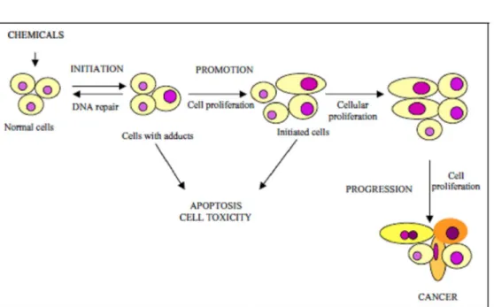
Changes in the genome’s structure occur across the three stages of neoplasic development (Simons 1995, Pitot 2001, Luch 2005). Changes in gene expression also take place during the promotion stage, with selec- tive proliferation of initiated cells and the development of pre-neoplastic cells (Grisham et al. 1984, Gutiérrez and Salsamendi 2001). During initiation and promo- tion, apoptosis and cell proliferation can occur at differ- ent rates, while remaining balanced. During progression, this balance is modified and from there malignancy arises (Mehta 1995) (Fig. 2).
Human life is led under very different conditions from these experimental procedures. Although the pro- cess of carcinogenesis is similar for man and experimen- tal animals, the different chemical compounds to which humans are exposed throughout their lives alter the speed of the process and the frequency of mutation, the speed of cell growth and the phenotypical expression of the changed genes. On the other hand, the individual’s sus- ceptibility and their defence mechanisms have their own interaction, which modifies each of the neoplasic stages.
INITIATION
The first stage of carcinogenesis has been labelled ini- tiation since 1947 (Beremblum and Shubik 1947). The conclusions reached from several experiments enabled the conclusion to be drawn that initiation is caused by irreversible genetic changes which predispose suscep- tible normal cells to malign evolution and immortality (Beremblum and Shubik 1947, Stenbäck et al. 1981, Butterworth et al. 1992, Mehta 1995, Dybing and Sanner 1999, Trosko 2001, 2003, Shacter and Weitzman 2002).
The initiated cell is not a neoplasic cell but has taken its first step towards this state, after successive genotypical and phenotypical changes have occurred (Trosko 2003). From a phenotypical perspective, the initiated cell is sim- ilar to the remaining cells. It undergoes mutations and these induce proliferation but not differentiation (Trosko 2001).
DNA damage has been well established as the event which kick-starts chemical carcinogenesis (Santella et al. 2005). DNA damage can be repaired by enzymatic mechanisms (Bertram 2001, Jeng et al. 2001, Shacter and Weitzman 2002). Cells which are proliferating have less time to repair the damaged DNA and remove covalent bonds that chemicals establish with the DNA – known as adducts (Heidelberger 1977, Richardson et al. 1986, Frowein 2000).
At this stage, the initiated cells can remain latent for weeks, months or years, or they can grow in an auto- nomous and clonal fashion (Scott et al. 1984, Dybing and Sanner 1999, Player et al. 2004). This initiation process ensures that cellular division remains symmetri- cal by creating two new initiated cells (Trosko 2003). The clonal expansion of initiated cells results from a mitogenic process caused by an increase in the number of new cells and apoptosis inhibition, which prevents ini- tiated cells from dying off (Trosko 2001).
The increase in DNA damage is specifically impor- tant to stem cells, because they survive for a long time and exist in several tissues (Potter 1978, Simons 1999, Trosko 2001, Williams 2001). In 1978, Potter explained that neoplasic cells could display a phenotype established between the embryonic aspect and the terminal differen- tiation, and that all neoplasic cells had monoclonal origin from a stem cell. By definition, stem cells are immortal cells until they differentiate, or death is induced. If we delay their differentiation they become initiated and ac- cumulate in tissues as clones of abnormal cells (Trosko 2003). Although stem cells are not identifiable in most tissues, it is believed that every tissue has a population of stem cells (Player et al. 2004).
Initiation is a fast, irreversible phenomenon and is transmitted to daughter cells (Farber 1984). Cell pro- liferation is essential for this stage, if cellular division occurs before DNA repair systems can act then the in- jury becomes permanent and irreversible. Initiation is an additive process, neoplasic development depends on the carcinogenic dose, increasing the dose increases the in- cidence and the multiplicity of resultant neoplasias and reduces the latent period of its manifestation. Not all cells of a living organism exposed to an initiator agent will be initiated even if they have suffered mutations, and the genes that regulate the terminal differentiation must
also be mutated (Farber 1984, Yuspa and Poirier 1988, Klaunig et al. 2000, Trosko 2001).
Spontaneously initiated cells exist in all living or- ganisms (Gomes-Carneiro et al. 1997, Trosko 2001). Ini- tiation can begin with spontaneous mutations, supported by normal occurrences such as DNA depurination and deamination. Errors in DNA replication are also asso- ciated with initiation. Although spontaneous initiation is less common than induced initiation, its existence has been confirmed by the occurrence of spontaneous neo- plasias in laboratory animals (Pitot and Dragan 1991, Gomes-Carneiro et al. 1997).
PROMOTION
The concept of promotion was introduced when chemi- cal substances with low carcinogenic activity were dis- covered, which were still able to induce the development of cancer under experimental conditions (Berem- blum and Shubik 1947).
Promoter compounds do not interact directly with DNA and unchain biological effects without being meta- bolically activated (Yuspa et al. 1983, Butterworth et al. 1992, Weisburger 1998, Williams 2001). These agents increase cell proliferation in susceptible tissues, con- tribute towards fixing mutations, enhance alterations in genetic expression and cause changes in cellular growth control (Mehta 1995, Gomes-Carneiro et al. 1997). On the other hand, these promoters may indirectly damage DNA by oxidation (Gutiérrez and Salsamendi 2001). At first, these occurrences were associated with epige- netic mechanisms, but nowadays it is widely agreed that promotion also involves genetic changes (Simons 1995, Hanahan and Weinberg 2000).
Promoters delay the natural inhibition of the quies- cent cells or in G0 by gap junctions (Barrett and Ander-
son 1993, Simons 1999, Bertram 2001, Trosko 2001). The promoters’ most important activity is mitogenesis – genotoxical and mutational actions are not necessary at this stage (Pitot and Dragan 1991). The promoter must be present for weeks, months and years in order to be effective and its effectiveness depends on its con- centration in the target tissue (Butterworth et al. 1992). Promotion is a reversible stage, after a promoter’s dis- appearance a regression in cell proliferation can occur, probably by apoptosis. It is a stage that can be moulded up by physiological factors and therefore limit the extent of experimental carcinogenesis. Some promoter agents are specific for a particular tissue, but others act simul- taneously upon several tissues (Yuspa et al. 1983, Scott et al. 1984, Yuspa and Poirier 1988, Gutiérrez and Sal- samendi 2001).
5 In studies of chemical carcinogenesis with pro- longed exposure and using high doses almost all of the promoter agents induce neoplasias without initiation (Pitot and Dragan 1991, Gutiérrez and Salsamendi 2001). Exposure to phenobarbital, benzene, asbestos, and ar- senic even without the previous application of initiator agents leads to neoplasic development (Melnick et al. 1996, Trosko 2001). This contradiction has two possible explanations: either the genotoxic effect was not iden- tified by mutagenicity and genotoxicity assays, or the initiated cells emerged spontaneously. In this last case we may consider that the promoter has an indirect effect – by increasing the frequency of cellular division it en- courages the appearance of errors in DNA replication, as well as mutations.
Not all cells exposed to promoters take part in the promotion stage, only cells which are stimulated to di- vide, that are undifferentiated, and have survived apop- tosis, can contribute to instability between growth and cell death and lead to the appearance of a malign neo- plasia (Trosko 2001).
PROGRE SSI ON
The sequence of lesions identified, via histopathology, between initiation and promotion are designated as pre- neoplastic lesions and/or benign neoplasias (Gutiérrez and Salsamendi 2001). Their transformation into ma- lign lesions is the last of the stages of carcinogenesis and is the most extended – it is labelled progression (Klaunig et al. 2000, Williams 2001). In progression, a neopla- sic phenotype is acquired through genetic and epigenetic mechanisms (Shacter and Weitzman 2002). During pro- gression, cell proliferation is independent from the pres- ence of stimulus (Lutz 2000, Gutiérrez and Salsamendi 2001).
Progression is characterised by irreversibility, ge- netic instability, faster growth, invasion, metastization, and changes in the biochemical, metabolical and mor- phological characteristics of cells (Pitot and Dragan 1991, Butterworth et al. 1998, Loeb 1998, Klaunig et al. 2000, Gutiérrez and Salsamendi 2001, Dixon and Kopras 2004).
Angiogenesis, as an epigenetic occurrence, is es- sential to neoplasic progression. The acquisition of an angiogenic phenotype precedes the development of char- acteristics that contribute to malignancy and its inhibition delays neoplasic development (Hawighorst et al. 2001).
ABSORPTION AND METABOLISM OF CHEMICAL CARCINOGENS
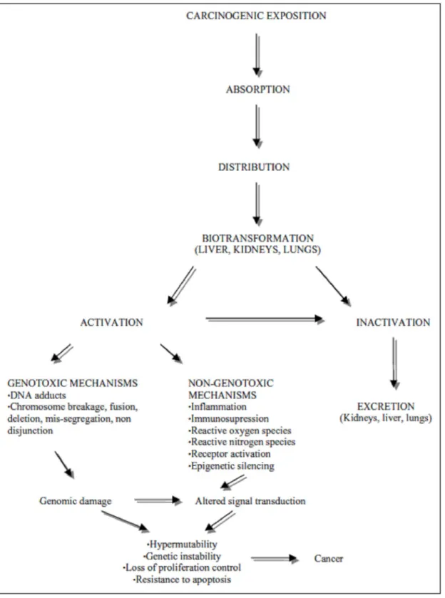
Following exposure, chemical carcinogens may be ab- sorbed in a number of ways (oral, inhalator, cutaneous, and injection) and distributed across several tissues (Con- noly et al. 1988). Absorption depends on the physico- chemical properties of the substance and can take place via passive or active transport. The substances absorbed orally pass through the liver and only then are they dis- tributed in the body; those absorbed in the lung are dis- tributed by the blood before reaching the liver at a later stage (King et al. 1995, van Leeuwen and Zonneveld 2001). Those carcinogenic compounds classified as di- rect act directly on DNA, but most require enzymatic con- version and are thus labelled as indirect or procarcino- gens (Sarasin and Meunier-Rotival 1976, Hayes 1995, Lai and Shields 1999, Klaunig et al. 2000, Oesch et al. 2000, Poirier et al. 2000, Luch 2005). Metabolic ac- tivation is controlled by phase I reactions, while phase II reactions protect the body through the transformation of activated compounds into inert products which are easily eliminated from the body (Fig. 3) (Hayes 1995, Bartsch and Hietanen 1996, Mostafa et al. 1999, Klaunig et al. 2000, Gonzalez and Kimura 2001, van Leeuwen and Zonneveld 2001, Park et al. 2005).
The performance of metabolic enzymes is essential for understanding chemical carcinogenesis and learning the differences between species as far as their suscep- tibility to neoplasic development is concerned (Sarasin and Meunier-Rotival 1976, Lai and Shields 1999, Guen- guerish 2000, 2001, Gonzalez 2001). The enzymes in phase I participate in the reactions of oxidation, reduc- tion and hydrolysis, and are classified as oxidoreductases (cytochrome P450 dependent monooxygenases, flavine monooxygenases, cyclooxygenases and alcohol dehy- drogenase) and hydrolases (epoxide hydrolases) (Hayes 1995, Garner 1998, Galati et al. 2000, Oesch et al. 2000, Garcea et al. 2003). Phase II enzymes participate in the conjugation and inactivation of chemical carcinogens and include transferases (glutathione S-transferases, N- acetyltransferases, UDP-glucuronosyltransferases, sul- photransferases) (Oesch et al. 2000, Guengerich 2000, Gonzalez 2001). Although these enzymes were origi- nally only thought to be involved in the detoxification stages of biotransformation, they can also contribute to the activation of certain procarcinogens in vivo (Luch 2005).
Metabolic activation occurs predominantly in the liver at the plain endoplasmic reticulum where the cy- tochrome P450 is more abundant, and to a lesser degree in the bladder, skin, gastrointestinal system, oesopha- gus, kidneys, and lungs (Bartsch and Hietanen 1996,
Mostafa et al. 1999, Guengerich 2001, van Leeuwen and Zonneveld 2001, Oda 2004). During this phase the cy- tochrome P450 mono-oxygenases introduces a reactive polar group into the carcinogenic, making it lipophylic. It then converts it into a powerful electrophilic prod- uct capable of establishing adducts with DNA (Straub and Burlingame 1981, Lai and Shields 1999, Galati et al. 2000, Park et al. 2005). Phase II reactions are catal- ysed by hepatic and extra hepatic, cytoplasmic and cy- tochromic enzymes, acting separately or joined together (Gonzalez 2001). Conjugation reactions enable these en- zymes to decompose the polar group in glucose, amino acids, glutathione and sulphate, which are less toxic metabolites that are more soluble in water and more eas- ily expelled by the urine and bile (Galati et al. 2000, Oesch et al. 2000, Gonzalez and Kimura 2001, van Leeuwen and Zonneveld 2001).
Peroxidations also occur parallel to metabolic reac- tions with the continuous production of reactive oxygen species (ROS) (Weisburger 1999, Klaunig et al. 2000, Ohshima et al. 2005). These radicals are associated with several chronic diseases including chemical carcinogen- esis (Klaunig et al. 2000). The ROS damage DNA, RNA, and proteins by chemical reactions such as oxidation, ni- tration/nitrosation and halogenation. This leads to an increase in mutations and alterations in the functions of important enzymes and proteins (Park et al. 2005). Sev- eral experiments have proved that chemical compounds, which create ROS in excess, encourage initiation, pro- motion and neoplasic progression through genotoxicity (Galati et al 2000, Shacter and Weitzman 2002). The impact of the ROS controlled by a cellular mechanism that operates at different levels: metabolism; reactions that maintain the redox balance in cells; transduction of the signal regulator of oxidation and DNA reparation (Bolt et al. 2004).
Park et al. (2005) says that the same enzyme may have the capacity to activate one chemical and deacti- vate another, all depending on its chemical structure. The specificity of the activation systems of different tis- sues regulate neoplasic development and is dependent on genetic polymorphism, which requires the expres- sion and distribution of the enzymes involved in phase I and II reactions, and the resulting susceptibility to can- cer development (Schut and Castonguay 1984, Hayes 1995, Henglster et al. 1998, Mostafa et al. 1999, Dybingand Sanner 1999, Gonzalez 2001, Gonzalez and Kimura 2001, Gutiérrez and Salsamendi 2001, Lutz 2002). Peo- ple with a high quantity of phase I and a low quantity of phase II enzymes have a higher probability of synthesis- ing intermediate compounds and exhibiting more DNA damage (Rojas et al. 2000). The previously described metabolic methods are
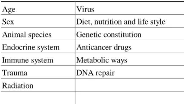
equally important for both humans and animals, although there exist qualitative and quantitative differences be- tween them. These have lead to incorrect interpreta- tions when animal models are used in the research and analysis of carcinogenic properties of chemical com- pounds (Guengerich 2000, Gonzalez 2001, Gonzalez and Kimura 2001). Several studies have been developed in order to evaluate the differences between several exogenous and endogenous factors on individual susceptibility to carci- nogenesis (Table I) (Barrett 1993, Bartsch and Hietanen 1996, Maronpot 1996, Lutz 1998, 1999, Ishikawa et al. 2001, Miller et al. 2001).
CARCINOGENIC CLASSIFICATION
Carcinogenic classification is by no means consensual (Butterworth and Bogdanffy 1999, Bolt et al. 2004). It is not easy to incorporate a carcinogenic compound into a certain group because the information obtained from different studies is increasingly complex (Pitot and Dragan 1991, Butterworth et al. 1992). Some authors classify them in function of their participation in each of the stages of carcinogenesis. In this way, incomplete carcinogens are mutagenic chemicals that instigate irre- versible DNA damage (Mirsalis et al. 1990, Pitot and Dragan 1991). A complete carcinogen displays pro- perties of both initiators and promoters simultane- ously depending on the dosage and exposure time (Pitot and Dragan 1991, Farmer 1994, Hasegawa et al. 1998, Trosko 2001).
Other authors classify chemical carcinogens in function of their mechanisms of action as being geno- toxic and non-genotoxic (mitogenic and cytogenic) (Cohen and Ellwein 1991, Butterworth et al. 1992, Nguyen-ba and Vasseur 1999, Klaunig et al. 2000, Williams 2001). The knowledge about the mechanism of action of non-genotoxic carcinogens is known to be inferior to that of genotoxic carcinogens.
Genotoxic carcinogens are complete carcinogens and qualitatively and quantitatively change a cell’s ge- netic information (Trosko 2001). They exhibit a direct analogy between their structure and activity, are muta- genic on in vitro assays, are active in high doses, and may affect several animal species, and damage different organs (Klaunig et al. 2000, Gutiérrez and Salsamendi 2001, Luch 2005). In high doses, they cause toxicity and cell proliferation, increasing DNA replication and influencing its carcinogenic activity (Cohen 1998). Fol- lowing transmembranar diffusion they are metabolized in electrophilic compounds that enter the nucleus and
7 interact with nucleophilic sites (DNA, RNA and pro- teins) changing their structural integrity and establish- ing covalent bonds known as adducts (Miller and Miller 1975, Straub and Burlingame 1981, Cohen et al. 1992, Ashby 1996, Weisburger 1998, Frowein 2000, Bertram 2001, Lutz 2001, Williams 2001, Baird and Mahadevan 2004). The formation of adducts constitutes the first crit- ical step of carcinogenesis and if these are not repaired before DNA replication then mutations may occur in the proto-oncogenes and tumour suppressor genes, which are essential for the initiation stage (Sobels 1975, Barrett and Wiseman 1987, Farmer 1994, Lutz 2001, Williams 2001, Li et al. 2005). The number of adducts formed by carcinogens is changeable and each of them may cause a specific damage to DNA (Straub and Burlingame 1981, Farmer 1994, Otteneder and Lutz 1999). Mu- tations linked to adducts can appear through deletion, frameshift, or by nucleotide substitution (Garner 1998). Mutations cause an undefined number of cell changes, translated into aberrant protein expression and in changes in cell cycle control. Adducts assume importance in chemical carcinogenesis because of the way they change DNA, possibly inducing an incorrect transcription and causing mutations of the new DNA chain. The existence of many adducts can break the DNA chain, causing mu- tation or loss of genetic material (Cohen 1995, Hayes and Pulford 1995, Trosko 2001). Adduct repair is coor- dinated by several enzymes and controlled by different genes. It can be done via the excision of bases, or nu- cleotides, recombined repair or mismatch repair (Farmer 1994, Moustacchi 1998, Miller et al. 2001, Hanawalt et al. 2003).
The identification of adducts suggests that chemical carcinogens are absorbed, metabolized and distributed by tissues, thus fleeing from the body’s detoxification and repair mechanisms (Garner 1998, Airoldi et al. 1999, Guengerich 2000). The identification and analysis of adducts can be carried out using marked radioactive car- cinogens, those most-commonly used are 14 C and tri- tium, each adduct can be identified by their 106 or 107
nucleotides (Garner 1998). However, the most used tech- niques are immunoassays with 32 P, gaseous chromatog-
raphy associated with mass spectrometry and HPLC as- sociated with fluorescent spectroscopy (Farmer 1994, Airoldi et al. 1999). There are also monoclonal and polyclonal antibodies available on the market which are used to identify adducts by immunohistochemistry (San- tella et al. 2005). There is a positive correlation between the quantity of adducts detected in animal models and
the number of neoplasias developed (Yuspa and Poirier
1988, Williams 2001, Baird and Mahadevan 2004). Non-genotoxic carcinogens act as promoters and do not need metabolical activation. They do not react di- rectly with DNA, do not raise adducts and show negative on mutagenicity tests carried out in vivo and in vitro (Butterworth et al. 1992, Melnick et al. 1996, Butter- worth and Bogdanffy 1999, Klaunig et al. 2000, Gon- zalez 2001, Williams 2001). These compounds modu- late growth and cell death, potentate the effects of geno- toxic compounds, do not show a direct correlation be- tween structure and activity, and their action is limited by their concentration. They are tissue- and species-specific (Farmer 1994, Melnick et al. 1996, Gomes-Carneiro et al. 1997, Butterworth and Bogdanffy 1999, Klaunig et al. 2000). Melnick et al. (1996) states that exposure to these compounds favours the synthesis of other sub- stances responsible for neoplasic development. These compounds promote effects on target cells which indi- rectly unchain neoplasic transformation or increase neo- plasic development from genetically changed cells (Wil- liams 2001). Non-genotoxic carcinogens are classified as cytotoxic and mitogenic in function of whether their activity is mediated by a receptor or not (Cohen 1991, Cohen et al. 1992, Butterworth and Bogdanffy 1999). Mitogenic compounds such as phorbol esters, dioxins, and phenobarbital induce cell proliferation in target tis- sue through interaction with a specific cellular receptor (Cohen et al. 1992). Cytotoxic carcinogens cause cell death in susceptible tissues followed by compensatory hyperplasia, taking chloroform as an example (Cohen et al. 1991, Butterworth et al. 1992, Klaunig et al. 2000). If the carcinogen dose is high, some cells cannot sur- vive. The more that nearby cells increase the number of cell divisions through regenerative procedures, the more likely it is that they will end up being prematurely recruited for the cell cycle and that the time available for reparation DNA will be inferior – this increases the probability of mutations occurring (Cohen 1991, Mel- nick et al. 1996). On the other hand, necrosed cells are destroyed by the immune system and ROS, reac- tive nitrogen species (RNS), and proteolytic enzymes are produced (Lutz 1998, Ohshima et al. 2005). When production of these ROS and RNS exceeds the cellular anti-oxidant capacity, it may cause oxidative damages to lipids, proteins, carbohydrates, and nucleic acids, leading to carcinogenesis and cell death (Ohshima et al. 2005). Mitogenic compounds need to be present in certain con-
centrations to promote their activity. Contrastingly, the action of non-cytotoxic compounds is independent of their concentrations (Butterworth et al. 1992, Butter- worth and Bogdanffy 1999).
Chemical carcinogens can be classified into several groups, on Table II we brought them together under the following headings: Group, compound, mechanism of action, and affected organs/cancer type.
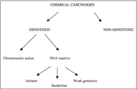
As we mentioned before, the classification of the carcinogenic compounds according to their mechanism of action continues to cause controversy. Bolt et al. (2004) propose the division of genotoxic compounds into two groups: those which react with DNA, and geno- toxic at a chromosomal level. Compounds, which react with DNA, are subdivided into three different groups: initiators (with unlimited doses), borderline, and weak genotoxic (they act by secondary mechanisms) (Fig. 4).
Chemical carcinogens can have additional synergic or antagonistic effects when simultaneously presented in different metabolic ways (Schmahl 1976, Lutz 2001). The synergy between smoking and exposure to asbestos favours lung cancer development as a consequence of chronic inflammation and compensatory cell prolifera- tion. This antagonism may be exemplified by the protec- tive action of fruit and vegetables in the modulation of individual susceptibility to neoplasic development (Lutz 2001, 2002).
EPIGENETIC MECHANISMS INVOLVED IN CHEMICAL CARCINGENESIS
The most well understood epigenetic mechanisms in- volve DNA methylation and histone acetylation, methy- lation, and phosphorylation (Fig. 5). Demethylation of promoter regions at the CpG sequences can lead to an over-expression of proto-oncogenes, and silencing of gene expression can occur as a result of hypermethy- lation, sometimes leading to chromosome condensation (Klaunig et al. 2000). There appears to be a relationship between DNA methylation and histone modifications; patterns of histone deacetylation and histone methyla- tion are associated with DNA methylation and gene si- lencing. Interestingly, these epigenetic changes in chro- matin can also alter the sensitivity of DNA sequences to mutation, thus rendering genes more susceptible to toxic insult (Dixon and Kopras 2004).
MOLECULAR TARGETS OF CHEMICAL CARCINOGENS The discovery of the ability of oncogenes to induce neo- plasic transformation when transfected into immortal- ized mouse cell lines, initially seemed to answer many
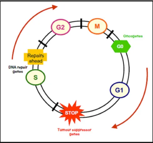
basic molecular questions about the molecular origins of cancer. However, it soon became clear that this was not the whole picture and that there existed other genes that could influence neoplasic transformation (Bertram 2001). There are several genes which intervene in car- cinogenesis – their identification revolutionised chem- ical carcinogenesis and oncology (Kinzler and Vogel- stein 1997, Bertram 2001). Out of all of these, proto- oncogenes, tumour suppressor genes and cell cycle regu- lator genes assume a particular importance (Mehta 1995, Nguyen-ba and Vasseur 1999, Klaunig et al. 2000). Un- like diseases such as cystic fibrosis or muscular dystro- phy, wherein mutations in one gene can cause disease, no single gene defect “causes” cancer. Mammalian cells have multiple safeguards to protect them against poten- tially lethal effects of cancer gene mutations, and only when several genes are defective does an invasive can- cer develop. Thus it is best to think of mutated cancer genes as contributing to, rather than causing, cancer (Vo- gelstein and Kinzler 2004). Neoplasic development re- quires errors in cellular defence mechanisms, which are controlled by checkpoints that may forbid the entry of cells with DNA damage into the cell cycle before DNA reparation occurs (blocked at G1 ) and the cell divides
(blocked at G2 ) (Fig. 6) (Khan et al. 1999, Khan and
Dipple 2000). The capacity of cells to evade the cel- lular defence mechanism has an undoubted contribution towards the carcinogenesis (Khan and Dipple 2000).
The tumour suppressor proteins p53; p21 and pRb play crucial roles in cellular protection, because they en- courage the blocking of cells at G1 (Khan et al.
1999). The loss of pRb protein function provokes an increase in the cell proliferation rate and an absence of termi- nal differentiation. p53 can interrupt the cell cycle at G1 and go on to repair DNA damage
(Melnick et al.
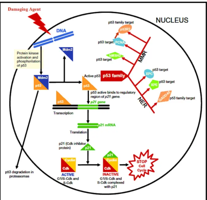
1993, Loeb 1998, Khan and Dipple 2000, Pritchard et al. 2003, Dixon and Kopras 2004). The most promi- nent and best-studied tumour suppressor is p53, if DNA is damaged then p53 can induce apoptosis in order to maintain the stability of the cells’ genome (Klaunig et al. 2000, Hanawalt et al. 2003, Babenko et al. 2006). The loss of p53 during carcinogenesis can predispose pre- neoplastic cells to accumulate additional mutations by blocking the normal apoptotic response to genetic dam- ages (Klaunig et al. 2000). The loss of p53 function activates proto-oncogenes and inactivates tumour sup- pressor genes therefore performing an exceptional role in chemical carcinogenesis (Luch 2005). The biological activity of p53 protein is dependent on its ability to bind transcriptional regulatory elements in DNA. The search for critical genes regulated by p53 led to the
9 discovery of the p21 gene. p21 acts as an inhibitor of cyclin-dependent kinases providing a functional link between p53 and cell cycle(Bertram 2001)
A common feature of all the known genetic cancer syndromes is that they are predisposed only to selective types of malignancy. However, many of the genes mu- tated in these syndromes are ubiquitously expressed, and influence seemingly universal processes such as DNA repair or cell cycle control (Chao and Lipkin 2006). DNA repair is a process which enables a cell to main- tain its genome fidelity. There are several routes towards DNA repair. For example, there is excision repair, which consists of both nucleotide excision repair (NER) and base excision repair (BER), mismatch repair (MMR), and double strand break (DSB) repair, as reviewed by Fried- berg (2003). Each pathway utilizes unique enzymatic mechanism. In this review we outline the DNA repair processes mediated by p53 family target genes (Fig. 7) once the p53 has been mutated in a very large fraction of tumours from nearly every possible source. In their role as genomic protectors, it is not surprising that the p53 family have a part to play in DNA repair (Fig. 7). The p53 family participate in NER by inducing the ex- pression of GADD45, xeroderma pigmentosum group E gene [XPE] and XPC (Hwang et al. 1999, Tan and Chu 2002, Adimoolam and Ford 2002). GADD45 has also been shown to interact with the core histones and facil- itate topoisomerase relaxing of chromatin (Carrier et al. 1999). Defective NER is associated with xeroderma pig- mentosum (XP), an autosomal recessive disorder charac- terized by excessive skin cancers caused by an extreme sensitivity to UV light (Harms et al. 2004).
The mismatch repair pathway is also influenced by the p53 family. p53 and p73 induce the expression of p53R2, a gene which is homologous with the R2 regula- tory subunit of ribonucleotide reductase (RNR) (Nakano et al. 2000). p53R2 functions in a non-specific manner to increase the pool of free dNTPs when the need for repair arises. Although p53R2 and R2 are similar, they differ in their N-terminal amino acid sequence and regu- lation. p53R2 is induced by p53 and p73, while R2 syn- thesis occurs during S phase. The p53R2 and R1 com- plex functions as an active RNR (Guittet et al. 2001). p53 upregulates two very important proteins along the MMR pathway: human MutS homologue 2 (hMSH2) and proliferating cell nuclear antigen (PCNA) (Scherer et al. 2000, Xu and Morris 1999). Mutations of hMSH2 result in hereditary nonpolyposis colorectal cancer, a colorectal cancer syndrome. hMSH2 functions in mismatch recognition and binds mismatched bases
(Lamers et al.
2000). PCNA, a cofactor for DNA polymerase δ, is an- other p53 target gene and has been shown to interact with hMSH2 to facilitate hMSH2 transfer to mismatched bases (Flores-Rozas et al. 2000).
Alterations in the ras gene have been identified in several neoplasias that have been chemically induced in rodents. Mutations of the ras gene exist in about 20% of human neoplasias located in the colon, breast, lung, and bladder (Pritchard et al. 2003). Analysis of the ras gene isolated from the DNA of these neoplasias reveals that changes in the sequence of nucleotides correspond to the places where carcinogens interact with DNA. Each chemical compound creates its own unique fingerprint on DNA (Robbins and Cotran 2005).
Some authors classify the genes involved in car- cinogenesis as caretaker and gatekeeper (Kinzler and Vogelstein 1997, Lai and Shields 1999). This classi- fication is based on their involvement in maintaining genome integrity and DNA repair, respectively (Lai and Shields 1999). The caretakers are responsible for main- tenance of genome stability. Mutations in the caretaker genes, which are considered to be typical tumour sup- pressors, compromise genome stability and, more specif- ically, increase the probability of mutation in the gate- keepers which include both tumours suppressor genes and oncogenes (Vogelstein and Kinzler 2004, Blagos- klonny 2005). Gatekeeper genes regulate neoplasic de- velopment by inhibiting its growth or killing it (Kinzler and Vogelstein 1997). In contrast, inactivity by caretaker genes does not support the starting phase of a neoplasia, instead favouring the genetic instability which results in an increase in mutations across all genes, including the gatekeeper. A neoplasia initiated by the inactivity of a gatekeeper gene can progress quickly as a consequence of its effect on genes that directly control cell death (Kinzler and Volgestein 1997).
EVALUATION OF CARCINOGENICITY
A major change in the field of carcinogenesis research has occurred over the last two decades with the develop- ment of analytical methods that are sensitive enough to detect background damage to DNA in healthy humans (Sharma and Farmer 2004). The control of responses to carcinogenesis through the application of several chem- ical, biochemical and biological techniques facilitates the identification of those basic mechanisms involved in neoplasic development (King et al. 1995, Maronpot and Boorman 1996). Experimental assays with labo- ratory animals, epidemiological studies and quick tests
enable the identification of carcinogenic compounds, the dissection of many aspects of carcinogenesis, and the es- tablishment of effective strategies to prevent the cancer which results from exposure to chemicals (Grisham et al.
1984, Butterworth et al. 1992, Maronpot and Boorman 1996, Airoldi et al. 1999).
IN VITRO ASS AYS OF CE LL TRANSFORMATI ON
In vitro models are used to study the molecular mecha- nisms inherent to the neoplasic transformation of normal cells (Guengerich 2000, Achanzar et al. 2002). These assays use prokaryotic and human cells, have differing levels of complexity, and can overcome the ethical as- pects related to animal experimentation (Masters 2000).
In 1970, a number of laboratory tests were devel- oped to evaluate the mutagenic power of different chemi- cal compounds, with the Ames test gaining particular dis- tinction. This test semi-quantitatively evaluates a chem- ical’s ability to induce mutations in Salmonella tiphy-
murium in a culture medium improved with microso-
matic enzymes (Ames 1984). Between 70 and 90% of known chemical carcinogens show positive results on the Ames test. Most mutagenic chemicals in vitro are car- cinogenic in vivo. Due to the high correlation that exists between mutagenecity and carcinogenicity, the Ames test is frequently used to evaluate the carcinogenic potential of chemicals. However, substances such as nitrosamines and beryllium do not strongly correspond to their results in the Ames test (Gonzalez 2001, Payne and Kemp 2003). It has been estimated that at least one hundred methods of in vitro testing the carcinogenic power of a compound have appeared over the last two decades.
Some scientists have questioned whether cells in culture maintain their bioactivation and detoxification mechanisms (Masters 2000, Gutiérrez and Salsamendi 2001). To validate the results obtained from these as- says it is important to check if these results occur under physiological conditions considered as normal. To over- come the advantages of these methods, and those previously mentioned regarding in vivo assays, new meth- ods were developed using human tissues and biological fluids to obtain specific biomarkers, which combined with the epidemiological studies gave results that are more reliable. These experiments are labelled as the molecular epidemiology of cancer or molecular dosime- try (Bondy 2004, Yang and Schlueter 2005).
IN VIVO ASS AYS OF CARCINOGE NESIS
Experimental models with animals have been used suc- cessfully for a number of decades. They have enabled us to understand diseases, to discover etiological fac-
tors and to test many treatments (Maronpot and Boor- man 1996). There are innumerable anatomic, physio- logical and biochemical resemblances between rodents and humans that justify their use in carcinogenicity test- ing (Maronpot and Boorman 1996, Balmain and Harris 2000). Results obtained from these studies permit the identification of the harmful carcinogenic compounds in the absence of real and credible human references and protect the public health (Huff 1992).
Current strategies to identify the carcinogenic po- tentiality of certain compounds include experimental protocols lasting a minimum of two years (Payne and Kemp 2003). These can stretch from 5 to 7 years if we take into account the posterior analysis of the results ob- tained via the different methods (Tennant et al. 1999). These assay groups of males and females, of mice and rats, are exposed to two or three doses of the agent be- ing tested while a non-exposed (control) group is also used (Weisburger 1999). The experiment has a previ- ously established duration and the animals that survive are sacrificed at the end of the experiment (van Leeuwen and Zonneveld 2001, Pitot 2001, Payne and Kemp 2003).
Animals are examined post-mortem in order to eval- uate the incidence of neoplasic development and other pathological changes. Statistical analysis is used to eval- uate if the neoplasic incidence is significantly different from the control group (Ito et al. 1992, Lutz 1998, Ca- margo et al. 1999, Tennant et al. 1999, Payne and Kemp 2003). On the cases in which the control animals do not show neoplasias, the results are considered significant if 10% of the animals exposed to the carcinogen develop neoplasias (Pitot 2001).
Carcinogenic assays on rodents identify potential carcinogens for humans. Achieving a positive result on a conventional essay indicates that there exists only a po- tential danger. Its meaning for human health will depend on other factors, some of which require additional stud- ies (Maronpot and Boorman 1996). The extrapolation of results obtained via experimental work with rodents is contested by the following arguments (Gaylor and Chen
1986, Huff 1992, Tennant et al. 1995, Haseman et al. 2001, Waddell 2002):
a) It has not been confirmed if rodent models are rep- resentative of carcinogenesis in humans.
b) The studies are too long.
c) The doses are too high and may cause a proliferative response in normal cells.
d) Many of the effects observed in animals have little importance for man.
11 e) The protective effects of the organism, metabolic
detoxification, and DNA repair cannot be taken into account once they are overwhelmed by exposure to high doses.
f) Synergic effects are not taken into account with other chemical compounds.
Based on data accumulated from experiments in re- cent years, and according to Gutiérrez and Salsamendi (2001), they provide the following factors which favour these assays:
a) All substances that revealed carcinogenic activity in humans, apart from rare exceptions, are also posi- tive in rodent assays.
b) Although many chemical carcinogens for animals do not cause cancer in humans, many of human carcinogens were discovered from assays in ani- mals such as: aflotoxins, diethylstilbestrol or vinyl chloride.
Molecular biology has provided new models with which to study carcinogenesis with the development of transgenic and knockout rodents. Some models have mutations in the ras proto-oncogenes and in the p53- suppressor gene (Sills et al. 2001, Pitot 2001). Animal models deficient in p53 protein and ras genes are more sensitive to the identification of genotoxic carcinogens (Sills et al. 2001). According to Pritchard et al. (2003), the utilization of transgenic models to identify carcino- genic compounds has the following advantages:
a) Tumours developed more quickly.
b) The assays are shorter, with a duration of 24 to 26 weeks.
c) Fewer animals are used.
d) Through genetic modification, it is possible to identify those mechanisms associated with neo- plasic development.
Although these models are promising, they also have limitations because they can exhibit metabolic al- terations, which are not consistently relevant to carcino- genesis. In addition, mutated genes can influence the nature of neoplasia that is developed, increasing the dif- ficulty of measuring the response in humans (Pritchard et al. 2003).
It is necessary to pay attention to the analysis of the results, because there is evidence which indicates that carcinogens can act through specific mechanisms.
The premise that those carcinogenic compounds experi- mentally tested are harmful for man is not always valid (Swenberg et al. 1992, Cohen and Lawson 1995). The results obtained using rodents act as back-up against any false negatives obtained through in vitro researches and can be used to prevent, or reduce, human exposure to a suspected carcinogen (Payne and Kemp 2003).
EPIDEMIOLOGIC AL ST UDIES
Epidemiological studies provide a great deal of informa- tion about exposure to those chemicals present in food, the environment and at work, but are limited as far as the identification of etiological factors are concerned, espe- cially in cases where neoplasic development results from the interaction of multiple agents (Garner 1998, Ten- nant 1998, Weinstein 1991). Epidemiological studies are retrospective and unless a large number of individuals are studied their sensitivity is reduced (Weinstein 1988, Tennant 1998).
Epidemiological techniques have been useful for identifying exposure to high carcinogenic concentra- tions. Yet, it is difficult to understand the individual con- tribution of a certain chemical within a complex situation like environmental contamination. Carrying out epi- demiological studies of a scientific nature is difficult for several reasons (Farmer 1994, Tennant 1998):
a) The difficulty in evaluating external and internal ex- position to chemicals.
b) The impossibility of simultaneously controlling ex- posure to other chemicals, and analysing the influ- ence of those environmental and physiological fac- tors that influence the evolution of the disease.
c) The latency period between initial exposure and cancer development.
Only in some cases, such as with tobacco smoke, does the epidemiological evidence of cause and effect be held beyond any doubt (Gutiérrez and Salsamendi 2001).
OTHER
MET HODS
The carcinogenic influence of a substance can be deter- mined using computer programmes that thoroughly sim- ulate man’s physiological and metabolic procedures and
relate them to the molecular configuration of the sub- stance being studied (Loew et al. 1985). These chemical properties are related to the molecular structure of chem- ical, physical, and toxicological properties (Barratt and Rodford 2001, Feng et al. 2003).
Statistical learning methods have recently been ex- plored as a new approach for genotoxicity prediction without any restrictions on the features of structures or types of molecules. Instead of focusing on specific struc- tural features or a particular group of related molecules, these methods classify molecules into genotoxic positive or non-genotoxic agents based on their general structural and physicochemical properties, regardless of their struc- tural and chemical types (Li et al. 2005).
Other available tests concern the use of protozoa cultures and the chorioallantoic membrane. The ciliated protozoan Tetrahymena pyriformis may be used in bioas- says to evaluate the cytotoxic impact of many chemical compounds (Bonnet et al. 2003). The chicken chorioal- lantoic membrane assay is used to study angiogenesis during tumour growth (Tufan and Satiroglu-Tufan 2005).
CONCLUSIONS
In summary, our objectives for this article were to re- view the current information available on chemical carci- nogenesis. Chemical carcinogenesis is a multistage and multicausal process in which normal cells become first initiated, then malignant and invasive. Each of these stages is exceedingly complex in itself. The acquisition of the capacity to survive and grow independently from other cells represents a crucial event in the mechanism of cancer development. Most of the morphological, bio- chemical and genetic changes currently observed should be considered as the expression of the adaptation of neo- plasic cells to survive in a familiar but hostile environ- ment. The prediction of chemical carcinogenicity is of great importance to human risk assessment.
ACKNOWLEDGMENTS
Grant support for this study was provide by Fundação para a Ciência e Tecnologia, Ministério da Ciência e Ensino Superior, Portugal (number 12453/2003).
RESUMO
A sociedade obtém numerosos benefícios da utilização de compostos químicos. A aplicação dos pesticidas, por exem- plo, permitiu obter alimento em quantidade suficiente para satisfazer as necessidades alimentares de milhões de pessoas, condição relacionada com o aumento da esperança de vida. Os benefícios estão, por vezes associados a desvantagens, os efeitos resultantes da exposição a compostos químicos enqua-
dram-se entre a morte imediata e um longo processo de car- cinogênese química. A carcinogênese química inclui três eta- pas definidas como iniciação, promoção e progressão. Cada uma delas caracteriza-se por transformações morfológicas e bioquímicas, e resulta de alterações genéticas e/ou epigenéti- cas. No grupo das alterações genéticas incluem-se mutações nos genes que controlam a proliferação celular, a morte celular e a reparação do DNA – i.e. mutações nos proto-oncogenes e genes supressores de tumor. Os fatores epigenéticos, também considerados como caracteres não genéticos, podem contribuir para a carcinogênese por mecanismos de silenciamento gênico. A utilização de diferentes metodologias possibilita o reconhe- cimento e a compreensão dos mecanismos básicos envolvidos no desenvolvimento do cancro. Ensaios experimentais com animais de laboratório, estudos epidemiológicos e alguns testes rápidos permitem identificar compostos carcinogênicos, ana- lisar os eventos envolvidos na carcinogênese e estabelecer es- tratégias para prevenir a exposição a estes agentes.
Palavras-chave: etapas da carcinogênese, avaliação de carci- nogeneicidade, carcinogênicos químicos, carcinogênese quí- mica.
REFERENCES
AC HANZAR WE, BR AMBILA EM, DIWAN BA, WEBBER
MM AND WAALKES MP. 2002. Inorganic arsenite- induced malignant transformation of human prostate epi- thelial cells. J Natl Cancer Inst 94: 1888– 1891.
ADIMOOLAM S AND FORD JM. 2002. p53 and DNA dam- ageinducible expression of the xeroderma pigmentosum group C gene. Proc Natl Acad Sci USA 99: 12985–12990.
AIROLDI L, PASTORE LLI R, MAGAGNOTTI C AND FANELLI
R. 1999. Carcinogen-DNA adducts as tools in risk assess- ment. Adv Exp Med Biol 472: 231–240.
AMES BN. 1984 The detection of environmental mutagens
and potential carcinogens. Cancer 53: 2034–2040. AS HBY J. 1996. Prediction of Rodent Carcinogenicity for 30
Chemicals. Environ Health Perspect 104S: 1101–1104. BABENKO VN, BASU MK, KONDRAS HOV FA, ROGOZIN
IB AND KOONIN EV. 2006. Signs of positive selection of somatic mutations in human cancers detected by EST sequence analysis. BMC Cancer 9: 26–36.
BAIRD WM AND MAHADEVAN B. 2004. The uses of
carci- nogen-DNA adduct measurement in establishing mecha- nisms of mutagenesis and in chemoprevention. Mutat Res
547: 1–4.
13 mouse and human cells: parallels and paradoxes. Carci- nogenesis 21: 371–377.
BARR ATT MD AND RODF ORD RA. 2001. The computational prediction of toxicity. Opin Chem Biol 5: 383–388. BARRETT JC. 1993. Mechanisms of multistep carcinogene-
sis and carcinogen risk assessment. Environ Health Per- spect 100: 9–20.
BARRETT JC AND ANDERS ON M. 1993. Molecular mecha-
nisms of carcinogenesis in humans and rodents. Mol Carcinog 7: 1–13.
BARRET JC AND WISEMAN RW. 1987. Cellular and molec- ular mechanisms of multistep carcinogenesis: relevance to carcinogen risk assessment. Environ Health Perspect 76: 65–70.
BARTSC H H AND HIETANEN E. 1996. The role of individual susceptibility in cancer burden related to environmental exposure. Environ Health Perspect 104: 569–577. BEREMBLUM I AND SHUBI K P. 1947. The role of croton oil
applications, associated with a single painting of a carcino- gen, in tumor induction of the mouse’s skin. Br J Cancer 1: 379–382.
BERTRAM JS. 2001. The molecular biology of cancer. Mol Aspects Med 21: 167–223.
BLAGOS KLONNY MV. 2005. Molecular theory of cancer. Cancer Biol Ther 4: 621–627.
BOLT HM, FOTH H, HENGSTLER JG AND DE GE N GH. 2004. Carcinogenicity categorization of chemicals-new aspects to be considered in a European perspective. Toxi- col Lett 151: 29–41.
BONDY M. 2004. Estimated risk in malignancy: the emerging
field of molecular epidemiology. Clin Adv Hematol Oncol 2: 147–151.
BONNET JL, DUSSER M, BOHATIER J AND LAFF OSSE J. 2003. Cytotoxic assessment of three therapeutic agents, cyclosporine-A, cisplatin and doxorubicin, with the cili- ated protozoan Tetrahymena pyriformis. Res Microbiol 154: 375–385.
BUTTERWORT H BE AND BOGDANFFY MS. 1999. A com-
prehensive approach for integration of toxicity and cancer risk assessments. Regul Toxicol Pharmacol 29: 23–36. BUTTERWORT H BE, POPP JA, CON OLLY RB AND GOLDS -
WORT HY TL. 1992. Chemically induced cell prolifera- tion in carcinogenesis. IARC Sci Publ 116: 279–305. BUTTERWORT H BE, TEMPLIN MV, CONSTAN AA, SPRAN -
KLE CS, WONG BA, PLUTA LJ, EVERITT JI AND
RECIO L. 1998. Long-term mutagenicity studies with
chloroform and dimethylnitrosamine in female lacI trans- genic B6C3F1 mice. Environ Mol Mutagen 31: 248–56. CAM AR GO JLV, SALVAD ORI DMF, ROCHA NS, BAEBIS AN
LF AND RIBEIRO LR. 1999. The detection of chemical carcinogens in an alternative medium-term bioassay. J Braz Ass Advan Science 51: 22–26.
CARRIER F ET AL . 1999. Gadd45, a p53-responsive stress
protein, modifies DNA accessibility on damaged chro- matin. Mol Cell Biol 19: 1673–1685.
CHAO EC AND LIP KIN SM. 2006. Molecular models for the tissue specificity of DNA mismatch repair-deficient carcinogenesis. Nucleic Acids Research 34: 840–852.
COHEN SM. 1991. Analysis of modifying factors in chemical carcinogenesis. Prog Exp Tumor Res 33: 21–40. COHEN SM. 1995. Role of urinary physiology and chemistry
in bladder carcinogenesis. Food Chem Toxicol 33: 715– 730.
COHEN SM. 1998. Cell proliferation and carcinogenesis. Drug Metab Rev 30: 339–
357.
COHEN SM AND ELLWEIN LB. 1991. Genetic errors, cell
proliferation, and carcinogenesis. Cancer Res 51: 6493–6505.
COHEN SM AND LAW S ON TA. 1995. Rodent bladder tumors do not always predict for humans. Cancer Lett 93: 9–16. COHEN SM, PURTILO DT AND ELLWEIN LB. 1991.
Ideas in pathology. Pivotal role of increased cell proliferation in human carcinogenesis. Mod Pathol 4: 371–382.
COHEN SM, GARLAND EM AND ELLWEIN LB. 1992. Can- cer enhancement by cell proliferation. Prog Clin Biol Res 374: 213–229.
CONN OLY RB, REITZ RH, CLEWE LL 3RD HJ AND ANDER -
SON ME. 1988. Pharmacokinetics, biochemical mecha- nism and mutation accumulation: a comprehensive model of chemical carcinogenesis. Toxicol Lett 43: 189–200.
COSTA M, YAN Y, ZHAO D AND SALNI KOW K.
2003.
Molecular mechanisms of nickel carcinogenesis: gene si- lencing by nickel delivery to the nucleus and gene acti- vation/inactivation by nickel-induced cells signalling. J Environ Monit 5: 222–223.
DEWHIRST MW, LORA -MIC HIELS M, VIGLI ANTI BL, DEWEY WC AND REPAC HOLI M. 2003. Carcinogenic
effects of hyperthermia. Int J Hyperthermia 19: 236–251. DIXON K AND KOPR AS E. 2004. Genetic alterations and
DNA repair in human carcinogenesis. Semin Cancer Biol 14: 441–448.
DRABLOS F ET AL . 1998. Studies of initiation and promo- tion of carcinogenesis by N-nitroso compounds. Cancer Lett 123: 185–191.
DYBDAHL M, FRENTZ G, VOGE L U, WALLIN H AND NEXO
BA. 1999. Low DNA repair is a risk factor in skin carci- nogenesis: a study of basal cell carcinoma in psoriasis patients. Mutat Res 433: 15–22.
DYBING E AND SANNER T. 1999. Species differences in chemical carcinogenesis of the thyroid gland, kidney and urinary bladder. IARC Sci Publ 147: 15–32.
FAR BER E. 1984. The multi-step nature of cancer develop- ment. Cancer Res 44: 4217–4223.
FARMER PB. 1994. Carcinogen adducts: use in diagnosis and risk assessment. Clin Chem 40: 1438–1443.
FENG J, LURATI L, OUYAN G H, ROBINSON T, WAN G Y, YUAN S AND YOUN G SS. 2003. Predictive
toxicology: benchmarking molecular descriptors and statistical meth- ods. J Chem Inf Comput Sci 43: 1463– 1470.
FLORES -ROZAS H, CLAR K D AND KOLODNER RD. 2000. Proliferating cell nuclear antigen and Msh2p-Msh6p in- teract to form an active mispair recognition complex. Nat Genet 26: 375–378.
FOULDS L. 1954. The experimental study of tumor progres- sion: a review. Cancer Res 14: 327–339.
FRIEDBERG EC. 2003. DNA damage and repair. Nature 421: 436–440.
FROW EIN J. 2000. Hypothesis: chemical carcinogenesis me-
diated by a transiently active carcinogen receptor. Cyto- genet Cell Genet 91: 102–104.
GALATI G, TENG S, MORIDANI MY, CHAN TS AND
O’BRIEN PJ. 2000. Cancer chemoprevention and apop- tosis mechanisms induced by dietary polyphenolics. Drug Metabol Drug Interact 17: 311–349.
GAR CEA G, DENNISON AR, STEWARD WP AND BERRY
DP. 2003. Chemoprevention of gastrointestinal malig- nancies. ANZ J Surg 73: 680–686.
GARNER RC. 1998. The role of DNA adducts in chemical carcinogenesis. Mutat Res 402: 67–75.
GAYLOR DW AND CHEN JJ. 1986. Relative potency of chem-
ical carcinogens in rodents. Risk Anal 6: 283–290. GOLKA K, KOPP S S AND MYSLAK ZW. 2004. Carcino-
genicity of azo colorants: influence of solubility and bio- availability. Toxicol Lett 151: 203–210.
GOME S -CARNEIRO MR, RI BEIRO -PINTO LF AND PAUM -
GARTTEN FJ. 1997. Environmental risk factors for gastric cancer: the toxicologist’s standpoint. Cad Saúde Pública 13 (Suppl): 27–38.
GON ZALE Z FJ. 2001. The use of gene knockout mice to unravel the mechanisms of toxicity and chemical carcino- genesis. Toxicol Lett 120: 199–208.
GON ZALE Z FJ AND KIMUR A S. 2001. Understanding the
role of xenobiotic-metabolism in chemical carcinogenesis using gene knockout mice. Mutat Res 477: 79–87.
GRISHAM JW, KAUFMANN WK AND KAUFMAN DG. 1984. The cell cycle and chemical carcinogenesis. Surv Synth Patho Res 1: 49–66.
GUENGERICH FP. 2000. Metabolism of chemical carcino-
gens. Carcinogenesis 21: 345–351.
GUENGERICH FP. 2001. Forging the links between metabol- ism and carcinogenesis. Mutat Res 488: 195–209.
GUITTET O, HAKANSS ON P, VOEVODS KAYA N, FRIDD S, GRAS LUND A, AR AKAWA H, NAKAMU R A Y AND
THELANDER L. 2001. Mammalian p53R2 protein forms an active ribonucleotide reductase in vitro with the R1 protein, which is expressed both in resting cells in response to DNA damage and in proliferating cells. J Biol Chem 276: 40647–40651.
GUTIÉRREZ JB AND SALS AMENDI AL. 2001. Fundamien- tos de ciência toxicológica. Diaz de Santos, Madrid, p. 155–177.
HANAHAN D AND WEINBERG RA. 2000. The hallmarks of
cancer. Cell 100: 57–70.
HANAWALT PC, FORD JM AND LLOYD DR. 2003. Func-
tional characterization of global genomic DNA repair and its implications for cancer. Mutat Res 544: 107–114. HARMS K, NOZE LL S AND CHEN X. 2004. The common
and distinct target genes of the p53 family transcription factors. Cell Mol Life Sci 61: 822–842.
HARTWI G A, ASMUSS M, EHLEBEN I, HERZER U, KOSTE -
LAC D, PELZER A, SCHWERDTLE T AND BURKLE A.
2002. Interference by toxic metal ions with DNA repair processes and cell cycle control: molecular mechanisms. Environ Health Perspect 110 (Suppl 5): 797–799. HASE GAWA R, FUTAKUC HI M, MIZOGUC HI Y, YAMA -
GUC HI T, SHIR AI T, ITO N AND LIJI NSKY W. 1998. Studies of initiation and promotion of carcinogenesis by N-nitroso compounds. Cancer Lett 123: 185–191. HASEMAN J, MELNICK R, TOMATIS L AND HUFF J. 2001.
Carcinogenesis bioassays: study duration and biological relevance. Food Chem Toxicol 39: 739–744.
HAW I GHORST T, VELASC O P, STREIT M, HONG YK, KYRIAKIDES TR, BROW N LF, BORNSTEIN P AND
DETMAR M. 2001. Thrombospondin-2 plays a protective role in multistep carcinogenesis: a novel host anti-tumor defense mechanism. EMBO J 20: 2631–2640.
HAYES RB. 1995. Genetic susceptibility and occupational
cancer. Med Lav 86: 206–213.
HAYES JD AND PULF ORD DJ. 1995. The glutathione S-
transferase supergene family: regulation of GST and the contribution of the isoenzymes to cancer chemoprotection and drug resistance. Crit Rev Biochem Mol Biol 30: 445– 600.
HEIDELBERGER C. 1977. Chemical carcinogenesis. Cancer
40: 430–433.
HENGSTLER JG, AR AND M, HERRERO ME AND OESC H
F. 1998. Polymorphisms of N-acetyltransferases, gluta- thione S-transferases, microsomal epoxide hydrolase and sulfotransferases: influence on cancer susceptibility. Re- cent Results Cancer Res 154: 47–85.
HUFF J. 1992. Chemical toxicity and chemical carcinogenesis. Is there a causal connection? A comparative morpholog-