Ercan YilmazI, Mehmet GulII, Rauf MelekogluIII, Isil KoleliIV
Immunhistochemical analysis of Nuclear Factor Kappa
Beta expression in etiopathogenesis of ovarian tumors
1Acta Cir Bras. 2018;33(7):641-650 Abstract
Purpose: To investigate the place of the transcription factor nuclear kappa B (NF-kB), which is a marker of chronic inflammation, in the etiology of the ovarian carcinoma.
Methods: NFkB analysis with the immunohistochemical method has been performed. To evaluate immunohistochemical NF-kB expression in the ovarian tissue, the H-score method. H-score = ∑ Pi (i+1), where ‘‘Pi’’ is the percentage of stained cells in each intensity category (0–100%) and ‘‘i’’ is the intensity indicating weak (i=1), moderate (i=2) or strong staining (i=3). Results: It has been seen that, the mean H score is statistically significantly higher in the patient group with serous and musinous adenocarcinoma diagnosis than the two other patient groups (p<0.005).
Conclusions: Factor nuclear kappa B is an important mediator that acts in the chronic inflammation. The highest expression rates are determined by the immunohistochemical method in the ovarian cancer group.
Key words:NF-kappa B. Ovarian Neoplasms. Inflammation. Etiology.
DOI: http://dx.doi.org/10.1590/s0102-865020180070000009
IAssociate Professor, Department of Obstetrics and Gynecology, Faculty of Medicine, Inonu University, Malatya, Turkey.
Manuscript writing.
IIFull Professor, Department of Histology and Embriyology, Faculty of Medicine, Inonu University, Malatya, Turkey.
Histopathological examinations.
IIIAssistant Professor, Department of Obstetrics and Gynecology, Faculty of Medicine, Inonu University, Malatya, Turkey.
Acquisition of data.
IVAssistant Professor, Department of Obstetrics and Gynecology, Faculty of Medicine, Inonu University, Malatya, Turkey.
apoptosis, angiogenesis, immune reaction, cellular adhesion and differentiation (NF-kB). NF-kB pathway plays an important role in the inflammation and carcinogenesis6. NF-kB
family consists of different subunits as p50, p65 (RelA), c-Rel, RelB, p50, and p52. The most prevalently found subtype in human cells and all other mammalian species is p65/ p50 heterodimer structure which is found bound to intracytoplasmic inhibitor protein (IkB). Following internal gene mutation and/ or external stimulation IkB kinase enzyme is activated and translocated into inactive NF-kB. As a consequence, it effects transcription genes with resultant stimulation of local and systemic inflammation leading indirectly to
carcinogenesis7.
In this study, our objective was to discuss the place of the NF-kB, which is an important factor of the cancer etiopathogenesis and indirectly contributes to the inflammation and inflammatory mediators, in the etiology of the ovarian cancer.
■
Methods
Patient selection
Patients presented to our clinic were divided into 3 groups based on histopathology results as benign, malignant and borderline cases. Group 1 consisted of patients diagnosed as serous cystadenoma, Group 2 comprised of cases with borderline ovarian tumors and Group 3 included patients with a diagnosis of serous cystadenocarcinoma. Ethics Committee Approval is done (2016/14).
Histopathologic diagnosis of ovarian tissues
When cyst composed of bland epithelium resembling fallopian tube epithelium or surface epithelium of ovary;
■
Introduction
Although ovarian cancer is the second
most frequently seen gynecologic tumor,
epithelial ovarian cancers have the highest
rates of mortality. Primary tumors of ovaries
originate from germ cells, sex chord, stromal
and superficial epithelial cells and ovarian tumors of epithelial origin constitute 95 % of the entire group1,2. Especially epithelial ovarian
cancers are diagnosed at a later stage when the tumor has already had distant metastases. This condition complicates treatment of the disease and decreases survival times below
the expected ones3. Borderline ovarian tumors
(BOT) is a definition independent from epithelial ovarian cancers, and it is firstly described in the year 1973 and constitutes nearly 15-20% of
epithelial ovarian cancers4.
Inflammation is the most important cause in the pathophysiology of cancer tissue.
Mediators released into the environment lead
to cellular DNA damage, inhibition of apoptosis and development of angiogenesis which results in growth and progression of tumor cells and formation of metastases. It is well known that interleukin-6 (IL-6), which is increasingly synthesized during the chronic inflammation process, stimulates angiogenesis, the main
factor in the local invasion and distant organ
metastasis. It has been demonstrated that this effect depends on the increase of mRNA synthesis induced by the vascular endothelial growth factor (VEGF). The levels of C-reactive protein (CRP) (another important mediator) increases also during the inflammation and the invasion capacity of the tumor cells, which consequently induces the cancer
development5.
serous cystadenoma was diagnosed. Serous cystadenoma has flat cyst lining and smooth outer surface. Cyst lining varies from pseudostratified tubal epithelium to single layer of flattened to cuboidal cells without atypia or mitoses. The serous borderline tumor was diagnosed when a cyst has fine, friable and exuberant papillary projections lining inner cyst walls and cells resemble those in benign serous tumors, extensive epithelial stratification, and tufting with a detachment of individual cells and cell clusters. But typically multilocular, cystic and solid with soft, friable papillae filling cyst cavity containing serous bloody fluid and hemorrhage and necrosis was seen in serous carcinoma. Invasive serous carcinoma was composed of neoplastic cells resembling those of fallopian tube epithelium and architecture varies from papillary to glandular to solid to complex papillary architecture.
The benign mucinous tumor was diagnosed when cyst lined by bland, nonstratified mucinous gastrointestinal-type epithelium. If glands lined by mild to moderately atypical gastrointestinal-type mucinous cells with variable nuclear stratification, tufting or filiform papillae;Mucinous Borderline Tumor was diagnosed. If cyst lined highly atypical gastrointestinal-type mucinous cells and confluent glandular expansile pattern of invasion with markedly crowded glands with little intervening stroma; Mucinous Carcinoma was diagnosed.
Ovarian samples for immunohistochemical labeling of NF-kappa B/65 (RelA) Ab-1
There are five different members of the NF-kB subunit family: p50, p65 (Rel A), c-Rel, RelB, p50, and p52. These proteins form homo- and heterodimers, with the classic NF-kB heterodimer containing p50 and p65. Of
these members of the NF-kB family, the p65 subunit is able to activate the transcription of target genes. Therefore, in the current study, a specific kit containing antibodies against the p65 subunit of NF-kB was used to detect the level of activated NF-kB in endometrial samples.
Samples for immunohistochemical assay were obtained four-micrometer paraffin sections on poly-L-lysine coated slides were dried in an oven for one h at 60°C. The sections were then dewaxed in xylene, rehydrated in ethanol and then incubated for 10 min in 3%
H2O2 to block endogenous peroxidase. After
washing in phosphate buffer saline (PBS), the sections were incubated for 8 min in Ultra V Block. The immunoreaction was performed for 60 min with ready to use NF KappaB/p65 (Rel A) Ab-1antibody (NeoMarkers, Labvision Corp., Fremont, CA, anti-NFkB p65, polyclonal PA 138279). After washing in PBS, slides were incubated with horse radish peroxidase (HRP) kit. Finally, the preparations were developed in AEC (3-amino-9-ethylcarbazole) chromogen counterstained with hematoxylin and mounted with aqueous-mount. To evaluate immunohistochemical NF-kB expression in the ovarian tissue, the H-score method introduced by Budwit-Novotny et al.8 was used. This
semiquantitative method consists of summing the percentages of positively stained cells and multiplying this by a weighted intensity of staining: H-score = ∑ Pi (i+1), where ‘‘Pi’’ is the percentage of stained cells in each intensity category (0–100%) and ‘‘i’’ is the intensity indicating weak (i=1), moderate (i=2) or strong staining (i=3).
Statistical analysis
whenever appropriate. Categorical variables were reported as a number and a percent. Normally distributed variables were compared by one-way ANOVA test, whereas Kruskal-Wallis test were used to compare the non-normally distributed variables between the groups. For multiple comparisons of groups, Tukey and Conover tests were used where appropriate.
■
Results
Our study participants were divided into three different groups based on their histopathology results. Group 1 consisted of 9 patients with the diagnosis of serous cystadenoma. Group 2 comprised of a total ten patients with a diagnosis of borderline ovarian tumor. In this group 9 patients were diagnosed as serous and the remaining patient was diagnosed as a mucinous borderline ovarian tumor. Group 3 contained eight patients diagnosed as serous carcinoma (n=6) and mucinous carcinoma (n=2).
The mean age of the patients was 52.03 ± 12.56 years and three groups did not differ significantly with respect to patient ages. All patients in Group 1 underwent a total abdominal hysterectomy and bilateral salpingo-oophorectomy (TAH+BSO). Ten patients diagnosed as borderline ovarian tumor underwent combined TAH+BSO (n=7), pelvic-paraaortic lymphadenectomy (PPLND) and omentectomy or unilateral salpingo-oophorectomy (USO), PPLND and contralateral ovarian biopsy (n=1) or USO, contralateral ovarian and multiple peritoneal biopsies and PPLND (n=1), while the remaining patient underwent only USO and multiple peritoneal biopsy procedures. All patients
were evaluated as FIGO stage 1 and during postoperative follow-up chemotherapy was not required.
Cytoreductive surgery (tumoral excision leaving <1 cm tumor tissue intact) was performed for 4 of 8 patients diagnosed as an epithelial ovarian tumor. Four patients in this group underwent TAH+BSO, PPLND, omentectomy and appendectomy (n=2), TAH+BSO, omentectomy, PPLND, peritonectomy and splenectomy (n=1) and TAH+BSO, PPLND and omentectomy (n=1) According to FIGO classification; the patients were categorized as Stage IIIC (n=7) and stage 1C (n=1). All of the patients received six courses of adjuvant paclitaxel and carboplatin treatment. Five patients diagnosed as epithelial ovarian tumor underwent intraoperative
hyperthermic chemotherapy (HIPEC).
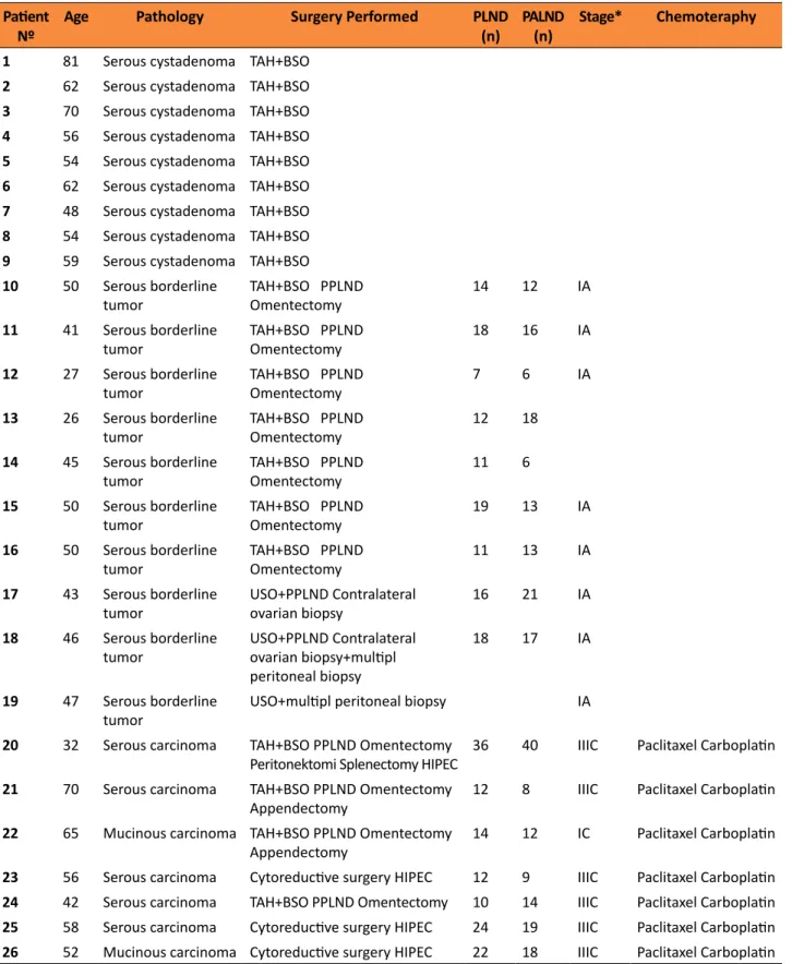
Characteristics of the patients participated in the study are summarized in Table 1. The assessment of immunostaining for NF-kB was made by using the H-score method. In the calculation of an H-score, a summation
of the percentage of area stained at each
Table 1 - Characteristics of the patients participating in the study. Patient
Nº
Age Pathology Surgery Performed PLND
(n)
PALND (n)
Stage* Chemoteraphy
1 81 Serous cystadenoma TAH+BSO
2 62 Serous cystadenoma TAH+BSO
3 70 Serous cystadenoma TAH+BSO
4 56 Serous cystadenoma TAH+BSO
5 54 Serous cystadenoma TAH+BSO
6 62 Serous cystadenoma TAH+BSO
7 48 Serous cystadenoma TAH+BSO
8 54 Serous cystadenoma TAH+BSO
9 59 Serous cystadenoma TAH+BSO
10 50 Serous borderline
tumor
TAH+BSO PPLND
Omentectomy 14 12 IA
11 41 Serous borderline
tumor
TAH+BSO PPLND
Omentectomy 18 16 IA
12 27 Serous borderline
tumor
TAH+BSO PPLND
Omentectomy 7 6 IA
13 26 Serous borderline
tumor
TAH+BSO PPLND
Omentectomy 12 18
14 45 Serous borderline
tumor
TAH+BSO PPLND
Omentectomy 11 6
15 50 Serous borderline
tumor
TAH+BSO PPLND
Omentectomy 19 13 IA
16 50 Serous borderline
tumor
TAH+BSO PPLND
Omentectomy 11 13 IA
17 43 Serous borderline
tumor
USO+PPLND Contralateral
ovarian biopsy 16 21 IA
18 46 Serous borderline
tumor
USO+PPLND Contralateral ovarian biopsy+multipl peritoneal biopsy
18 17 IA
19 47 Serous borderline
tumor
USO+multipl peritoneal biopsy IA
20 32 Serous carcinoma TAH+BSO PPLND Omentectomy
Peritonektomi Splenectomy HIPEC 36 40 IIIC Paclitaxel Carboplatin
21 70 Serous carcinoma TAH+BSO PPLND Omentectomy
Appendectomy 12 8 IIIC Paclitaxel Carboplatin
22 65 Mucinous carcinoma TAH+BSO PPLND Omentectomy
Appendectomy 14 12 IC Paclitaxel Carboplatin
23 56 Serous carcinoma Cytoreductive surgery HIPEC 12 9 IIIC Paclitaxel Carboplatin
24 42 Serous carcinoma TAH+BSO PPLND Omentectomy 10 14 IIIC Paclitaxel Carboplatin
25 58 Serous carcinoma Cytoreductive surgery HIPEC 24 19 IIIC Paclitaxel Carboplatin
26 52 Mucinous carcinoma Cytoreductive surgery HIPEC 22 18 IIIC Paclitaxel Carboplatin
TAH+BSO. total abdominal hysterectomy+bilateralsalpingo-oophorectomy; PPLND. pelvic-paraaortic lymphadenectomy; PLND. pelvic
lymphadenectomy; PALND. paraaortic lymphadenectomy; USO. unilateral salpingo-oophorectomy; HIPEC. hyperthermic intraperitoneal
chemotherapy; cytoreductive surgery. surgical procedure performed leaving less than 1 cm margin of tumoral tissue intact; Stage*. Based
Table 2 - Intergroup comparison of H scores, p<0.05 was accepted as the level of statistical significance.
Patient Groups H score p value
Group 1 (Serous cystadenoma, n=9) 127.7± 48.4
Group 2 (Serous-mucinous borderline tumor, n=10) 166.0 ± 95.5
Group 3 (Serous-mucinous cystadenocarcinoma, n= 8) 311.2 ± 80.2 <0.001
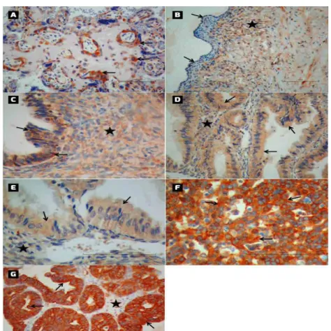
When the images from the light microscope of the immunohistochemical
results of our study were examined, it was seen that cytoplasmic and/nuclear NF-kB expression
was more apparent in the group diagnosed with ovarian cancer compared to the other two groups (Figure 1).
Figure 1 – A. Positive Control;Human plasenta. Immunohistochemical positive immunoreactivity of NF-kB
(arrow). IHC, x20. B. Serous cystadenoma; Epithelial (arrows) and stromal (aster) immunohistochemical
positive immunoreactivity of NF-kB. IHC, x20. C. Serous cystadenoma;Epithelial (arrows) and stromal (aster)
immunohistochemical positive immunoreactivity of NF-kB. IHC, x40. D. Borderline; Epithelial (arrows) and
stromal (aster) immunohistochemical positive immunoreactivity of NF-kB. IHC, x20. E. Borderline; Epithelial
(arrows) and stromal (aster) immunohistochemical positive immunoreactivity of NF-kB. IHC, x40. F.
■
Discussion
Tumors developing from ovarian tissue have been classified as germ cell, sex chord
stromal and epithelial cell tumors, epithelial
ovarian cancers have become an important health problem, and because they are diagnosed at a late stage when they had distant organ metastases and being the most fatal female genital system cancer, they threaten patient health significantly. In the whole world, nearly 2/3 of the newly diagnosed 240.000
cases lose their lives8. However, borderline
ovarian tumors consist 15-20% of all epithelial ovarian tumors. They were firstly defined by International Federation of Gynecology and Obstetrics (FIGO) in the year 1973 and classified as a tumor with low malignant potential. Although its definition bases on totally histopathological findings, epithelial cell proliferation without stromal invasion is the gold standard in the establishment of the diagnosis of the borderline tumor9.
Although many different causes have been described in the etiology of ovarian cancer, chronic inflammation is an important factor in the etiology of both epithelial and also borderline ovarian tumors. Regular cycles of ovulation cause chronic inflammation which increases cytokine secretion leading to an acceleration of cellular mitosis, apoptotic errors, and development of malignant transformation10. In many literature
studies significantly higher levels of some proinflammatory cytokines (i.e., interleukin 2-4-6-12 and 13) have been detected in follicular fluids of patients diagnosed as ovarian tumor when compared with the control group11.
However, the mechanism in which increased cytokine secretion induces carcinogenesis in ovarian tissue has not been clarified yet. This challenging condition requires demonstration of different intracellular mechanisms which explain the etiological role of inflammation in
carcinogenesis. The importance of estrogen in the etiology of ovarian carcinoma should not be forgotten. In their study, Perniconi et al.12
determined that dysmorphological changes emerged in the ovarian superficial epithelium in rats, which received high dose estrogen. It was also demonstrated that inflammatory mediators were responsible for the effect of estrogen. The increased mitogenic activity stimulated the synthesis of the growth factors and cytokines. Bcl-2, c-Myc proto-oncogene, α-TGF and IL-6 levels increased during the estrogen-depended increase of mitosis. These mediators were also responsible for tumorigenesis due to the stimulation of the cell proliferation and apoptosis.
To this end, in recent years mostly studied chronic inflammation mediator is nuclear transcription factor-kappa beta (NF-kB) which at the same time controls cellular proliferation, apoptosis, angiogenesis, immune reaction, cellular adhesion and differentiation (NF-kB)13. NF-kB pathway plays a very important
role in inflammation and carcinogenesis14.
NF-kB family consists of different subunits as p50, p65 (RelA), c-Rel, RelB, p50, and p52. The most prevalently found subtype in human cells and all other mammalian species is p65/ p50 heterodimer structure which is found bound to intracytoplasmic inhibitor protein (IkB). Following internal gene mutation and/ or external stimulation IkB kinase enzyme is activated, translocated into inactive NF-kB. As a consequence, it effects transcription genes with resultant stimulation of local and systemic inflammation leading indirectly to
carcinogenesis7.
NF-kB activation at a cellular level was demonstrated in cases with Hodgkin’s lymphoma which displayed extranodal
spread16. Nagel et al.18 demonstrated higher
NF-kB activity in cases with hairy cell leukemia
17. In another study where 33 patients
with colorectal cancer, increased NF-kB activation was demonstrated in patients with established diagnosis of malignancy. Zhao et al.19 demonstrated increased NF-kB activation
in patients diagnosed as papillary thyroid carcinoma.
When studies investigating gynecologic tumors and NF-kB expression are reviewed, publications supporting our study have striked our attention. In a study which consisted of 63 patients diagnosed as epithelial ovarian cancer, a direct correlation was detected between increased NF-kB expression, chemoresistant disease and lymph node metastasis20. Chen
et al.21 compared outcomes of 411 patients
with epithelial ovarian tumor and healthy 438 patients and demonstrated significantly higher NF-kB expression in the tissues of the patients with established diagnosis of malignancy. Although the presence of a viral effect in the etiology of cervical cancer has been indisputably accepted, in recent years the significant role of NF-kB expression in the development of cervical cancer has been also demonstrated. In a study where 240 cervical cancer tissue and 290 normal cervical tissue specimens were investigated, higher NF-kB expression has been demonstrated in patients with the diagnosis of cervical cancer22. Nakahara
et al.23 displayed that contamination with
Human Papilloma Virus (HPV) type-16 which is the most important agent in the etiology of cervical cancer increases NF-kB expression which consequently stimulates persistence of HPV infection. Review of the studies which investigated the correlation between NF-kB and endometrial cancer have yielded similar results. Various studies have demonstrated
that IL-6 cytokine which induces cellular proliferation, exerts this effect by indirectly enhancing NF-kB expression24. In a study where
NF-kB expression was investigated in in vitro cell cultures using Western blot technique, increased NF-kB expression was observed in
cancer cells25.
The importance of the chronic inflammation and of the cytokines and inflammatory markers, which are increasingly secreted from the inflammatory cells, is well known. These cytokines cause direct DNA damage and, in addition, contribute to the tumorigenesis due to the indirect inhibition of the apoptosis and stimulation of angiogenesis. In the estrogen-dependent ovarian carcinoma, the levels of the inflammatory markers increase under the influence of estrogen. The importance of these mediators is well known in the etiology of the ovarian carcinoma, which is considered as a secondary development to the dysmorphology that emerges at the cellular level in the superficial epithelium.
■
Conclusions
Factor nuclear kappa B is an important mediator that acts in the chronic inflammation. The highest expression rates are determined by the immunohistochemical method in the ovarian cancer group.
■
References
1. Chandrashekhara SH, Triveni GS, Kumar R.
Imaging of peritoneal deposits in ovarian cancer: a pictorial review. World J Radiol. 2016; :513-7. doı: 10.4329/wjr.v8.i5.513. 2. Cannistra SA. Cancer of the ovary. N Eng
J Med. 2004;351:2519-29. doı: 10.1056/ NEJMra041842.
1999–2007 by country and age: results of EUROCARE−5 a population-based study. Lancet Oncol. 2014;15:23–34. doı: 10.1016/ S1470-2045(13)70546-1.
4. Cosyns S, Polyzos NP, Carprieaux M, Carprieaux M, Tournaye H, De Sutter P.
The role of appendectomy as part of the treatment of a mucinous borderline ovarian tumor. Eur J Gynaecol Oncol. 2016;37:167-70. PMID: 27172739.
5. Kim DK, Oh SY, Kwon HC, Lee S, Kwon KA, Kim BG. Kim SH, Jang JS, Kim MC, Kim KH, Han JY,
Kim HJ. Clinical signifi cances of preoperative serum interleukin-6 and C-reactive protein level inoperable gastric cancer. BMC Cancer. 2009;9:155-9. doı: 10.1186/1471-2407-9-155. 6. Karin M, Cao Y, Greten FR, Li ZW. NF-kappaB
in cancer: from innocent bystander to major culprit. Nat Rev Cancer. 2002;2:301–10. doı: 10.1038/nrc780.
7. Chen Y, Lu R, Zheng H, Xiao R, Feng J, Wang H, Gao X, Guo L. The NFKB1 polymorphism
(rs4648068) is associated with the cell proliferation and motility in gastric cancer. BMC Gastroenterol. 2015;15:1-12. doı: 10.1186/s12876-015-0243-0.
8. Ataseven B, Chiva LM, Harter P,
Gonzalez-Martin A, du Bois A. FIGO stage IV epithelial
ovarian, fallopian tube and peritoneal cancer revisited. Gynecol Oncol. 2016;1-11. doı: 10.1016/j.ygyno.2016.06.013.
9. Tropé CG, Kaern J, Davidson B. Borderline ovarian tumors. Best Pract Res Clin Obstet Gynaecol. 2012;26:325-36. doı: 10.1016/j. bpobgyn.2011.12.006.
10. Clendenen TV, Lundin E, Zeleniuch-Jacquotte A, Koenig KL, Berrino F, Lukanova
A, Lokshin AE, Idahl A, Ohlson N, Hallmans
G, Krogh V, Sieri S, Muti P, Marrangoni A,
Nolen BM, Liu M, Shore RE, Arslan AA.
Circulating inflammation markers and risk of epithelial ovarian cancer. Cancer Epidemiol Biomarkers Prev2011;20:799-810. doı: 10.1158/1055-9965.EPI-10-1180.
11. Wang YQ, Jin C, Zheng HM, Zhou K, Shi BB,
Zhang Q, Zheng FY, Lin F. A novel prognostic
inflammation score predicts outcomes in patients with ovarian cancer. Clin Chim Acta. 2016; 456:163-9. doı: 10.1016/j. cca.2016.03.013.
12. Perniconi SE, Simões Mde J, Simões Rdos
S, Haidar MA, Baracat EC, Soares JM Jr.
Proliferation ofthe superficial epithelium
of ovaries in senile female rats following oraladministration of conjugated equine estrogens. Clinics (Sao Paulo). 2008;63:381-8. PMID: 18568250.
13. Chen CD, Sawyers CL. NF-kappa B activates prostate-specific antigen expression and is upregulated in androgen-independent prostate cancer. Mol Cell Biol. 2002;22:2862– 70. PMID: 11909978.
14. Karin M, Cao Y, Greten FR, Li ZW. NF-kappa B in cancer: from innocent bystander to major culprit. Nat Rev Cancer. 2002;2:301–10. doı: 10.1038/nrc780.
15. Mosialos G. The role of Rel/NF-kappa B proteins in viral oncogenesis and the regulation of viral transcription. Semin Cancer Biol. 1997;8:121–9. doı: 10.1006/ scbi.1997.0063.
16. Dolcet X, Llobet D, Pallares J, Matias-Guiu X. NF-kB in development and progression of human cancer. Virchows Arch. 2005;446:475-82. doı: 10.1007/s00428-005-1264-9.
17. Nagel S, Ehrentraut S, Meyer C, Kaufmann M, Drexler HG, MacLeod RA. NFkB is
activated by multiple mechanisms in hairy cell leukemia. Genes Chromosomes Cancer. 2015;54:418-32. doı: 10.1002/gcc.22253. 18. Hai Ping P, Feng Bo T, Li L, Nan Hui Y, Hong
Z. IL-1β/NF-kb signaling promotes colorectal cancer cell growth through miR-181a/PTEN axis. Arch Biochem Biophys. 2016;604:20-6. doı: 10.1016/j.abb.2016.06.001.
19. Zhao S, Wang Q, Li Z, Ma X, Wu L, Ji H, Qin G..
LDOC1 inhibits proliferation and promotes apoptosis by repressing NF-κB activation in papillary thyroid carcinoma. J Exp Clin Cancer Res. 2015;34: 1-12. doı: 10.1186/ s13046-015-0265-z.
20. Shuang T, Wang M, Zhou Y, Shi C.
Over-expression of nuclear NF-κB1 and c-Rel correlates with chemoresistance and
prognosis of serous epithelial ovarian
cancer. Exp Mol Pathol. 2016;100:139-44. doı: 10.1016/j.yexmp.2015.11.030.
21. Chen LP, Cai PS, Liang HB. Association of
the genetic polymorphisms of NFKB1 with susceptibility to ovarian cancer. Genet Mol Res. 2015;14:8273-82. doı: 10.4238/2015. July.27.15.
of cervical carcinoma in HPV-infected postmenopausal women from rural area. Tumour Biol. 2015;36:6265-76. doı: 10.1007/s13277-015-3312-7.
23. Nakahara T, Tanaka K, Ohno S, Egawa N, Yugawa T, Kiyono T. Activation of NF-κB by human papillomavirus 16 E1 limits E1-dependent viral replication through degradation of E1. J Virol. 2015;89:5040-59. doı: 10.1128/JVI.00389-15.
24. Che Q, Liu BY, Wang FY, He YY, Lu W, Liao
Y, Gu W, Wan XP. Interleukin 6 promotes endometrial cancer growth through an autocrine feedback loop involving ERK-NF-κB signaling pathway. Biochem Biophys Res Commun. 2014;446:167-72. doı: 10.1016/j. bbrc.2014.02.080.
25. Lundqvist J, Yde CW, Lykkesfeldt AE. 1α,25-dihydroxyvitamin D3 inhibits cell growth and NFκB signaling in tamoxifen-resistant breast cancer cells. Steroids. 2014;85:30-5. doı: 10.1016/j.steroids.2014.04.001.
Correspondence: Ercan Yilmaz
Department of Obstetrics and Gynecology, Faculty of Medicine
Inonu University, Malatya Turkey Phone: +0905369556180
Received: Mar 08, 2018 Review: May 05, 2018 Accepted: June 04, 2018
Conflict of interest: none Financial source: none
1Research performed at Histology and