EDUARDA SOFIA LOPES COSTA
ANTICANCER EFFECTS OF THE SEAWEED COMPOUNDS
FUCOXANTHIN AND PHLOROGLUCINOL, ALONE AND IN
COMBINATION WITH 5-FLUOROURACIL IN COLON CELLS
Dissertação de Candidatura ao grau de Mestre em Ciências do Mar – Recursos Marinhos submetida ao Instituto de Ciências Biomédicas de Abel Salazar da Universidade do Porto
Orientadora – Doutora Alice Fernanda Abreu Ramos Categoria – Investigadora Post-Doc Afiliação – Centro Interdisciplinar de
Investigação Marinha e Ambiental
Coorientador – Professor Doutor Eduardo Jorge Sousa Rocha Categoria – Professor Catedrático Afiliação – Instituto de Ciências Biomédicas Abel Salazar
AKNOWLEDGMENTS
I would like to thank:
Doctor Alice Ramos, my supervisor, for all the help, support, and affection you gave me. Your support during this stage of my academic life was outstanding and extended far beyond the academy. Without your working ability, scientific knowledge and constant presence this thesis would not be possible.
My co-supervisor, Professor Doctor Eduardo Rocha, a science enthusiast who is able to inspire people, for giving me the opportunity to work in this project and to have integrated a wonderful research team.
Professor Doctor Daniela Gargiulo, for your friendship and for her wise guidance.
All my laboratory colleagues, especially Mariana Abreu and Diana Pádua.
My co-worker, Sofia Fernandes.
And especially I would like to thank my family.
This work was partially funded by the ICBAS and by the project MARBIOTECH (reference NORTE-07-0124-FEDER-000047), co-financed by the North Portugal Regional Operational Programme (ON.2 – O Novo Norte), under the National Strategic Reference Framework (NSRF), through the European Regional Development Fund (ERDF).
A
BSTRACT
On a global scale, colorectal cancer is considered to be the third most common, and the second most common cause of deaths related with cancer both in men and women. The incidence of colorectal cancer cases has been rising at an alarming rate, particularly in regions with a western lifestyle.
Currently, treatment for colorectal cancer involves the combination of surgery with chemotherapy, by administration of cytotoxic drugs and radiation. 5-fluorouracil (5-Fu), an antimetabolite, is one of the most commonly used cytotoxic drugs in colorectal treatment. Unfortunately, resistance to 5-Fu may emerge during treatment due to several biological mechanisms, namely over expression of thymidylate synthase (TS) and alterations in the apoptotic pathway. One of the strategies to overcome drug resistance is the combination with other drugs and/or natural compounds.
The chemical and biological diversity of the marine environment is incalculable, making this a reservoir of natural bioactive compounds. Many of these products exhibit structural and chemical characteristics that are not found in natural terrestrial compounds. With this in mind, the research of new natural products with high efficacy against tumour cells and no apparent cytotoxicity against normal cells has gained a prominent place in the scientific community. Marine algae are a magnificent source of bioactive compounds which demonstrate a broad range of biological activities such as antioxidant, antimicrobial, anti-inflammatory, and anticancer activities. The biological activities of fucoxanthin, a marine carotenoid, and phloroglucinol, a polyphenolic compound, have been explored in diverse studies, including cytotoxic effects against cancer cell lines.
In the present study, the anticancer effects of fucoxanthin and phloroglucinol alone or combined with 5-Fu were assessed on two colorectal cancer cell lines (HCT-116 and HT-29) resorting to anti-proliferative, apoptotic, and genotoxic assays. Also, to assess the specificity of the anticancer effects, a normal colon cell line (CCD-Co18) was also tested. Our results showed that 5-Fu decreased cell viability in a dose-dependent manner and induced DNA damage in both cell lines, with HCT-116 cells presenting increased sensitivity to 5-Fu. Furthermore, fucoxanthin decreased cell viability more efficiently than 5-Fu alone and when in co-incubation it enhanced the cytotoxic effect of 5-Fu in the cancer cell line, without toxic effects to normal cells, which could be a promising alternative for resistant colorectal cancer cell lines.
Phloroglucinol alone also decreased cell viability and in combination increased the sensitivity to 5-Fu in both cancer cell lines, especially in the p53 mutant HT-29 cancer cell line. Nonetheless, at lower concentrations phloroglucinol inhibits the effects of 5-Fu in HCT-116 cells, which is not a desired effect during a cancer treatment.
Both natural compounds, fucoxanthin and phloroglucinol enhance the efficacy of 5-Fu in certain conditions. However, further studies are required to clarify the mechanisms involved in order to become a possible strategy for some cases of 5-Fu-resistant colon cancer.
R
ESUMO
À escala global, o cancro do coloretal é considerado o terceiro tipo de tumor maligno mais comum, e o segundo responsável pelo maior número de mortes relacionadas com o cancro, tanto em homens como em mulheres. Assistimos a um aumento na incidência de casos de cancro coloretal, de uma forma alarmante, principalmente em regiões com um estilo de vida caraterístico de países desenvolvidos.
Atualmente, o tratamento para o cancro coloretal passa pela combinação de cirurgia com quimioterapia, através da administração de fármacos citotóxicos e do uso de radiação. O 5-fluorouracilo (5-Fu), um anti-metabolito, é um dos compostos citotóxicos mais utilizados no tratamento do cancro coloretal. Infelizmente, durante o tratamento com 5-Fu poderá surgir resistência ao mesmo devido a diversos mecanismos biológicos, nomeadamente expressão elevada da timidilato sintase e alterações na via apoptótica. A combinação com outras fármacos e/ou com compostos naturais é uma das estratégias utilizadas para ultrapassar a resistência a fármacos.
A diversidade química e biológica do ambiente marinho é incalculável, tornando-o um reservatóritornando-o de ctornando-omptornando-osttornando-os naturais bitornando-oativtornando-os. Muitas destas mtornando-oléculas exibem caraterísticas estruturais e químicas que não são encontradas nos compostos naturais terrestres. Tendo isto em consideração, a pesquisa de novos produtos naturais com elevada eficiência contra células tumorais e aparentemente sem citotoxicidade contra células normais têm ganho um lugar de destaque dentro da comunidade científica. As algas marinhas são uma fonte magnífica de compostos bioativos que demonstram uma ação vasta de atividades biológicas, entre as quais atividade antioxidante, antimicrobiana, anti-inflamatória e antitumoral. As atividades biológicas da fucoxantina, um “carotenóide marinho”, e do floroglucinol, um composto fenólico, que também pode ter origem em organismos marinhos (em particular em algas castanhas), têm sido exploradas em diversos estudos, inclusive os seus efeitos citotóxicos em linhas celulares de cancro.
No presente estudo foram aferidos os efeitos antitumorais da fucoxantina e do floroglucinol, isolados ou em combinação com o 5-Fu, em duas linhas celulares de cancro coloretal (HCT-116 e HT-29) recorrendo a ensaios anti-proliferativos, apoptóticos e de genotoxicidade. A especificidade do efeito antitumoral foi também avaliada utilizando uma linha celular normal de colon (CCD-Co18). Os nossos
resultados mostraram que o 5-Fu, nas duas linhas celulares de cancro testadas, diminuiu a viabilidade celular de uma forma dose-dependente com a capacidade de induzir danos no DNA, sendo as células HCT-116 mais sensíveis ao 5-Fu. Além disso, a fucoxantina diminuiu a viabilidade celular mais eficientemente do que o 5-Fu isolado e quando em co-incubação potenciou o efeito citotóxico do mesmo, sem citotoxicidade para as células normais, tornando-se uma alternativa promissora a ser utilizada em linhas celulares resistentes. O floroglucinol isolado diminuiu também a viabilidade celular e em co-incubação aumentou a sensibilidade de ambas as linhas celulares para o 5-Fu, especialmente da linha celular HT-29 com p53 mutante. Contudo, em concentrações mais baixas inibiu o efeito do 5-Fu nas células HCT-116, condição que não é pretendida durante um tratamento oncológico.
Ambos os compostos naturais, fucoxantina e floroglucinol em certas condições potenciam a eficiência do 5-Fu. Contudo, é necessário a realização de mais estudos de modo a clarificar os mecanismos envolvidos e assim tornar-se numa possível estratégia terapêutica para certos casos de cancro do coloretal resistentes a 5-Fu.
T
ABLE OF CONTENTS
AKNOWLEDGMENTS... iiiAbstract ...v
Resumo ... vii
Table of contents ... ix
List of abbreviations... xi
CHAPTER I - INTRODUCTION...13
1.
Cancer ...15
1.1
A brief introduction ... 15
1.2
Cancer statistics... 16
1.2.1
Colorectal carcinoma... 16
1.3
Cancer biology ... 18
1.3.1
Genomic instability ... 18
1.3.2
Cell proliferation ... 19
1.3.3
Apoptosis... 20
1.4
Colorectal cancer ... 22
1.5
Therapeutic approaches ... 25
1.5.1
5-Fluorouracil (5-Fu)... 26
1.6
Marine natural compounds ... 27
1.7
Marine algae ... 28
1.7.1
Fucoxanthin... 29
1.7.2
Phloroglucinol... 30
1.8
Methodologies available to screen anticancer activity ... 31
1.8.1
Cell viability/proliferation assays ... 32
1.9
Evaluation of the possible action mechanisms ... 33
1.9.1
Genotoxicity evaluation by comet assay ... 33
1.9.2
Apoptosis... 34
1.9.3
Western blot ... 34
CHAPTER II – ANTICANCER EFFECTS OF THE SEAWEED COMPOUNDS FUCOXANTHIN AND PHLOROGLUCINOL, ALONE AND IN COMBINATION WITH
5-FLUOROURACIL IN COLON CELLS...43
References ...73
CHAPTER III - CONCLUSIONS AND FUTURE PERSPECTIVES ...77
L
IST OF ABBREVIATIONS
5-Fu – 5-fluorouracil
ACE – Angiotensin I-converting enzyme
APAF1 – Apoptotic protease activating factor 1 APC – Adenomatous polyposis coli
BCA – Bicinchoninic Acid BRCA – Breast cancer gene BrdU – Bromodeoxyuridine
CD-DST - collagen gel droplet-embedded culture drug sensitivity test CIMP – CpG island methylator phenotype
CIN – Chromosomal instability
DAPI – 4’, 6-diamidino-2-phenylindole DISC – Death-inducing signaling complex DMEM – Dulbecco’s Modified Eagle Medium DMSO – Dimethyl sulfoxide
DNA – Deoxyribonucleic acid
EDTA – Ethylenediaminetetraacetic acid EGFR – Epidermal growth factor receptor EMT – Epithelial-mesenchymal transition ET-743 – Trabectedin
FADD – Fas-associated death domain protein FBS – Fetal Bovine Serum
FdUMP – Fluorodexoyuridine monophosphate FdUTP – Fluorodeoxyuridine triphosphate FUTP – Fluorouridine triphosphate
HCT-116 – Human colorectal carcinoma cell line
HEPES – N-(2-hydroxyethyl)piperazine-N’-(2-ethanesulfonic acid) HNPCC – Hereditary Non-Polyposis Colorectal Cancer
HT-29 – Human colon adenocarcinoma cell line IGF-1 – Insulin-like growth factor-1
LOH – Loss of heterozygosity
MAPK – Mitogen-activated protein kinase MSI – Microsatellite instability
mTOR – Mammalian target of rapamycin
MTT – 3-(4,5-dimethylthiazol-2-yl)-2,5-diphenyltetrazolium bromide NaCl – Sodium Chloride
PI3K – Phosphatidylinositol 3-kinase PMSF – Phenylmethanesulfonylfluoride PBS – Phosphate buffer saline
PS – Phosphatidylserine
RIPA – Radioimmunoprecipation assay RPMI – Roswell Park Memorial Institute TKRs – Tyrosine kinase receptors TNF-α - Tumour necrosis factor-α
TNFR1 – Tumour necrosis factor receptor 1 TP53 – Tumour Protein p53 gene
TS – Thymidylate synthase
1. C
ANCER
1.1 A brief introduction
Cancer is the general name for a group of diseases associated to an uncontrolled cell growth and invasiveness. In fact, more than a hundred different types of cancer have been described (Hanahan & Weinberg, 2000).
There is a general assumption that cancer is a modern age disease. However, that is not entirely true since there are early descriptions of cancerous lesions reported in an ancient Egyptian textbook on trauma surgery (3000 BC). This textbook is known as the Edwin Smith Papyrus and described eight cases of breast tumours (Hajdu, 2011). However, the origin of the word “cancer” is attributed to Hippocrates (460-370 BC) that used the terms carcinos and carcinoma to report non-ulcer forming and ulcer-forming tumours (Sudhakar, 2010).
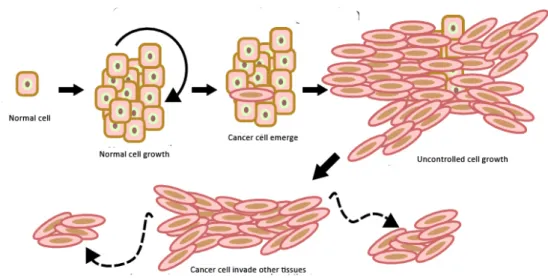
Generally, the growth of a cancer cell is different from the normal cell. While a normal cell grows and eventually dies at one point, cancer cells can avoid death and continue to grow and form new abnormal cells (Fig. 1). Another aspect that distinguishes a cancer cell from a normal cell is its ability to invade other tissues, where they are able to proliferate (Cooper, 2000).
Figure 1 - Cancer cell growth.
Adapted from: http://www.unc.edu/depts/our/hhmi/hh-mift_learning_modules/cancermodule/pages/cancer.html
There are two types of risk factors in cancer, the external factors, representing the cause of 90-95% off all cancers, and internal factors. They can act together, or in sequence, in order to start or to promote a carcinogenic condition. Alcohol consumption, smoking, eating habits, physical inactivity, environmental pollution are examples of external factors (Jemal et al., 2011). Hormonal dysfunctions, immune condition of an individual and mutations count as internal factors (Anand et al., 2008).
1.2 Cancer statistics
Over the past decades, a drastic increase in cancer cases in developed and developing countries has been witnessed. Cancer is accountable for causing more deaths than AIDS, tuberculosis and malaria all together (American Society Cancer, 2011).
According to Globocan project, that provides contemporary estimates of incidence, mortality and prevalence from the major types of cancer, there were 14.1 million new cases of cancer in 2012. In the same year, the number of deaths due to cancer was 8.2 million and it is estimated that 32.6 million people have cancer (Ferlay et al., 2015).
The most common cancer worldwide in 2012 was lung cancer, contributing to 13% of the total number of new cases diagnosed, followed by breast cancer (women only). Colorectal cancer was the third most common cancer with nearly 1.4 million of new cases (Globocan, 2012).
1.2.1 Colorectal carcinoma
Colorectal cancer is considered one of the major causes of morbidity and mortality on a global scale. It affects men and women in the same proportion, representing 9.4% in men and 10.1% in women off all incident cancers (Haggar & Boushey, 2009).
In terms of geographic incidence, colorectal cancer is mainly a condition of developed countries that counts with 54% of colorectal cancer cases. Oceania and Europe have the highest incidence and Africa and Asia the lowest (Fig. 2) (Globocan, 2012).
Figure 2 - Estimated colorectal cancer incidence worldwide in 2012 by age-standardized rates per 100,000 individuals.
Adapted from Globocan, 2012.
There are convincing evidences that a western life-style is related to an increase risk of colorectal cancer disease – namely consumption of red meat and processed food, alcohol abuse, chronic smoking, high caloric intake, low vegetable and fruits intake, and physical inactivity are seen as risk factors of colorectal cancer. Another important risk factor in the case of colorectal cancer is the hereditary factor or a history of inflammatory bowel disease (Haggar et al., 2009). On the other hand, physical activity and consumption of dietary fiber, like vegetables and fruits, are linked to cancer prevention (Key et al., 2004).
Awareness and anticipated diagnosis are key factors in an increased survival rate of any oncological patient. Several screening tests have been developed to detect colorectal cancer in early phases. The most common tests are high-sensitivity fecal occult blood tests (FOBT), that checks for tiny amounts of blood in feces that can be from bleeding polyps or tumours; and colonoscopy, in which the rectum and entire colon are examined using a colonoscope and any abnormal growths in the colon and the rectum can be removed (National Cancer Institute, 2014). Jarvinen and colleagues demonstrated that screening colonoscopy at 3-year intervals decreases mortality by about 65% in Hereditary Non-Polyposis Colorectal Cancer (HNPCC) families (Jarvinen et al., 2000)
1.3 Cancer biology
Douglas Hanahan and Robert A. Weinberg advocate that during development of human tumours, the cells acquire a set of biological capabilities referred as hallmarks. They include sustaining proliferative signaling, evading growth suppressors, escape from apoptosis, enabling replicative immortality, sustained angiogenesis, tissue invasion and metastasis, inflammation, avoiding immune destruction, and deregulating cellular energetics. Genomic instability is now considered another important hallmark and is responsible for generating genetic diversity (Hanahan & Weinberg, 2011).
1.3.1 Genomic instability
Genomic instability corresponds to an increased propensity of cell genome to accumulate alterations during the cell cycle. Ordinarily, it is associated with the failure of parent cells to accurately duplicate the genome and precisely distribute the genomic material among the daughter cells (Kramer et al., 2002; Shen, 2011). During the cell cycle there are four significant mechanisms that ensure genomic stability: (1) high-fidelity of DNA replication in the S-phase, (2) accurate distribution of chromosomes among daughter cells during mitosis, (3) error-free repair of sporadic DNA damage through the cell cycle, and (4) cell cycle progression and checkpoint control (Shen, 2011). Changes to the normal function of these mechanisms leads to genetic alterations that provide advantages to these cells allowing them to proliferate, survive and spread. Generally, genetic alterations can be divided into: (1) microsatellite instability (MSI), (2) CpG island methylator phenotype (CIMP) and (3) chromosomal instability (CIN) (Grady, 2004).
Precise repair of sporadic DNA damage is one of the major mechanisms that allows the maintenance of genomic stability throughout the cell cycle. A DNA damage signaling network regulates the activity of repair pathways and their coordination with several biological functions such as cell death. Dysregulation of this efficient network may lead to genomic instability. Furthermore, many of the chemotherapeutic drugs work by causing DNA damage to cancer cells, so it is imperative that the DNA repair mechanisms performs properly without compromising the treatment outcome (Shen, 2011).
A question quite explored by scientists is if genomic instability is the cause or the consequence of cancer, and how it influences the progression of hereditary and sporadic cancers. Loeb suggested that cancer cells must acquire some form of intrinsic
genomic instability, a “mutator phenotype” (Loeb, 1991). This mutator hypothesis states that genomic instability is present in precancerous lesions, driving to tumour development by increasing the spontaneous mutation rate in caretaker genes.
Germline mutations targeting DNA repair genes, such as BRCA 1 or BRCA 2, are constant in the genome of patient with hereditary cancer. Thus, a single event such as the loss of the remaining wildtype allele would lead to genomic instability and tumour progression (O’Donovan & Livingston, 2010).
On the contrary, in sporadic cancers the activation of growth signaling pathways due to mutations in oncogenes seems to induce loss of heterozygosity (LOH) and genomic instability involving DNA replication stress. Specific genomic regions, called common fragile sites, are prone to breakage under DNA replication stress. In human precancerous lesions in which oncogenes have been activated, genomic instability preferentially affects common fragile sites (Negrini et al., 2010).
1.3.2 Cell proliferation
Under normal conditions cell proliferation is defined by the balance between the number of new cells and the number of cells lost through apoptosis or differentiation. Thus, cell proliferation is a tightly regulated process with the contribution of a wide number of molecules and interconnected pathways, responsible for maintaining the number of cells of an adult tissue constant (Guo & Hay, 1999)
Cell growth and proliferation can be controlled by the production and release of growth-promoting signals that normally bind to a cell-surface receptor, such as tyrosine kinase receptors (TKRs). TKRs activity is responsible for the recruitment of certain molecules that activate intracellular signaling pathways associated with cell proliferation, differentiation, and others cellular processes (Schlessinger, 2000). Cancer cells can achieve a sustaining proliferative state by disrupting this homeostatic condition trough the dysregulation of TKrs and/or their downstream signal transduction pathways, by increasing growth factors production and also by stimulating production of growth factors by normal cells (Hanahan & Weinberg., 2011).
Epidermal growth factor receptor (EGFR) and Ras-Raf-MEK-ERK signaling commonly known as the mitogen-activated protein kinase (MAPK) pathway, is a well-known signal transduction pathway in cell biology. One of the main functions of MAPK pathway is to transduce signals that will be received by the cell nucleus, leading to the activation of specific genes involved in cell growth, division, and differentiation (Molina & Adjei., 2006). Dysregulation of this pathway leads to an uncontrolled cellular growth and has been reported in several human cancers (Santarpia et al., 2012).
20 Another intracellular signaling pathway activated by TKRs and with an important role in tumorogenesis is Phosphatidylinositol 3-kinase (PI3K), protein kinase Akt, and mammalian target of rapamycin (mTOR) also designated as PI3K/AKT/mTOR signaling pathway (Vara et al., 2004).
1.3.3 Apoptosis
Apoptosis or programmed cell death is a highly regulated and active mechanism that occurs in response to physiological or pathological stimuli. Apoptosis occurs normally during development and aging, acting like a homeostatic mechanism to maintain cell populations in tissues (Renehan et al., 2001).
Apoptotic cells display a set of unique morphological characteristics that distinguish them from normal cells such as condensation and fragmentation of DNA, blebbing of the membrane, formation of apoptotic bodies and rapid phagocytosis by neighbouring cells without promoting inflammatory response (Kerr et al., 1972; Saraste & Pulkki, 2000; Elmore, 2007).
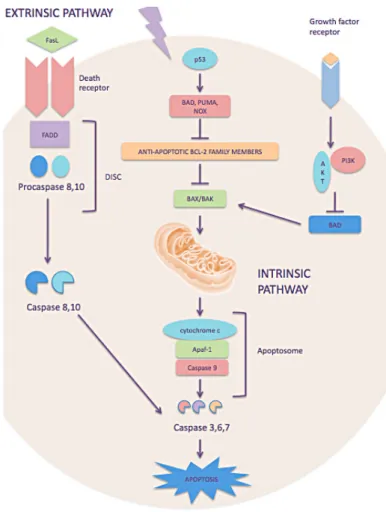
Figure 3 - The two major pathways of apoptosis: extrinsic or intrinsic pathway. Adapted from Ashkenazi, 2008.
A wide variety of stimuli can induce the activation of the two major pathways of apoptosis, the extrinsic or death receptor pathway and the intrinsic or mitochondrial pathway (Fig. 3) (Elmore, 2007).
The extrinsic pathway is initiated by the activation of death receptors (e.g. FAS and TNFR1), located on the cell surface, by extracellular death ligands (such as FasL or tumour necrosis factor-α (TNF-α)). Activation of death receptors results in the recruitment of adaptor proteins, such as the Fas-associated death domain protein (FADD), which in turn recruits and aggregates several molecules of procaspases -8 and -10, forming a complex that is designated as DISC (death-inducing signaling complex). The DISC formation results in auto-activation of pro-caspase 8, promoting activation of other caspases such as caspase-3 , -6 and -7, known as effectors caspases, provoking further caspase activation events that culminate in substrate proteolysis and apoptosis (Fulda & Debatin, 2007; Taylor et al., 2008; Fardilha & Cruz e Silva, 2012).
The intrinsic pathway is mediated by several stimuli such as excessive oncogene activation, cell stress, or DNA damage that are recognized by several proteins namely the tumour suppressor TP53, that activates one or more members of the only protein family (such as BAD, BID, PUMA, NOXA). The proapoptotic BH3-only (BIK, HRK, BIM, BAD, BID, PUMA, NOX, BMF), antiapoptotic BCL-2 (BCL-2, BCL-XL BCL-W, MCL1, BCL2A1) and proapoptotic BCL-2 proteins (BAX, BAK and BOK) are known as the B-Cell Lymphoma (BCL-2) family proteins and the ratio between them is a crucial regulator of apoptosis. An increase of BH3-only protein family expression overcomes the inhibitory effects of the antiapoptotic BCL-2 proteins and promotes the assembly of BAK-BAX oligomers forming pore in the mitochondrial outer membranes (BAX-BAK channels). These events promote changes in the inner mitochondrial membrane that results in the opening of the mitochondrial permeability transition pore, loss of the mitochondrial transmembrane potential and release of pro-apoptotic proteins, such as cytochrome c, into the cytosol. The association of cytochrome c, apoptosis protease activating factor 1 (APAF1) and procaspase-9 constitutes a proteic complex designated as apoptosome. Active caspase-9, in the apoptosome, propagates a proteolytic cascade of further caspase activation, such as caspase-3, -6 and -7, that leads to death cell (Taylor et al., 2008; Fardilha & Cruz e Silva, 2012).
The tumour cells feature a series of maneuvers to limit or escape apoptosis. The most common is the loss of TP53 that acts as damage sensor and consequently inactivates apoptosis (Fridman & Lowe, 2003). Overexpression of antiapoptotic
proteins such as BCL-2, BCL-XL BCL-W, MCL1, BCL2A1, or a downregulation of proapoptotic proteins (BAX, BAK and BOK) and also BH3-only proteins that promote apoptosis (BIK, HRK, BIM, BAD, BID, PUMA, NOX, BMF) may also destabilize the normal mechanism of apoptosis (Adams & Cory, 2007; Taylor et al., 2008; Lomonosova & Chinnadurai, 2008).
1.4 Colorectal cancer
The colon and the rectum are parts of the large intestine, which is the last part of the digestive system. The main function of the large intestine is to temporarily store the waste of digestion and allow subsequent elimination. The colon is divided into four segments: ascending colon, transverse colon, descending colon and sigmoid colon (Fig. 4).
During digestion, food goes through the stomach and small intestine into the colon. The stool or waste matter goes through the colon by means of peristalsis, first in a liquid state and ultimately in a solid form. As stool moves through the colon, water and nutrients are absorbed. When the descending colon becomes full of stool, it directs them into the rectum for future elimination (National Cancer Institute, 2014).
Figure 4 - Colon division in four segments – ascending colon, transverse colon, descending colon and sigmoid colon.
The progressive reabsorption of water that occurs in the colon leads to a prolonged permanence and exposure of several substances of the stool with the intestinal tract cells. Besides, intestinal tract cells suffer further damage due to the friction created by the removal of water, which induces a strong cell replacement and consequently increases the number of cell divisions. A prolonged exposure of substances allied to an intense cell division rate could be responsible for long-term accumulation of genetic changes (Junqueira & Carneiro, 2013).
In 1990, Fearon and Volgestein proposed the “adenoma-carcinoma sequence”, a multistep genetic model of colorectal carcinogenesis that is characterized by specific mutations in signal transduction pathways (Fig. 5).The first event, and the most common mutation in colorectal cancer, is the inactivation of a tumor suppressor gene known as the adenomatous polyposis coli (APC) (Fearon & Volgestein, 1990). In the colon, the Wnt pathway is responsible for endoderm formation and for the crypt development, maintenance, and proliferation. It is also involved in the process of epithelial to mesenchymal transition and invasion.
Activation of Wnt pathway occurs when β-catenin, an oncoprotein, migrates to the nucleus and activates genes that are involved in cellular growth. The APC/β-catenin complex is responsible for controlling the levels of β-APC/β-catenin protein through its degradation (MacDonald et al., 2009). Mutation in the APC gene results in a constant activation of Wnt pathway leading to events of uncontrolled proliferation that result in aberrant crypt foci (Goss & Groden, 2000).
Figure 5 - Multistep genetic model of colorectal carcinogenesis. Adapted from Pino and Chung, 2010.
As previously mentioned, mitogenic-activate protein kinases, best known as MAPKs, are serine-threonine kinases that mediate intracellular signaling associated
with cell proliferation, differentiation, death and other cellular activities. The MAPK pathway is activated by several extracellular signals, such as oxidative stress, and intracellular stimuli including cytokines, hormones, among others (Kim & Choi, 2010).
The following event on this multistep genetic model is oncogenic mutation of RAS, an oncogenic small GTPase, which activates the MAPK signaling pathway. Mutations of KRAS, one of the three isoforms of RAS, leads to a permanently active state that allows the cell to evade apoptosis and acquire a growth advantage (Calcagno et al., 2008). This type of mutation is detected in over a third of human colorectal cancers (Bos et al., 1987).
PI3K/AKT/mTOR signaling pathway regulates relevant cellular functions, such as proliferation, growth and survival (LoPicollo et al., 2008). Over activation of PI3K and AKT is considered a key factor for tumour progression (Karakas et al., 2006). One third of colorectal cancers have this pathway constitutively activated due to somatic mutations on PI3K (Markowitz & Bertagnolli, 2009).
During the carcinogenesis process, progression to larger adenomas and carcinomas requires mutations in TP53 gene (located on chromosome 17p) (Schwitalla et al., 2013). The encoded protein p53 is a well-known tumor suppressor protein that has several functions such as induction of cell cycle arrest, apoptosis, senescence, and DNA repair in response to cellular stress. In most tumors, the two p53 alleles are inactivated, usually by a combination of a missense mutation and a 17p chromosomal deletion (Baker et al., 1989).
Another genetic alteration that occurs in colorectal cancer is TGFBR2 (transforming growth factor β receptor II) mutations. Transforming growth factor beta (TGF-β) are multifunctional peptides that regulate proliferation, differentiation, adhesion and migration by transducing their signal through combinations of transmembrane receptors and their downstream effectors, SMAD proteins. In this particularly case, TGF-β works as an inhibitor of intestinal epithelial cell proliferation and inducer of apoptosis. In cancer, the TGF-β signaling pathway is changed by mutations in the receptors and in the intracellular mediators (e.g. Smad4 and Smad2) (Xu & Attisano, 2000).
The acquisition of genomic instability, along with this series of events, is recognized as a crucial feature on tumour development. An imbalance in chromosome number, subchromosomal genomic amplifications, and a high frequency of loss of heterozygosity (LOH) come as a result of chromossomal instability (CIN). The CIN phenotype is a hallmark of solid cancers and is observed in 65%-70% of sporadic colorectal cancers (Pino & Chung, 2010).
1.5 Therapeutic approaches
There are several therapeutic approaches that can be used in the treatment of a neoplastic disease such as surgery, radiation, chemotherapy, hormone therapy, immunotherapy and targeted therapy. Concerning colorectal cancer, surgery allied to chemotherapeutic drug administration is the most widely used treatment.
Hormones and anti-hormones (substances capable of blocking the action of a certain hormone) are often applied in the treatment of breast and prostate cancer, since hormonal environment is crucial for tumor expansion. Hormones and anti-hormones are seen mainly as effective in mitigating symptoms (Cancer Research UK). Lin and colleagues proposed that Gucy2c, an intestinal cell receptor, and its ligands, guanylin and uroguanylin, could act as possible candidates for anti-colorectal cancer therapy. Guanylin and uroguanylin expression is associated with the transition of a proliferative state to a differentiate state along the crypt-villus axis and, in colorectal cancer are among the most ordinarily lost gene products. Furthermore, Gucy2c receptors acts as a tumour suppressor through AKT inhibition, promoting epithelial homeostasis (Lin et al., 2010). Another study evaluated the up-take of menopausal hormones such as estrogen and progestin, and the risk of colorectal cancer in women. The results showed that a sequential regimen use of estrogen plus progestin was associated with a higher reduced risk of colorectal cancer (Johnson et al., 2009).
Immunotherapy resorts to biological agents - immunomodulators that mimic some of the natural signals involved in cell growth control or acts indirectly in healthy cells. Immunomodulators are administered to patients to imitate or influence the natural immune response. Anticancer immunotherapy for colorectal cancer has been highlighted in the scientific community. Several immunotherapeutic approaches such as cancer vaccines and adaptative cell therapy are currently under investigation in order to minimize toxicity in colorectal patients (Xiang et al., 2013).
Targeted cancer therapies resort to drugs that are able to block the growth and spread of cancer by interfering with specific molecules involved in the growth, progression and spread of cancer. Unlike chemotherapeutic drugs, targeted drugs are designed to act specifically in cancer cells and not in healthy cells although some side effects can emerge (Gerber, 2008). For example, angiogenesis is the process of making new blood vessels. During tumor progression angiogenesis is activated and remains on, providing the tumor with enough blood supply to continue growing. Drugs such as angiogenesis inhibitors stop tumors by blocking the formation of new blood vessels, through inhibition of vascular endothelial growth factor (VEGF) proteins or the
VEGF receptors. Bevacizumab (Avastin) and ramucirumab (Cyramza) are examples of angiogenesis inhibitors (Rosen, 2005).
At the level of chemotherapy, the antineoplastic agents, also called cytotoxic agents, are used in the treatment of malignant neoplasms, normally in situations in which surgery or radiotherapy proved powerless or, as a complementary treatment. In most cases, chemotherapy results in a combination of drugs in order to achieve a cytotoxic effect in a heterogeneous population of neoplastic cells, preventing the development of resistant clones. In an attempt that the combination of drugs is ideal, it is necessary to take into account the several aspects: 1) each of the drugs must show activity against the tumor in question; 2) should display different mechanisms of action; 3) cross-resistance between the intervenient must be minimized; 4) toxic effects should differ for each drug used (Cancer Network, 2005).
For example, the ABVD is a known combination of cytotoxic compounds consisting of Doxorubicin (Adriamicin), Bleomycin, Vinblastine and Dacarbazine. Among these compounds we can mention the Bleomycin, a generic name given to a group of antibiotics isolated from Streptomyces verticillus, with anti-tumor activity that selectively inhibit the synthesis of DNA (Galm et al., 2005). Also, Vinblastin, an important alkaloid that behaves as a strong proliferation inhibitor through the disruption of microtubules function (Panda et al., 1996). However a crucial aspect to be taken into consideration is the adverse effects of cytotoxic compounds in humans. It is important to create a balance between the therapeutic effect and the level of toxicity (Ma & Lu, 2011).
1.5.1 5-Fluorouracil (5-Fu)
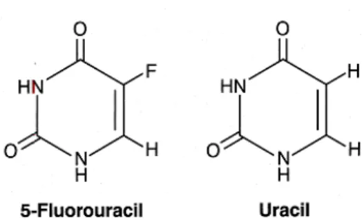
The fluoropyrimidine, 5-fluorouracil (5-Fu), is one of the most commonly used chemotherapeutic drugs and it is used to treat many types of cancer such as breast cancer, colorectal cancer, head and neck cancers. Usually, it is combined with other cancer drugs or with radiotherapy. 5-fluorouracil is an analogue of uracil, with a fluorine atom at the C-5 position in place of hydrogen, and it is classified as an antimetabolite (Fig. 6) (Bracht et al., 2010). Antimetabolite drugs work by inhibiting fundamental biosynthetic processes, or by integration into DNA and RNA, compromising their normal function (Duci, 2015).
Figure 6 - Chemical structure of fluorouracil and uracil.
5-Fu is converted intracellularly into three active metabolites: fluorodeoxyuridine monophosphate (FdUMP), fluorodeoxyuridine triphosphate (FdUTP) and fluorouridine triphosphate (FUTP). These are responsible for induction of RNA and DNA damage, through incorporation of its metabolites into nucleic acids, and inhibition of the nucleotide synthetic enzyme thymidylate synthase (TS) and consequently inhibition of DNA synthesis (Longley et al., 2003).
Like other chemotherapeutic drugs, some unwanted effects may emerge due to the treatment with fluorouracil. Side effects in the gastrointestinal tract are considered the most severe. Stomatitis and esophagopharyngitis, diarrhea, anorexia, nausea and emesis are commonly seen during treatment. Resistance of 5-Fu is another undesirable effect and may emerge due to several mechanisms, namely over expression of TS and alterations in apoptotic pathway (Zang et al., 2008). Because of these important limitations, combination with other drugs has been emerged as a new strategy.
1.6 Marine natural compounds
The research of bioactive compounds in different marine organisms (e.g. marine algae, sponges, corals) as potential antitumor agents has been highly exploited in the last decades. The chemical and biological diversity of the marine environment is incalculable, making this a reservoir of natural bioactive compounds. Many of these products exhibit structural and chemical characteristics that are not found in natural terrestrial compounds. The intense concentration of species in certain habitats makes these highly competitive and complex organisms. For example, sessile organisms such as corals or sponges are in constant struggle for having an appropriate fixation space, and by adopting certain mechanisms that can be chemical, structural and behavioral
they can guarantee their survival (Giangrande et al., 2014). In view of this extremely competitive situation a large proportion of species have developed chemical adaptations that enable them to defend themselves against predation and also represent an advantage in situations of competition. Generally, these adjustments are chemical designated by secondary metabolites and cover a large diversity of compounds such as terpenoids, sugars, alkaloids, steroids, among others (Simmons et al., 2005).
In 2007, trabectedin (ET-743) a marine alkaloid isolated from the Caribbean tunicate (Ecteinascidia turbinata) obtained marketing authorization from the European Commission for the treatment of advanced soft tissue sarcoma, and in 2009 it received marketing authorization for the treatment of platinum-sensitive ovarian cancer (D’Incalci and Galmarini, 2010). Another example of a marine compound approved as a therapeutic drug in leukemia is Cytostar-U, a synthetic pyrimidine nucleoside based on spongothymidine isolated from a sponge (Tethya crypta) (Löwenberg et al., 2011).
1.7 Marine algae
Algae are unicellular or pluricellular organisms that contain chlorophyll. In a simple way, algae can be classified into two major groups, microalgae and macroalgae. Microalgae are found in benthic and littoral habitats and also throughout the ocean waters as phytoplankton. Blue green algae, dinoflagellates, diatoms are included in this group. The group of macroalgae or seaweeds includes green (Chlorophyta), brown (Phaeophyta) and red (Rodhophyta) algae and has been founded in the littoral zone (El Gamal, 2010).
Several species of macroalgae have been used as human and animal food since very early times, especially in China and Japan, such as Nori (Porphyra spp.), wakame (Undaria pinnatifida) and kombu (Laminaria japonica). In this region, they also have been used to treat many diseases that show iodine deficiency (e.g. hyperthyroidism, goiter and Basedow’s disease).
Regarding chemical composition, 70 to 90% of the weight of fresh macroalgae is water, containing also considerable amounts of polysaccharides such as fucoidans present in brown macroalgae, that are responsible for about 45-75% of its dry weight. This is followed by proteins that make up 7-35% of macroalgae dry weight and fats, less than 5% (Lopes, 2014). Another important compound found in macroalgae is carotenoids, which are essential components in photosynthesis and exhibit a powerful antioxidant activity (Takaichi, 2011). Polyphenols, also designated by phlorotannins,
algae (Iwai, 2008). Iodine, calcium, phosphorous, magnesium, iron, sodium, potassium and chlorine are the main mineral elements presents and they are accountable for several macroalgae health benefits. Furthermore, macroalgae are also rich in vitamin A, vitamin C and vitamins from B complex (Burtin, 2003).
Studies on the bioactivities of marine algae have showed several health-promoting effects such as antioxidant, antimicrobial, anti-inflammatory and anticancer effects in several biological models (Raja et al., 2013).
1.7.1 Fucoxanthin
Fucoxanthin, a marine carotenoid, can be found in marine brown macroalgae (Phaeophyceae) and microalgae diatoms (Bacillariophyta).The chemical structure of fucoxanthin includes an allelic bond and some oxygenic functional groups, such as epoxy, hydroxyl, carbonyl and carboxyl groups (Fig. 7) (Peng et al., 2011).
Figure 7 - Chemical structure of fucoxanthin and its deacetylated metabolite, fucoxanthinol.
Sugawara and colleagues, reported that dietary fucoxanthin is hydrolyzed to fucoxanthinol, the major fucoxanthin deacetylated metabolite, in the intestinal tract by lipase and cholesterol esterase. Then, fucoxanthinol is taken up by the intestinal cells and is secreted into the blood circulation system in mammals (Sugawara et al., 2002). Several studies have shown that fucoxanthin and/or fucoxanthinol displays a huge number of beneficial effects in human health. Antioxidant, anti-inflammatory, anticancer, anti-obesity, antidiabetic effects are examples of known fucoxanthin bioactivities.
Apoptotic events in cancer cells are a promising method to control and treat cancer. Hosokawa and colleagues have demonstrated that fucoxanthin decreased cell viability in Caco-2, HT-29 and DLD-1 colorectal cancer cell lines. Furthermore, it
induces DNA fragmentation, a typical characteristic of apoptotic cells, and suppresses the levels of Bcl-2 protein. The Caco-2 cell line when treated with a combination of fucoxanthin and troglitozone at 3.8 µM and 10 µM respectively, showed a decreased in cell viability. However, the compounds alone and at the same concentrations were not able to affect the viability of Caco-2 cell line (Hosokawa et al., 2004).
In a recent study, the anticancer effects of fucoxanthin and fucoxanthinol in six colorectal cancer cell lines and in twenty cases of colorectal cancer specimens, resected surgically from patients, was investigated using the CD-DST technique. The CD-DST mimics the environment of the human body through the use of three-dimension collagen gel, and has been used in clinical practice to select effective chemotherapeutic drugs. The results were obtained in the form of T/C (%), where T is the absorbance of cells which stained by neutral red treated with carotenoids and C is the absorbance of non-stained cells. Fucoxanthin and fucoxanthinol showed anticancer effects in a dose-dependent manner in colorectal cancer cell lines and in the colorectal cancer specimens. Another relevant aspect was that the median of T/C (%) of the 20 samples subjected to fucoxanthinol at 10 µM was similar to a treatment with 5-FU and paclitaxel in a standard dose used in clinical practice (Takahashi et al., 2015).
One of the most exciting aspects of fucoxanthin is the fact that it is apparently a safe compound in terms of mutagenicity. Beppu and colleagues, observed the incidence of micronucleus and mutagenicity of fucoxanthin in bone marrow cells of mice that were orally administered for 24h. The results for micronucleus and mutagenicity were found to be negative (Beppu et al., 2009).
1.7.2 Phloroglucinol
Phlorotannins, a subgroup of tannins found only in brown algae, are oligomeric compounds of phloroglucinol units (Fig. 8). Six kinds of phlorotannins can be isolated of Ecklonia species, such as phloroglucinol (1,3,4-trihydroxybenzene), triphlorethol-A (an open-chain trimer of phloroglucinol), eckol (a closed-chain trimer of phloroglucinol), dieckol (a hexamer), phlorofucofuroeckol (a pentamer) and 6,6’-bieckol (a hexamer) (Kang et al., 2006).
Ecklonia cava is a brown algae, from the family of Laminariceae that grows in subtidal zone in water depth from 5 to 20 meters. It can be found in many warm temperate regions such as China, Japan and Korea (seaweed industry). This brown algae exhibits many biological activities that can be useful in the medical field. For example, a study conducted by Wijesinghe and colleagues, showed that extracts of Ecklonia cava are able to inhibit angiotensin I-converting enzyme (ACE) activity. This
inhibition is essential in the treatment of hypertension (Wijesinghe et al., 2011). Chaudhuri and colleagues evaluated the effect of phloroglucinol derivatives against gastric ulceration in mice, and concluded that phloroglucinol derivatives were able to attenuate gastric ulceration due to their antioxidant activity (Chaudhuri et al., 2011).
Figure 8 - Chemical structure of phloroglucinol.
Phloroglucinol is one of the phloratannins that have been assessed in some anticancer studies. A recent study evaluated the anticancer effects of phloroglucinol on apoptosis and insulin-like growth factor-1 receptor (IGF-1R) signaling in HT-29 human colon cancer cells. The results showed that cells treated with phloroglucinol exhibited fragmented nuclei and condensed chromatin, characteristic of an apoptotic cells. Phloroglucinol also reduced the expression of IGF-1R inhibiting cancer cell proliferation (Kang et al., 2014). The same investigators, but in a different study, observed that phloroglucinol inhibits cell growth and induces apoptosis in HT-29 colon cancer cells via both extrinsic and intrinsic cell death signaling pathways. It was observed that protein expression levels of Fas, FADD and caspase-8 were higher in cells treated with phloroglucinol. In the intrinsic signaling pathway, phloroglucinol induces a decrease in Bcl-2 and Bcl-xL expression (anti-apoptotic proteins) and an increase in Bax and Bad expression (pro-apoptotic proteins) (Kang et al., 2014).
1.8 Methodologies available to screen anticancer activity
In the field of drug development, evaluation of the anticancer activity, side effects in normal cells and elucidation of the mechanisms are the first step to be taken to evaluate the potential activity of the compound, before starting the long period of clinical trials and marketing permission.
When a compound is found and presented as a candidate therapeutic agent it is crucial that it doesn’t display mutagenic effects in healthy cells. A mutagen can interact
directly with the DNA molecule or indirectly through formation of reactive compounds that can act with other components of the cell, or both situations. If the damage caused by the agent is not liable to repair, the cellular response can occur in two possible ways: cell death or cell survival with mutations, the latter resulting in changes of the genetic code transmitted to the daughters cells (Słoczyńska et al., 2014).
Nowadays, several methodologies are available to perform screening tests with several advantages and limitations associated. The use of cancer cell lines as a model for these screening tests has been extensively used and also normal cell lines appear as relevant models to assess specificity. López-Lázaro proposed a basic protocol that includes preclinical in vitro and in vivo testing to assess if a drug is a possible candidate for clinical trials. The preclinical in vitro tests evaluate the anticancer activity of a candidate drug in cancer cells from a particular tissue and in nonmalignant cells from a variety of tissues, and compares it with the anticancer activity of a standard drug under the same conditions. This strategy allows to find a drug capable of killing cancer cells without affecting nonmalignant cells which is the main objective in cancer research (López-Lázaro, 2015).
1.8.1 Cell viability/proliferation assays
The first evaluation of a drug action is usually done through the determination of its effect on cell growth rates. Cell viability or proliferation can be determined, depending on what factor is being measured, in four different ways: metabolic activity, DNA synthesis, antigens related with cell proliferation and ATP concentration (Biocompare, 2012).
The colorimetric MTT assay is a simple and fast method that assesses cell viability or proliferation. It is considered one of the most popular and versatile tests to determine cytotoxicity associated with metabolic activity. Viable cells with an active metabolism are able to convert MTT (3-(4,5-dimethylthiazol-2-yl)-2,5-diphenyltetrazolium bromide) into formazan crystals. The formazan crystals accumulate as an insoluble precipitate inside the cells. They are also present near the cell surface and in the culture medium. The crystals can be extracted and dissolved with DMSO (an organic solvent), allowing its quantification by reading absorbance at 570 nm in a spectrophotometer. Higher absorbance values correspond to a higher number of living cells in the sample (Mosmann, 1983).
Other methods to assess proliferation through DNA synthesis measurement are frequently used. An example is BrdU assay in which a modified nucleotide, such as bromodeoxyuridine (BrdU) can be incorporated into cellular DNA during cell
proliferation. BrdU being a pyrimidine analog will be incorporated by proliferating cells, in place of thymidine into the newly synthesized DNA. BrdU presence can be tracked using antibody probes responsible for developing color that will be assessed by absorbance values or fluorescence (Crane & Bhattacharya, 2013).
1.9 Evaluation of the possible action mechanisms
1.9.1 Genotoxicity evaluation by comet assay
The comet assay is a simple, versatile and quick assay to detect DNA damage in individualized cells. It can be applied to proliferative cells but also to non-proliferating cell population. This aspect is clearly important since these types of cells and tissues are the first to come into contact with potential toxic substances (Kassie et al., 2000)
In 1984, Ostling and Johanson presented the first protocol of comet assay. Generally, the protocol consisted of soaking isolated cells in agarose followed by lysis in a highly saline solution and then performing an electrophoresis in neutral conditions to allow the migration of DNA loops. This approach allows detecting, specifically, double strand breaks in the DNA. Singh and colleagues (1988) carried out for the first time the comet assay on alkaline conditions (pH> 13). This modification made the test able to detect not only double breaks in the DNA but also simple strand breaks, alkali-labile sites, cross-links and incomplete excision-repair sites. More recently, the introduction of repair enzymes after lysis allowed identifying different damages that initially are not strand breaks such as oxidized bases. The most commonly used enzymes are endonuclease III, FPG and AlkA that detect oxidized pyrimidines, purines and modified alkyl purine, respectively (Collins, 2008).
The assessment of damage can be done by two ways: through visual observation of cells by assigning a score of 0 to 4 which corresponds to the intensity of fluorescence in the tail or using software that automatically measures several parameters namely tail intensity. An increase of the fluorescence intensity in the tail of the comet means that the number of breaks is increased and so more damage exist (Gyori et al., 2014).
1.9.2 Apoptosis
Nuclear condensation assays provide a convenient approach for analysis of apoptosis by microscopy. During apoptosis several morphological characteristics appear, such as condensation and fragmentation of DNA, blebbing of the membrane and formation of apoptotic bodies (Elmore, 2007). Using a nuclear and chromosome counterstain, like DAPI, cells with condensed nuclei can be counted.
Another assay that is used to determine apoptotic cells is annexin-V. In an initial phase of apoptosis, cells loose plasma membrane asymmetry and the membrane phospholipid phosphatidylserine (PS) normally found in the inner leaflet of the plasma membrane is translocated to the outer leaflet. Annexin-V is a Ca2+-dependent
phospholipid-binding protein that shows high affinity for PS. Thus, exposed PS in the external cellular environment will be subject to annexin-V binding (Walton et al, 1997). Using annexin-V conjugated with fluorochromes allows detection of apoptotic cells using flow cytometry or microscopy.
1.9.3 Western blot
In response to external stimulus (potential anticancer drug) several proteins involved in survival and death pathways can be inactivated or activated. An example is the activation of several cytosolic proteases known as caspases, e.g. caspases -8, -9 and -3 in response to an apoptotic event (Taylor et al., 2008).
Western blot, or immunoblotting, was introduced by Towbin and colleagues in 1979, and is widely used to separate and identify proteins. In a simplistic manner, the identification of a specific protein is accomplished in three steps: (1) separation by size (molecular weight), (2) transfer to a solid support, and (3) marking target protein, using a suitable primary and secondary antibody (Mahmood & Yang, 2012).
The aim of this study was to assess the in vitro anticancer bioactivity of two algae compounds, fucoxanthin and phloroglucinol, alone and in co-incubation with 5-Fu on two colorectal cancer cell lines (HCT-116 and HT-29), ressorting to preclinical in vitro tests. Also, to assess the specificity of the anticancer effects, a normal colon cell line (CCD-Co18) was tested.
R
EFERENCES
Adams, J. M., & Cory, S. (2007). The Bcl-2 apoptotic switch in cancer development and therapy. Oncogene, 26(9), 1324–1337.
Anand, P., Kunnumakara, A. B., Sundaram, C., Harikumar, K. B., Tharakan, S. T., Lai, O. S., Sung, B., Aggarwal, B. B. (2008). Cancer is a preventable disease that requires major lifestyle changes. Pharmaceutical Research, 25(9), 2097–2116.
Baker, S. J., Fearon, E. R., Nigro, J. M., Hamilton, S. R., Preisinger, a C., Jessup, J. M., Vogelstein, B. (1989). Chromosome 17 deletions and p53 gene mutations in colorectal carcinomas. Science (New York, N.Y.), 244(4901), 217–221.
Beppu, F., Niwano, Y., Sato, E., Kohno, M., Tsukui, T., Hosokawa, M., & Miyashita, K. (2009). In vitro and in vivo evaluation of mutagenicity of fucoxanthin (FX) and its metabolite fucoxanthinol (FXOH). The Journal of Toxicological Sciences, 34(6), 693– 698.
Biocompare. Smith, C. Cell Proliferation Assays: Methods for Measuring Dividing Cells. Retrieved August 13, 2015, from http://www.biocompare.com/Editorial-Articles/117892-Cell-Proliferation-Assays/
Bos, J. L., Fearon, E. R., Hamilton, S. R., Vries, M. V., van Boom, J. H., van der Eb, A. J., & Vogelstein, B. (1987). Prevalence of ras gene mutations in human colorectal cancers. Nature, 327(6120), 293–297.
Bracht, K., Nicholls, a M., Liu, Y., & Bodmer, W. F. (2010). 5-Fluorouracil response in a large panel of colorectal cancer cell lines is associated with mismatch repair deficiency. British Journal of Cancer, 103(3), 340–346.
Burtin, P. (2003). Nutritional Value of Seaweeds. Electronic Journal of Environmental, Agricultural and Food Chemistry, 2(4), 1579 – 4377.
Calcagano, S. R., Li, S., Colon, M., Kreinest, P. A., Thompson, E. A., Fields, A. P., & Murray, N. R. (2008). Oncogenic K-ras promotes early carcinogenesis in the mouse proximal colon. International Journal of Cancer, 76(6), 1358–1375.
Cancer Network. Takimoto, C. H., & Calvo, E. Principles of oncologic pharmotherapy. Retrieved June 6, 2015, from http://www.cancernetwork.com/articles/principles-oncologic-pharmacotherapy
Chaudhuri, S. R., Modak, A., Bhaumik, A., & Swarnakar, S. (2011). PHLOROGLUCINOL DERIVATIVES AS POTENTIAL ANTI- METALLOPROTEINASE-9, 2(4), 237–252.
Collins, A. R., Oscoz, A. A., Brunborg, G., Gaivão, I., Giovannelli, L., Kruszewski, M., Smith, C. C., Stetina, R. (2008). The comet assay: topical issues. Mutagenesis, 23(3), 143–51.
Cooper, GM. (2000). The Cell: A Molecular Approach. 2nd edition. Sunderland (MA): Sinauer Associates.
Crane, A., & Bhattacharya, S. (2013). The Use of Bromodeoxyuridine Incorporation Assays to Assess Corneal Stem Cell Proliferation. In B. Wright & C. J. Connon (Eds.), Corneal Regenerative Medicine SE - 4 (Vol. 1014, pp. 65–70).
Duci, S. B. (2015). Justification of the topical use of pharmacological agents on reduce of tendon adhesion after surgical repair. SM Journal of Orthopedics, 1(2), 2–4.
D’Incalci, M., & Galmarini, C. M. (2010). A review of trabectedin (ET-743): a unique mechanism of action. Molecular Cancer Therapeutics, 9(8), 2157–2163.
El Gamal, A. a. (2010). Biological importance of marine algae. Saudi Pharmaceutical Journal, 18(1), 1–25.
Elmore, S. (2007). Apoptosis: A review of Programmed Cell Death. Toxicol Pathol, 29(6), 997–1003.
Fardilha, M., & Cruz e Silva, O. A. B. (2012). O Essencial em Sinalização Celular. Edições Afrontamento, Lda.
Fearon, E. R., & Vogelstein, B. (1990). A genetic model for colorectal tumorigenesis. Cell, 61(5), 759-67
Ferlay, J., Soerjomataram, I., Dikshit, R., Eser, S., Mathers, C., Rebelo, M., Parkin, D. M., Forman, D., Bray, F. (2015). Cancer incidence and mortality worldwide : Sources, methods and major patterns in GLOBOCAN 2012, 386.
Fulda, S., & Debatin, K.-M. (2006). Extrinsic versus intrinsic apoptosis pathways in anticancer chemotherapy. Oncogene, 25(34), 4798–4811.
Fridman, J. S., & Lowe, S. W. (2003). Control of apoptosis by p53. Oncogene, 22(56), 9030–9040.
Galm, U., Hager, M. H., Van Lanen, S. G., Ju, J., Thorson, J. S., & Shen, B. (2005). Antitumor Antibiotics: Bleomycin, Enediynes, and Mitomycin. Chemical Reviews, 105(2), 739–758.
Gerber, D. E., & Gerber, D. E. (2008). Targeted therapies: a new generation of cancer treatments. American Family Physician, 77(3), 311–9.
Giangrande, A., Licciano, M., Schirosi, R., Musco, L., & Stabili, L. (2014). Chemical and structural defensive external strategies in six sabellid worms (Annelida). Marine Ecology, 35(1), 36–45.
Globocan 2012 : Estimated Cancer Incidence, Mortalitty and Prevalence Worldwide in 2012 Retrieved June 21, 2015, from http://globocan.iarc.fr/Pages/ fact_sheets_cancer. aspx
Goss, K. H. & Groden, J. (2000) Biology of the adenomatous polyposis coli tumor suppressor. Journal of Clinical Oncology, 18, 1967-79.
Grady, W.M. (2004). Genomic instability and colon cancer. Cancer Metastasis Rev, 23(1-2), 11-27.
Guo, M., & Bruce, A. H. (1999). Cell proliferation and apoptosis. Cell Biology, 11, 745– 752.
Gyori, B. M., Venkatachalam, G., Thiagarajan, P. S., Hsu, D., & Clement, M.-V. (2014). OpenComet: An automated tool for comet assay image analysis. Redox Biology, 2, 457–465.
Hajdu, S. I. (2011). A note from history: landmarks in history of cancer, part 1. Cancer, 117(5), 1097-1102.
Haggar, F. A., Boushey, R. P., & Ph, D. (2009). Colorectal Cancer Epidemiology : Incidence , Mortality , Survival , and Risk Factors. Clinics in Colon and Rectal Surgery, 6(212), 191–197.
Hanahan, D., & Weinberg, R. A. (2000). Hallmarks of cancer. Cell, 100(1), 646–74. Hanahan, D., & Weinberg, R. A. (2011). Hallmarks of cancer: the next generation. Cell, 144(5), 646–74.
Hosokawa, M., Kudo, M., Maeda, H., Kohno, H., Tanaka, T., & Miyashita, K. (2004). Fucoxanthin induces apoptosis and enhances the antiproliferative effect of the PPARγ ligand, troglitazone, on colon cancer cells. Biochimica et Biophysica Acta (BBA) - General Subjects, 1675(1–3), 113–119.
Iwai, K. (2008). Antidiabetic and Antioxidant Effects of Polyphenols in Brown Alga Ecklonia stolonifera in Genetically Diabetic KK-Ay Mice. Plant Foods for Human Nutrition, 63(4), 163–169.
Järvinen, H. J., Aarnio, M., Mustonen, H., Aktan-Collan, K., Aaltonen, L. A., Peltomäki, P., De La Chapelle, A., Mecklin, J. P. (2000). Controlled 15-year trial on screening for colorectal cancer in families with hereditary nonpolyposis colorectal cancer. Gastroenterology, 118(5), 829–834.
Jemal, A., Bray, F., Center, M. M., Ferlay, J., Ward, E., & Forman, D. (2011) Global cancer statistics. CA Cancer J Clin, 61(2), 69-90.
Johnson, J. R., Lacey, J. V, Lazovich, D., Geller, M. a, Schairer, C., Schatzkin, A., & Flood, A. (2009). Menopausal hormone therapy and risk of colorectal cancer. Cancer Epidemiology, Biomarkers & Prevention : A Publication of the American Association for Cancer Research, Cosponsored by the American Society of Preventive Oncology, 18(1), 196–203.
Junqueira, L. C., & Carneiro, J. (2013). Histologia Básica – Texto e Atlas (12th ed.). Guanabara Koogan.
Kalluri, R., & Weinberg, R. a. (2009). Review series The basics of epithelial-mesenchymal transition. Journal of Clinical Investigation, 119(6), 1420–1428.
Kang, K. a., Lee, K. H., Chae, S., Zhang, R., Jung, M. S., Ham, Y. M., … Hyun, J. W. (2006). Cytoprotective effect of phloroglucinol on oxidative stress induced cell damage via catalase activation. Journal of Cellular Biochemistry, 97(3), 609–620.
Kang, M.-H., Kim, I.-H., & Nam, T.-J. (2014a). Phloroglucinol induces apoptosis through the regulation of insulin-like growth factor 1 receptor signaling pathways in human colon cancer HT-29 cells. International Journal of Oncology, 1036–1042.
Kang, M.-H., Kim, I.-H., & Nam, T.-J. (2014b). Phloroglucinol induces apoptosis via apoptotic signaling pathways in HT-29 colon cancer cells. Oncology Reports, 1341– 1346.
Karakas, B., Bachman, K. E., & Park, B. H. (2006). Mutation of the PIK3CA oncogene in human cancers. British Journal of Cancer, 94(4), 455–459.
Kassie, F., Parzefall, W., & Knasmüller, S. (2000). Single cell gel electrophoresis assay: a new technique for human biomonitoring studies. Mutation Research, 463(1), 13–31.
Key, T. J., Schatzkin, A., Willett, W. C., Allen, N. E., Spencer, E. A., & Travis, R. C. (2004). Diet , nutrition and the prevention of cancer. Public Health Nutrition, 7(1A), 187–200.
Kerr, J. F. R., Wyllie, A. H., & Currie, A. R. (1972). Apoptosis: A Basic Biological Phenomenon with Wide-ranging Implications in Tissue Kinetics.British Journal of Cancer, 26(4), 239–257.
Kim, E. K., & Choi, E. J. (2010). Pathological roles of MAPK signaling pathways in human diseases. Biochimica et Biophysica Acta, 1802(4), 396–405.
Krämer, a, Neben, K., & Ho, a D. (2002). Centrosome replication, genomic instability and cancer. Leukemia : Official Journal of the Leukemia Society of America, Leukemia Research Fund, U.K, 16(5), 767–775.
Lin, J. E., Li, P., Snook, A. E., Schulz, S., Dasgupta, A., Hyslop, T. M., Gibbons, A. V., Marszlowics, G., Pitaria, G. M., Waldman, S. A. (2010). The hormone receptor GUCY2C suppresses intestinal tumor formation by inhibiting AKT signaling. Gastroenterology, 138(1), 241.
Loeb, L. a. (1991). Mutator phenotype may be required for multistage carcinogenesis. Cancer Research, 51(12), 3075–3079.
Longley, D. B., Harkin, D. P., & Johnston, P. G. (2003). 5-Fluorouracil: Mechanisms of Action and Clinical Strategies. Nature Reviews. Cancer, 3(5), 330–338.
Lopes, G. (2014). Seaweeds from the Portuguese Coast: Chemistry, Antimicrobial and Anti-inflammatory Capacity.
López-Lázaro, M. (2015). Two preclinical tests to evaluate anticancer activity and to hel validate drug candidates for clinical trials. Oncoscience, 2(2), 91-98.
Lopiccolo, J., Blumenthal, G. M., Bernstein, W. B., & Dennis, P. a. (2008). Targeting the PI3K/Akt/mTOR pathway: effective combinations and clinical considerations. Drug Resist, 11(301), 32–50.
Lomonosova, E., & Chinnadurai, G. (2008). BH3-only proteins in apoptosis and beyond: an overview. Oncogene, 27 Suppl 1(S1), S2–S19.
Löwenberg, B., Pabst, T., Vellenga, E., van Putten, W., Schouten, H. C., Graux, C., Ossenkoppele, G. J. (2011). Cytarabine dose for acute myeloid leukemia. The New England Journal of Medicine, 364(11), 1027–1036.