Universidade de Lisboa
Faculdade de Medicina de Lisboa
Effects of chemotherapy and radiotherapy
in rectal cancer: significance of different
cellular outcomes in tumor behavior
Joana Maria Tato Ribeiro da Costa
Doutoramento em Ciências Biomédicas
Especialidade de Ciências Biopatológicas
Universidade de Lisboa
Faculdade de Medicina de Lisboa
Effects of chemotherapy and radiotherapy
in rectal cancer: significance of different
cellular outcomes in tumor behavior
Joana Maria Tato Ribeiro da Costa
Tese orientada pelo
Prof. Doutor Luís Marques da Costa
Tese co-orientada pela
Prof.
aDoutora Sandra Casimiro
Doutoramento em Ciências Biomédicas
Especialidade de Ciências Biopatológicas
Todas as afirmações efetuadas no presente documento são da exclusiva
responsabilidade do seu autor, não cabendo qualquer responsabilidade à
Faculdade de Medicina de Lisboa pelos conteúdos nele apresentados.
A impressão desta dissertação foi aprovada pelo Conselho Científico da
Faculdade de Medicina de Lisboa em reunião de 28 de Outubro de 2014
O trabalho descrito na presente tese foi desenvolvido no Instituto de
Medicina Molecular, Faculdade de Medicina de Lisboa sob orientação do
Professor Doutor Luís Marques da Costa e sob co-orientação da Professora
Doutora Sandra Cristina Cara de Anjo Casimiro. Este trabalho foi
financiado
por
uma
bolsa
de
doutoramento
individual
(SFRH/BD/45219/2008) da Fundação para a Ciência e a Tecnologia.
“Pensar é mais interessante que saber, mas menos interessante que olhar.”
Johann Goethe
I
AGRADECIMENTOS
Muitas são as pessoas a quem eu quero dirigir o meu agradecimento, pelos seus contributos tão essenciais para que eu chegasse até aqui.
Em primeiro lugar, gostaria de agradecer aos meus orientadores, Prof. Doutor Luís Costa e Prof. Doutora Sandra Casimiro, por toda a ajuda que me deram na orientação deste trabalho.
Obrigada pela confiança que depositaram em mim quando me atribuíram este projeto e pela oportunidade de desenvolver o meu trabalho nesta unidade. Obrigada também por tudo o que me ensinaram, pela liberdade que me deram de poder pensar de forma independente e por aceitarem as minhas ideias.
Durante os últimos cinco anos adquiri a perceção de que o nosso trabalho pode efetivamente fazer a diferença. O convívio com a realidade clinica, com os doentes e os seus tratamentos, a noção de que cada amostra é valiosa e que é cedida por cada paciente porque acredita no nosso trabalho. Saber que trabalhamos para melhorar as condições dos doentes, dá um sentido diferente à ciência que fazemos todos os dias. Não vejo maior responsabilidade do que esta. Obrigada por me mostrarem tudo isto.
Como não podia deixar de ser, o agradecimento seguinte vai para todos os doentes do hospital de dia de oncologia do Hospital de Santa Maria. Muito obrigada a todos aqueles que consentiram que colhesse amostras biológicas e que permitiram a realização de todo os estudos in vivo que apresento nesta tese.
Agradeço também a todos os profissionais de saúde do hospital de dia de oncologia. É graças ao empenho conjunto e diário de médicos, enfermeiros, auxiliares e administrativos, que os nossos projetos de investigação de translação avançam. Um agradecimento especial e partícula à Dra. Margarida Matias e à Dra. Mafalda Casa-Nova pela enorme ajuda que me deram na elaboração das bases de dados e na seleção dos doentes. Gostaria também de agradecer à Dra. Emília Oliveira e ao técnico Pedro Gonçalo Rodrigues do serviço de Anatomia Patológica pela indispensável ajuda com as amostras de arquivo.
II
Agradeço também aos membros do meu comité de tese: Prof. Doutor João Ferreira, Prof. Doutor Afonso Fernandes e Prof. Doutora Joana Desterro, por todas as críticas científicas sempre construtivas e as trocas de ideias tão importantes no progresso de um projeto de doutoramento. Um agradecimento especial ao Prof. Doutor Afonso Fernandes, por todas as horas passadas no microscópio e pela clareza e rigor em tudo o que faz e ensina. Muito obrigada.
À Faculdade de Medicina de Lisboa e ao Instituto de Medicina Molecular pelas excelentes condições de trabalho e pela grande comunidade científica, tão estimulantes e importantes para o crescimento científico de um estudante de doutoramento.
À Fundação para a Ciência e Tecnologia pelo financiamento (SFRH/BD/45219/2008). A todos os meus colegas que pertencem ou já pertenceram à Unidade de Investigação em Oncologia Clínica. Obrigada à Teresa, ao Ricardo, à Ana Cristina, ao Mário, à Diana e à Sara, todos excelentes colegas e companheiros de trabalho.
Aos meus amigos e parceiros na longa caminhada deste doutoramento: Irina, Pedro, Andreia, Ana Margarida, Ana Pires, Raquel, Dinora, Marisa, Ana Jesus, Sandra Martins, Silvia, Ram, Virgínia e Daniel. Dizem que a amizade não se agradece, retribui-se. Por isso agradeço-vos muito toda a ajuda e o carinho com que sempre me trataram e espero estar à altura de retribuir tudo aquilo que vocês me deram.
À minha querida Carolina, a pessoa mais generosa e amiga que conheço. Mesmo estando longe está sempre perto.
Para último guardo os agradecimentos mais importantes, à minha família. Aos meus avós, aos meus pais e à minha irmã, nada no mundo é mais importante para mim do que vocês. Este trabalho só foi possível porque vos tive sempre do meu lado e o vosso apoio incondicional. A vocês eu devo tudo o que sou. É imensurável o amor, orgulho e gratidão que sinto por todos. Sempre juntos, sempre unidos.
III
TABLE OF CONTENTS
AGRADECIMENTOS ... I TABLE OF CONTENTS ... III Index of Illustrations/figures... V Index of tables ... VI Index of supplementary FIGURES ... VI Units of measurement and Abbreviations ... VII Summary ... XIII Resumo ... XV Scope of the thesis ... XVI
1 GENERAL INTRODUCTION ... 1
1.1 Incidence and classification of colorectal cancer ... 3
1.2 Rectal Cancer ... 5
1.2.1 Treatment of locally advanced primary rectal cancer ... 5
1.2.2 Prognostic and predictive factors ... 7
1.3 5-fluorouracil (5-FU) ... 8
1.3.1 Mechanism of action ... 8
1.3.2 Chemo-resistance to 5-fluorouracil ... 11
1.3.3 5-FluorouraciI pharmacokinetics ... 12
1.4 Cellular senescence ... 12
1.4.1 Signaling pathways mediating senescence ... 14
1.4.1.1 p53 and pRb, two master regulators of senescence ... 16
1.4.1.2 The Arf-p53- p21WAF1 pathway. ... 18
1.4.1.3 The p16INK4a-pRb pathway ... 20
1.4.2 Senescence-associated heterochromatic foci ... 21
1.4.3 Senescence-associated secretory phenotype ... 22
1.4.4 Markers of cellular senescence ... 26
1.4.5 Significance of cellular senescence in cancer ... 28
1.5 Aim of the thesis ... 33
2 THERAPY-INDUCED CELLULAR SENESCENCE INCREASES INVASIVENESS AND CHEMOSENSITIVITY IN RECTAL CANCER ... 35
IV
2.2 Introduction ... 38
2.3 Material and Methods... 40
2.4 Results ... 45
2.4.1 Low-dose 5-FU induces cellular senescence in HCT 116 colorectal cancer cells ... 45
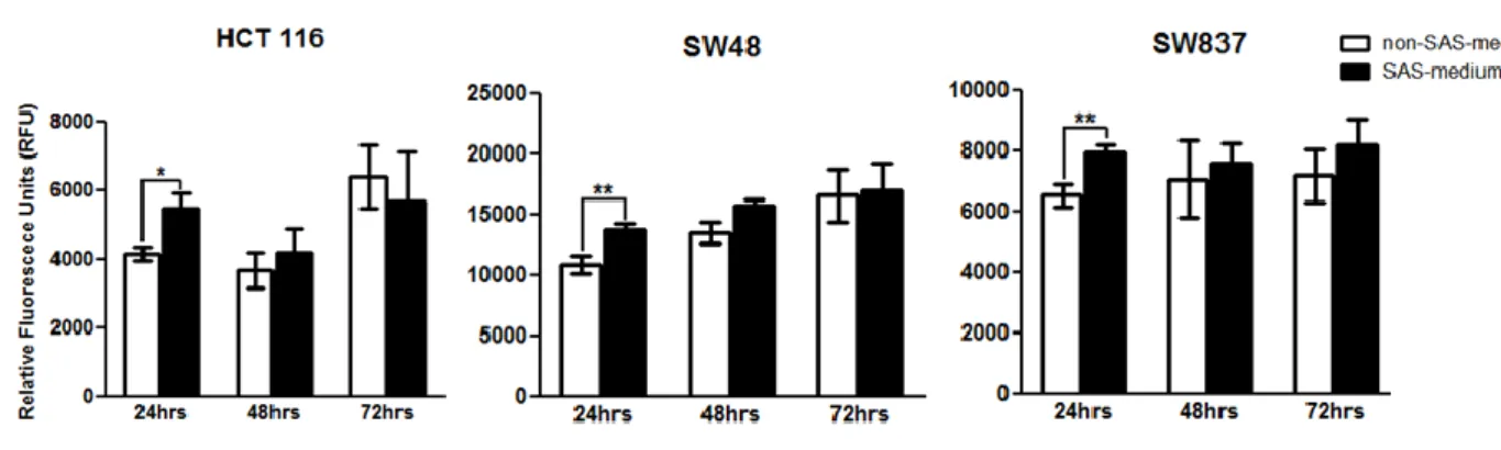
2.4.2 The secretome of senescent HCT 116 cells stimulates the proliferation of non-senescent cells .. 46
2.4.3 The secretome of senescent colon cancer cells promotes epithelial-to-mesenchymal transition and increased invasiveness ... 48
2.4.4 The secretome of senescent colon cancer cells increases the chemosensitivity to 5-FU ... 51
2.4.5 Neoadjuvant chemotherapy promotes emergence of senescence and EMT in human rectal cancers 53 2.5 Discussion ... 58
3 THE IMPACT OF DRUG-INDUCED CELLULAR SENESCENCE IN RECTAL CANCER RELAPSE: A RETROSPECTIVE STUDY ... 61
3.1 Introduction ... 63
3.2 Materials and Methods ... 65
3.3 Results and Discussion ... 68
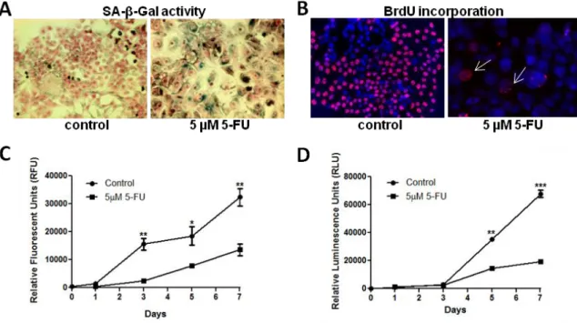
3.3.1 β-galactosidase is increased in 5-FU-induced HCT 116 senescent cells ... 68
3.3.2 β-galactosidase immunostaining associates with SA-β-gal activity in frozen rectal tumors ... 69
3.3.3 Positive β-gal/p16INK4a cells are heterogeneously found among rectal cancer tissue specimens 70 3.3.4 There is no correlation between senescence markers p16INK4a or p21WAF1 and cancer relapse .... 71
4 GENERAL DISCUSSION ... 75
4.1 General discussion ... 77
V
INDEX OF ILLUSTRATIONS/FIGURES
Illustration 1- Conceptual framework used for the investigation of the effects of cellular senescence in rectal cancer. ... XVII
Figure 1.1- Representative scheme of current guidelines for the treatment of locally
advanced rectal cancer. ... 5
Figure 1.2- Representative scheme of 5-Fluorouracil metabolism. ... 9
Figure 1.3- Mechanism of thymidylate synthase (TS) inhibition by 5-Fluorouracil. ... 10
Figure 1.4- The DNA damage response. ... 15
Figure 1.5- Structure of the INK4a/ARF and INK4b loci. ... 18
Figure 1.6- Signal transduction pathways mediating senescence. ... 20
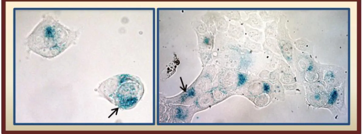
Figure 1.7- Representative images of senescent cells with distinct morphological features in vitro. ... 26
Figure 2.1- Low-dose 5-FU induces cellular senescence in HCT 116 colon cancer cells... 45
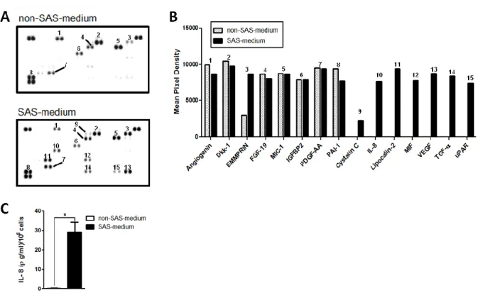
Figure 2.2- Cytokine screening array identifies secreted compounds by 5-FU-induced senescent HCT 116 colon cancer cells. ... 47
Figure 2.3- The secretome of senescent HCT 116 cells stimulates the proliferation of non-senescent cells. ... 48
Figure 2.4- The secretome of senescent colon cancer cells induces epithelial-to-mesenchymal transition and increases invasiveness. ... 49
Figure 2.5- The secretome of senescent colon cancer cells increases the chemosensitivity of proliferating cells to 5-FU. ... 51
Figure 2.6- Neoadjuvant chemotherapy promotes emergence of senescence and EMT in human rectal cancers. ... 55
Figure 3.1- β-galactosidase expression is increased in senescent HCT116 cells. ... 68
Figure 3.2- SA-β-gal activity co-localizes with β-galactosidase expression in frozen rectal cancer samples. ... 69
VI
INDEX OF TABLES
Table 1.1- TNM Classification system for colorectal cancers [8]. ... 4 Table 2.1- Patient and tumor characteristics ... 54 Table 3.1- Patient and tumor characteristics and treatment factors (n=35) ... 72 Table 3.2- Logistic regression analysis to correlate independently p16INK4a and p21WAF1 immunohistochemical scores with relapse (n=35). ... 73
INDEX OF SUPPLEMENTARY FIGURES
Supplementary Figure S2.1- Cytokine screening array and EMT induction by SAS-medium obtained from doxorubicin-induced senescent HCT 116 colon cancer cells... 50 Supplementary Figure S2.2- The secretome from colon cancer cells induced into senescence by doxorubicin increases chemosensitivity of proliferating HCT cells to 5-FU. ... 52 Supplementary Figure S2.3- Representative sequential frozen sections of human rectal cancer tissue selected for isolation of senescent-positive and senescent-negative epithelial cell populations by laser microdissection. ... 56 Supplementary Figure S2.4- EMT-related genes are poorly expressed in senescent cells. ... 57
VII
UNITS OF MEASUREMENT AND ABBREVIATIONS
(In alphabetic order)% Percent C Degree Celsius µg Microgram µM Micro molar µm Micrometer 53BP1 p53-Binding Protein 1 5-FU 5-fluorouracil
9-1-1 RAD9–RAD1–HUS1 Protein Complex Ang Angiogenin
AREG Amphiregulin
Arf Alternative Reading Frame ATM Ataxia-Telangiectasia Mutated
ATR Ataxia Telangiectasia and Rad3-related BRCA1 Breast Cancer 1
BrdU Bromodeoxyuridine CDC25 Cell Division Cycle 25 Cdk Cyclin Dependent Kinase Cells/ml Cells per Milliliter
CH2THF 5,10-Methylenetetrahydrofolate
CHK2 Checkpoint Kinase 2 CM Conditioned Medium CO2 Carbon dioxide
Cpss Steady-State Serum Concentrations CRC Colorectal cancer
VIII
CRT Chemoradiotherapy
CT Chemotherapy
CT+ Bev Chemotherapy plus Bevacizumab CT+PBO Chemotherapy plus Placebo
DAPI Fluorescent Stain - 4',6-diamidino-2-phenylindole DCR2 Decoy Receptor 2
DDR DNA Damage Response
DEC1 Basic Helix–loop–Helix Transcription Factor DFS Disease Free Survival
DHFU Dihydrofluorouracil
Dkk-1 Dickkopf-related protein 1 DNA Deoxyribonucleic Acid DNase I Deoxyribonuclease I
DPD Dihydropyrimidine Dehydrogenase DSBs Double Strand Breaks
dTMP Deoxythymidine Monophosphate dUMP Deoxyuridine Monophosphate ECM Extracellular Matrix
EDTA Ethylenediamine Tetraacetic acid EGFR Epidermal Growth Factor Receptor EGTA Ethylene Glycol Tetraacetic Acid ELISA Enzyme-Linked Immunosorbent Assay EMT Epithelial-Mesenchymal Transition
EORTC European Organization for Research and Treatment of Cancer ESMO European Society for Medical Oncology
IX
FdUMP Fluorodeoxyuridine Monophosphate FdUTP Fluorodeoxyuridine Triphosphate FFPE Formalin-Fixed Paraffin-Embedded FGF-19 Fibroblast Growth Factor 19 FGF7 Fibroblast Growth Factor 7 FUdR 5-Fluoro-2'-Deoxyuridine FUTP Fluorouridine Triphosphate
GAPDH Glyceraldehyde 3-phosphate dehydrogenase GI Gastrointestinal
GROα Growth Regulated Oncogene-alpha H3K9me3 Trimethylation of Lysine 9 in Histone 3 HCT 116 Homo sapiens Colorectal Carcinoma Cell Line HGF Hepatocyte Growth Factor
HIRA Histone Repressor A
HP1γ Heterochromatin Protein 1-γ HPF Hepatocyte Growth Factor
IARC International Agency for Research on Cancer IC50 Half Maximal Inhibitory Concentration IF Immunofluorescence
IGFBP Insulin-like Growth Factor Binding Protein IGFBP2 Insulin-like Growth Factor-Binding Protein 2 IgG Immunoglobulin G
IHC Immunohistochemistry IL Interleukin
X
KO Knockout
LCN2 Lipocalin-2
M Molar
MDC1 Mediator of DNA Damage Checkpoint 1 MDM2 E3 Ubiquitin-Protein Ligase
MEFs Mouse Embryonic Fibroblasts Mg/m2/day Milligram per Square Meter per Day MgCl2 Magnesium chloride
MIC 1 Macrophage Inhibitory Cytokine 1 MIF Macrophage Migration Inhibitory Factor ml Milliliter
mM Milimolar
mm Millimeter
MMPs Matrix Metalloproteinases MRI Magnetic Resonance Imaging
MRN MRE11–RAD50–NBS1 Protein Complex mRNA Messenger Ribonucleic Acid
Neo.CRT Neoadjuvant Chemoradiotherapy
nm Nanometer
Non-SAS Conditioned medium from non-senescent cells -medium
NSABP R‑03 National Surgical Adjuvant Breast and Bowel Project R-03 OS Overall Survival
PAI-1 Plasminogen Activator Inhibitor-1 PBS buffer Phosphate Buffered Saline
XI
PCNA Proliferating Cell Nuclear Antigen PCR Polymerase Chain Reaction
PDGF-AA Platelet-Derived Growth Factor AA PFA Paraformaldehyde
PFS Progression Free Survival pg/mL Pico gram per Milliliter pH Power of Hydrogen PML Promyelocytic Leukemia pRb Retinoblastoma Protein RNA Ribonucleic Acid
RNase A Ribonuclease A RPA Replication Protein A RT Radiotherapy
RT Room Temperature RT-PCR Reverse Transcriptase PCR RT-qPCR semi-quantitative real-time PCR
SAHF Senescence-Associated Heterochromatic Foci SAS Senescence-Associated Secretome
SAS-medium Conditioned medium from senescent cells SASP Senescence-Associated Secretory Phenotype SA-β-Gal Senescence-Associated β- Galactosidase SEM Standard Error Mean
SSB Single Strand Break
TGF-α Transforming Growth Factor alpha TIS Therapy-Induced Senescence
XII
TMP Thymidine Monophosphate
TOPBP1 Topoisomerase-II-Binding Protein 1 TP Thymidine Phosphorylase
TRG Tumor Regressing Grade TS Thymidylate Synthase TTP Time to Progression
TUNEL Terminal Deoxynucleotidyl Transferase dUTP Nick End Labeling U/ml Units per milliliter
uPAR Urokinase-Plasminogen Activator Receptor Uv Ultraviolet
VEGF Vascular Endothelial Growth Factor WHO World Health Organization
X-Gal 5-bromo-4-chloro-3-indolyl β-d-Galactopyranoside γH2AX Phosphorylation of the histone variant H2AX on Ser139 μg/ml Microgram per Milliliter
XIII
SUMMARY
Rectal cancers comprise 35% of all diagnosed colorectal cancers (CRC), being the third most common among gastrointestinal cancers. Despite the benefits of neoadjuvant chemoradiotherapy (CRT), 5-fluorouracil (5-FU)-based regimens plus radiotherapy (RT), 15-20% of patients suffer from relapse. Pathological staging remains the most important prognostic factor in rectal cancer and the search for new prognostic and predictive biomarkers is fundamental. DNA damaging agents and ionizing radiation used in the therapy of human cancers may induce senescence of cancer cells. Senescent cells exhibit a secretory phenotype that can affect cancer cell behavior and, eventually, clinical prognosis. In this work
we hypothesize that neoadjuvant CRT-induced cellular senescence may affect rectal cancer relapse. To experimentally test our hypothesis, we cultured colon cancer cells induced into
senescence by exposure to 5-FU or doxorubicin. SAS-media were enriched in IL-8, TGF-α, VEGF, cystatin C, LCN2, MIF, EMMPRIN, and uPAR, and exerts a positive effect on the proliferation of cycling colon and rectal cancer cells. SAS-medium was capable of paracrine induction of epithelial-to-mesenchymal (EMT) transition in colon and rectal cancer cell lines, of increased cell invasion in vitro, and of increased chemosensitivity to 5-FU. Moreover, we found that in rectal cancer samples from patients treated with neoadjuvant CRT tumor cell niches enriched for senescent cells bookmark regions of increased expression of EMT-related genes (slug, snail, vimentin) when compared to nearby senescent-null control regions. We provide evidences that therapy-induced senescent cancer cells influence the tumor microenvironment by promoting EMT via short range interactions. Next, to relate neoadjuvant CRT-induced cellular senescence with rectal cancer relapse, we retrospectively studied rectal cancers from 35 patients treated with neoadjuvant therapy. Data showed no correlation between the senescence markers p16INK4a and p21WAF1 and relapse, and it was not possible to validate a method for senescence detection in formalin-fixed and paraffin-embedded (FFPE) samples. Altogether, our findings showed that secretomes from senescent colon cancer cells may induce effects with opposite prognostic value. Prospective studies shall clarify whether, after neoadjuvant therapy, the presence of senescent cells add prognostic power on cancer recurrence and patient survival.
Keywords: Therapy-induced cellular senescence; rectal cancer; neoadjuvant chemotherapy; senescence-associated secretory phenotype; 5-fluorouracil
XV
RESUMO
O cancro do reto representa cerca de 35% de todos os tumores colo-retais (CCR) diagnosticados, sendo o terceiro mais comum entre os tumores gastrointestinais. Apesar da melhoria significativa no contexto da terapia neoadjuvante, 5-fluorouracil (5-FU) em concomitância com radioterapia (RT), as taxas de recidiva permanecem elevadas, cerca de 15-20%. O estadiamento histo-patológico permanece o principal fator de prognóstico sendo fundamental a pesquisa de novos biomarcadores de prognóstico. No tratamento da doença oncológica, a terapêutica com citostáticos e radiação pode induzir senescência cujo fenótipo secretor é conhecido porsenescence-associated secretory phenotype (SASP), capaz de modificar o micro-ambiente e
contribuir para a progressão tumoral. Este trabalho teve por base a hipótese de que a senescência celular induzida pela quimioterapia (QT) pode estar correlacionada com a recidiva em doentes com cancro do reto em estadio avançado. Para testar experimentalmente esta hipótese foi induzida senescência em células de cancro do cólon (HCT 116) por exposição ao 5-FU ou doxorrubicina. A análise dos meios condicionados obtidos (SAS-media) revelou a presença dos compostos IL-8, MIF, VEGF, uPAR, EMMPRIN, cistatina C, lipocalina-2 e TGF-α, que se verificou estimularem a proliferação de células de cancro do cólon e reto não senescentes. SAS-medium induziu também um aumento significativo da expressão de marcadores moleculares de transição epitélio-mesênquima (TEM), do potencial invasivo em células tumorais não senescentes, e da quimio-sensibilidade, o que sugere um duplo efeito nas células tumorais não senescentes. Em amostras humanas de tumores do reto verificamos um aumento da expressão de marcadores mesenquimais em zonas tumorais associadas à presença de células senescentes, constituindo uma forte evidência de que a senescência induzida pela QT influencia de facto o micro-ambiente tumoral, promovendo a TEM. Para determinar uma possível correlação entre a presença de células senescentes e a recidiva no cancro do reto, realizámos um estudo retrospetivo envolvendo 35 amostras de doentes submetidos a terapia neoadjuvante. Os dados obtidos não permitiram estabelecer uma correlação entre os marcadores de senescência p16INK4a e p21WAF1 e a recidiva, não tendo sido possível validar um método que consideremos adequado para a identificação de células senescentes em amostras fixadas. Em conclusão, os dados obtidos neste trabalho sugerem que o secretoma de células epiteliais tumorais pode induzir fenómenos com valor prognóstico opostos. Estudos prospetivos futuros irão contribuir para clarificar o papel da senescência celular na definição de novos critérios de prognóstico e identificação de novos alvos terapêuticos.
Palavras-chave: Senescência celular induzida pela terapia; cancro do reto; quimioterapia neoadjuvante; secretoma associado à senescência; 5-fluorouracil.
XVI
SCOPE OF THE THESIS
This thesis is organized in four chapters. In Chapter 1 the background, significance, and aims of this study are presented. Rectal cancer epidemiology and clinical management; the mechanism of action of 5- fluorouracil; and the molecular basis and significance of cellular senescence, were reviewed according to the literature. Together, this review comprises the theoretical fundaments that support our hypothesis that therapy-induced cellular senescence can be related with rectal cancer relapse. In Chapters 2 and 3 are presented and discussed the results obtained during the course of this work (Illustration
1). Chapter 2 refers to a manuscript submitted to Clinical & Experimental Metastasis
(CLIN-S-14-00188, submitted on September 11, 2014), entitled “Therapy-induced cellular senescence has a dual effect in rectal cancer increasing invasiveness and chemosensitivity”, by Joana Tato-Costa, Sandra Casimiro, Teresa Pacheco, Ricardo Pires, Afonso Fernandes, Irina Alho, Pedro Pereira, Paulo Costa, Henrique Bicha Castelo, João Ferreira, and Luís Costa. Chapter 3 describes a clinicopathologic study conducted to elucidate if cellular senescence could be correlated with relapse in rectal cancer. Finally, in Chapter 4 it is presented a final discussion of the key findings from the previous chapters and of the potential translational relevance of senescence in the management of advanced rectal cancer.
Disclaimer: I hereby declare that I majorly contributed for the conception and design,
development of methodology, acquisition of data, analysis and interpretation of data and writing, review and/or revision of each group of results and chapters of this thesis.
XVII
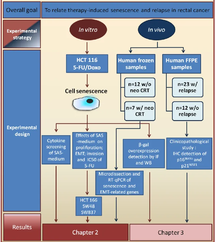
Illustration 1- Conceptual framework used for the investigation of the effects of cellular senescence in rectal cancer. HCT 116; SW48 – colon cancer cell lines and SW837 - rectal cancer cell line, all acquired from ATCC; SAS-medium – culture medium enriched of the Senescence-Associated Secretome; EMT - Epithelial-Mesenchymal Transition; W/O – Without; W/ - With; CRT –Chemoradiotherapy; RT-qPCR - Semi-quantitative real-time PCR; β-gal - β-galactosidase; IF – Immunofluorescence; WB – Western blot; FFPE - Formalin-Fixed, Paraffin-Embedded; IHC – Immunohistochemistry.
1
1 GENERAL
INTRODUCTION
3
1.1 Incidence and classification of colorectal cancer
Colorectal cancer (CRC) is the third most common form of cancer worldwide. In 2012, according to the GLOBOCAN Project, sponsored by the International Agency for Research on Cancer (IARC) and World Health Organization (WHO), CRC represented 9.7% of the world's new cancers with 1.36 million new cases diagnosed [5].
In Portugal, in the same period, 7 129 new cases were diagnosed with CRC, corresponding to 14.5% of all diagnosed cancers. Here, CRC was the second most common cancer affecting women (after breast cancer) and men (after prostate cancer), and had the highest mortality rate (15.7%) [5].
Staging of the disease dictates the therapeutic strategies. According to international guidelines from the European Society for Medical Oncology (ESMO), the preoperative staging of CRC is based on the clinical and radiological evaluation of the tumor growth and distant spread [6, 7]. Following surgery, the pathologic TNM classification is based on the evaluation of size and extent of the primary tumor (ranging from T0 to T4), the presence of regional lymph nodes involvement (N0 to N2), and the existence of distant metastasis (M0 or M1) (Table 1.1) [8].
CRC can occur at different locations within the colon or rectum. Rectal cancer is the third most common among gastrointestinal (GI) cancers, and represents approximately 35% of all CRC cancers [6]. Rectal cancers are located below 12 cm from the anal verge as measured with a rigid rectoscope, or below the pelvic promontory as visualized by X-ray, computed tomography, magnetic resonance imaging (MRI) or during surgery [7]. Colon and rectal cancers have several features in common and are epidemiologically gathered as CRC. However, the surgical and therapeutic management of colon and rectal cancers is different. This thesis and following chapters will focus on rectal cancer.
4
Table 1.1- TNM Classification system for colorectal cancers [8].
TNM system
T- Primary tumor
Tx- Primary tumor cannot be assessed T0- No evidence of primary tumor Tis- Carcinoma in situ: intraepithelial or invasion of lamina propria
T1- Tumor invades submucosa T2- Tumor invades muscularis propria T3- Tumor invades subserosa or into non-peritonealized pericolic or perirectal tissue T4- Tumor directly invades other organs or structures and/or perforates visceral peritoneum
T4a- Tumor perforates visceral
peritoneum
T4b- Tumor directly invades other
organs or structures
N- Regional lymph
nodes Nx- Regional lymph nodes cannot be
assessed
N0- No regional lymph nodes metastasis
N1- Metastasis in 1-3 regional lymph nodes N1a- Metastasis in 1 regional lymph
node
N1b- Metastasis in 2-3 regional lymph
nodes
N1c- Tumor deposit(s), i.e., macro or
microscopic nests or nodules in the subserosa, or in non-peritonealized pericolic or perirectal soft tissue without regional lymph nodes metastasis
N2- Metastasis in 4 or more regional lymph nodes
N2a- Metastasis in 4-6 regional lymph
nodes
N2b- Metastasis in 7 or more regional
lymph nodes
M- Distant metastasis M0- No distant metastasis
M1- Distant metastasis M1a- Metastasis confined to 1 organ M1b- Metastasis in more than 1 organ
5
1.2 Rectal Cancer
1.2.1 Treatment of locally advanced primary rectal cancer
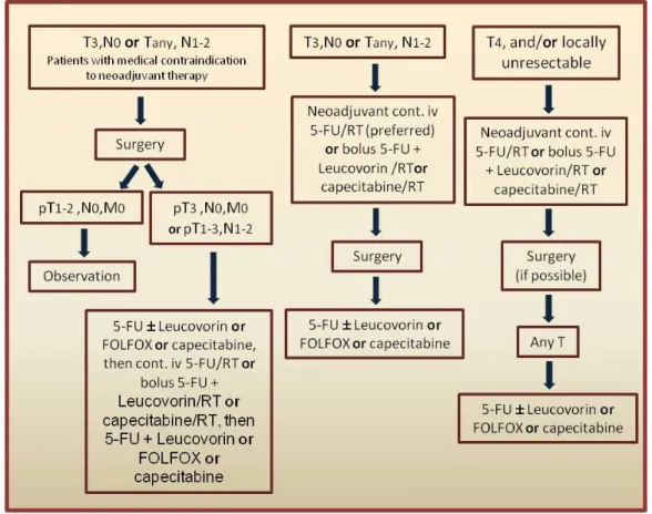
The standard of care treatment for locally advanced rectal cancer (stage T3/T4 and/or N positive) comprises preoperative (neoadjuvant) chemoradiotherapy (CRT), followed by surgery and postoperative (adjuvant) chemotherapy (CT) (Figure 1.1).
Figure 1.1- Representative scheme of current guidelines for the treatment of locally advanced rectal cancer.
5-FU -5-fluorouracil; Leucovorin- Folinic acid; FOLFOX- Chemotherapy regimen comprising folinic acid (Leucovorin), 5- fluorouracil (5-FU) and oxaliplatin; RT- radiotherapy; cont. iv- continuous intravenous infusion. Adapted from [9].
New surgical techniques have substantially improved local-regional control and overall survival1 (OS), being total mesorectal excision the gold standard procedure, with local failure rates below 10% and better prevention of tumor cells spread at surgery [10].
1
Overall survival (OS) - The length of time those patients are alive since either the date of diagnosis or the start of treatment for a disease.
6
However, and despite surgical and therapeutic advances, 15 to 20% of patients with locally advanced rectal cancer will suffer from local or distant relapse, mainly to the liver, lung and distant lymph nodes. In consequence, about one third of the diagnosed patients will die within 5 years after treatment [11].
In 2006, results from The European Organization for Research and Treatment of Cancer (EORTC) 22921 multicentre clinical trial, enrolling 1011 patients with primary T3/T4 rectal cancers, suggested that a pre-surgical treatment with radiotherapy (RT) combined with radiosensitive CT could significantly improve downsizing and downstaging of tumors and decrease local recurrence rates, when compared to RT alone (57.1% versus 42.4%) [11]. Results from the National Surgical Adjuvant Breast and Bowel Project R-03 (NSABP R‑03) trial, comparing the effects of neo plus adjuvant CRT in patients with locally advanced rectal adenocarcinoma, showed that the five year disease free survival2 (DFS) was significantly improved for patients treated with neo plus adjuvant therapy (65% versus 53%, p = 0.011). In addition, the five year OS for patients treated with neo plus adjuvant therapy was significantly higher in comparison with patients with only post-operative therapy (75% versus 66%, p = 0.065) [12].
Different studies have demonstrated that neoadjuvant CRT not only improves surgical outcomes, but also reduces tumor burden and RT-related toxicity, and increases radiosensitivity [13, 14].
The advent of targeted therapy with target-specific monoclonal antibodies, namely cetuximab and bevacizumab, in combination with CT expanded the treatment options and further improved clinical outcomes. However, these improvements are so far only reported in cases of metastatic CRC [15].
Cetuximab is a monoclonal antibody that binds to Epidermal Growth Factor Receptor (EGFR) and blocks ligand-induced phosphorylation of the downstream effectors of the pathway. The overexpression or up-regulation of EGFR is associated with poor survival in
2
Disease-free survival (DFS) - the period of time after primary treatment in which the patient does not show any signs or symptoms of the disease.
7
CRC and occurs in 60 to 80 % of CRCs. Upon activation, EGFR activates an important signaling pathway that regulates cell differentiation, proliferation, migration, angiogenesis, and apoptosis, all of which become deregulated upon over expression of the receptor. A clinical trial where was compared the combination of cetuximab and irinotecan (the semi-synthetic topoisomerase I inhibitor camptothecin used as CT drug) with cetuximab in monotherapy in metastatic CRC cancer patients showed that the response rate in the combination-therapy group was significantly higher than that in the monotherapy group (22.9% vs. 10.8%, p=0.007). The median time to progression3 (TTP) was significantly improved in the first group (4.1 vs. 1.5 months, P<0.001) although without significant differences in median OS (8.6 versus 6.9 months, p=0.48). Cetuximab has proved to be clinically significant when given in combination with irinotecan in patients with metastatic disease [16].
Bevacizumab is a monoclonal antibody against Vascular Endothelial Growth Factor (VEGF), an important regulator of physiologic and pathologic angiogenesis. Bevacizumab has antiangiogenic effects and currently being evaluated in clinical trials as a treatment for several cancers. In a phase 3 clinical trial the addition of bevacizumab to a standard combination of three CT drugs (CT+bev) improved OS among patients with metastatic CRC when compared to CT plus placebo (CT+PBO) (20.3 versus 15.6 months, p<0.001). The median progression free survival4 (PFS) was 10.6 months vs. 6.2 months (p<0.001) and the corresponding rates of response were 44.8% vs. and 34.8% (p=0.004) [17].
1.2.2 Prognostic and predictive factors
The major prognostic factors in rectal cancer are: clinical staging before surgery and pathologic tumor staging after surgery, including: the presence of residual tumor; the number of positive lymph nodes; and tumor differentiation grade. In addition, vascular and nodal invasion; the number of positive lymph nodes; perineural growth; and invasion of adjacent organs, are also important prognostic factors [18-20].
3
Time to progression (TTP) - the period of time from the date of diagnosis or the start of treatment for a disease until the progression of the disease.
4
Progression-free survival (PFS) - the period of time between treatment initiation and tumor progression or death from any cause.
8
The pathologic response assessed by the tumor regressing grade (TRG) after pre-operative CRT, has also been considered a valuable tool to predict prognosis and long-term survival [21]. This system is based on the delong-termination of the amount of viable tumor cells versus fibrosis, ranging from TRG4 (when no viable tumor cells are detected) to TRG0 (when fibrosis is completely absent). Despite some evidences that indicate TRG as an independent prognostic factor and predictor for long-term outcome after preoperative treatment [22], only pathological staging (TNM) has been validated in multi-institutional prospective studies and remains the main prognostic tool for rectal cancer. Mutational profiling of specific genes has also been reported as useful in predicting treatment response and survival in patients with metastatic CRC. Mutation(s) in K-ras, PIK3CA, and BRAF showed to be highly associated with treatment response to targeted therapy with monoclonal antibodies anti-EGFR as well as to the clinical outcome of the patients [23]. The most clinically relevant example relates with K-ras mutations and poor responses to EGFR monoclonal antibodies like cetuximab. In fact, patients with mutations in K-ras are unlikely to benefit from anti-EGFR therapy and are significantly associated with resistance to cetuximab (p< 0.001) and a lower OS (10.1 versus 14.3 months in patients without mutation; p = 0.026) [24].
1.3 5-fluorouracil (5-FU)
1.3.1 Mechanism of action
5-fluorouracil (5-FU) has been used for more than 40 years to treat cancer and is still recommended as a first-line anticancer drug for patients with locally advanced rectal cancer in both neo and adjuvant treatment setting [25]. Since 5-FU can act as a radio sensitizer at the cellular level, it has been extensively used concomitantly with radiation [26].
5-FU is a fluoropyrimidine antimetabolite most frequently administrated by continuous intravenous (Iv) infusion (225–300 mg/m2/day), or alternatively in its oral form – Capecitabine [13].
9
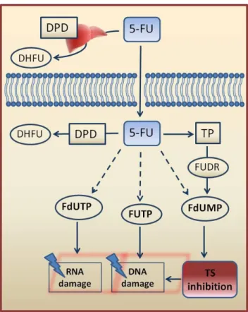
5-FU rapidly enters the cells where is converted into three active metabolites: fluorodeoxyuridine monophosphate (FdUMP), fluorodeoxyuridine triphosphate (FdUTP), and fluorouridine triphosphate (FUTP), which have different effects. FdUMP inhibits the action of thymidylate synthase (TS), FUTP disrupts RNA synthesis, and FdUTP is directly misincorporated into DNA (Figure 1.2) [27].
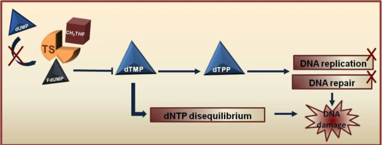
TS is a nucleotide synthetic enzyme, that catalyses the reductive methylation of deoxyuridine monophosphate (dUMP) to deoxythymidine monophosphate (dTMP) with the reduced folate 5,10-methylenetetrahydrofolate (CH2THF) as the methyl donor,
creating the only de novo source of thymidine monophosphate (TMP), which is necessary
Figure 1.2- Representative scheme of 5-Fluorouracil metabolism.
5-FU is taken up into cells and converted into three main active metabolites: fluorodeoxyuridine triphosphate (FdUTP), fluorouridine triphosphate (FUTP) and fluorodeoxyuridine monophosphate (FdUMP). An alternative activation pathway involves the thymidine phosphorylase (TP) catalysed conversion of 5-FU to fluorodeoxyuridine (FUDR), which is then phosphorylated and converted into FdUMP. 5-FU can inhibit RNA synthesis in a pathway that involves its transformation into FdUTP, can cause DNA damage by directly misincorporation of FUTP and by FdUMP binding to the nucleotide-binding site of thymidylate synthase (TS), inhibiting DNA synthesis. Dihydropyrimidine dehydrogenase (DPD)-mediated conversion of 5-FU to dihydrofluorouracil (DHFU) is the rate-limiting step of 5-FU catabolism in normal and tumor cells. Up to 80% of administered 5-FU is metabolized by DPD in the liver. Adapted from [3].
10
Figure 1.3- Mechanism of thymidylate synthase (TS) inhibition by 5-Fluorouracil.
Thymidylate synthase (TS) catalyses the conversion of deoxyuridine monophosphate (dUMP) to deoxythymidine monophosphate (dTMP) with 5,10-methylene tetrahydrofolate (CH2THF) as the methyl donor. In the presence of the 5-FU active metabolite FdUMP it binds to the nucleotide-binding site of TS and forms a stable ternary complex with TS and CH2THF, blocking access of dUMP and inhibiting dTMP synthesis. This results in deoxynucleotide (dNTP) disequilibrium and DNA damage. Adapted from [3].
for DNA replication and repair [28, 29]. Due to its crucial importance to DNA replication, TS is an important target for fluoropyrimidine drugs, such as 5-FU.
TS contains a nucleotide-binding site and a binding site for CH2THF. The 5-FU metabolite
FdUMP binds covalently to the active site of TS forming a stable ternary complex with the enzyme and with CH2THF, blocking the binding of the normal substrate dUMP thereby
inhibiting dTMP synthesis. As a consequence, there is a depletion of dTMP which induces deoxynucleotides disequilibrium, restricting proper DNA replication and repair mechanisms, resulting in DNA damage and inducing cell-cycle arrest (Figure 1.3) [3, 30].
Other relevant enzymes involved in 5-FU metabolism are dihydropyrimidine dehydrogenase (DPD) and thymidine phosphorylase (TP) (Figure 1.2). DPD is the rate-limiting enzyme in 5-FU catabolism which converts 5-FU to dihydrofluorouracil (DHFU). Over 80% of the administered 5-FU is normally catabolised primarily in the liver, where DPD is abundantly expressed [31]. TP function is related with an indirect metabolization pathway of 5-FU to FdUMP by 5-fluoro-2'-deoxyuridine (FUdR).
11
1.3.2 Chemo-resistance to 5-fluorouracil
Poor response rates to CT are mainly due to the acquisition of resistance mechanisms. Chemo-resistance is believed to be the main cause of treatment failure in around 90% of patients with metastatic malignant diseases [32]. Therefore, it is important to characterize the biological factors that are involved in these mechanisms since they are directly correlated with treatment responses.
Regarding 5-FU, the expression of TS influences its therapeutic efficacy. In gastric cancer and CRC, data showed that patients whose tumors did not respond to treatment had the mean TS protein level significantly higher (14.5 versus 1.36, p< 0.01) comparing with samples from patients with responsive disease. The same result was achieved concerning the expression levels of TS messenger RNA (mRNA) (0.17 versus 0.60 arbitrary units, p< 0.01). Thus high expression and protein levels were correlated with lack of response to 5-FU [33]. In accordance, other studies show that low tumoral TS expression improves the efficacy of 5-FU based therapies, since TS expression was significantly associated with OS (p= 0.002) [34, 35].
TS protein expression is regulated at the post-transcriptional level, by inhibition of a negative feedback mechanism in which ligand-free TS binds to its own mRNA inhibiting its own translation. When stably bound to FdUMP, TS can no longer bind to the mRNA, maintaining a constant level of free enzyme [27, 36]. This suggests that the amount of FdUMP and the acute increase in TS expression (that facilitates the recovery of the enzyme activity) in response to cytotoxic agents, may play a role in the development of an important resistance mechanism [33].
Also in CRC, low levels of DPD mRNA expression were found in all 5-FU responders compared with higher levels in the 5-FU non-responders. Furthermore, few studies also demonstrated that high levels of TP mRNA also correlates with resistance to 5-FU [37]. However, there is no correlation between TS, DPD, and TP expression values in CRC, indicating that they are independent predictive markers of 5-FU response [38].
12
1.3.3 5-FluorouraciI pharmacokinetics
Regarding 5-FU pharmacokinetics, parameters like steady-state concentrations (Cpss) and total body clearance have been studied in order to determine the benefits in terms of clinical response and toxicity obtained by prolonging the drug delivery period and/or increasing the blood concentration [39].
Following a continuous drug infusion, the concentration of the drug in the plasma will increase until the rates of drug administration and drug elimination are equal. When the plasma concentration is constant Cpss is reached.
Cpss of 5-FU in plasma, measured in patients after continuous 5-FU infusion of 200 mg/m2/day ranged from 0,39 µM to 66,3 µM [40, 41]. This fact is extremely important, since it has been reported that standard CT regimens, are not only cytotoxic but also able to induce cellular senescence, a state of irreversible proliferative arrest [42, 43]. This means that at low levels CT drugs can have different effects on the tumor cells.
1.4 Cellular senescence
Senescence derives from the Latin word senescere that means ‘to grow old’. When applied to life sciences, senescence was first defined as a set of deteriorative processes resulting from biological aging, where an organism progressively accumulates changes in their molecular and cellular structure, consequently altering their metabolism and resulting in death [44, 45]. However, the biological concept of senescence has evolved substantially.
In 1961 Hayflick and Moorhead [46] described for the first time that normal somatic cells have limited proliferation ability, and that cells that initially proliferate at a normal rate gradually lose the ability to proliferate despite optimal culturing conditions. These results were later confirmed, leading to the hypothesis that cellular senescence is the cell mechanism responsible for the finite life time of human diploid cells [47].
The process observed by Hayflick is named replicative or cellular senescence, and occurs due to the progressive shortening of the telomeres in proliferating cells, leading to a proliferation limit. The first report suggesting that telomere shortening is the major mechanism responsible for replicative senescence came in early 1990s [48] and it is
13
currently the only known endogenous mechanism of senescence induction. The majority of normal cells, from all vertebrate species, undergo replicative senescence, although the number of cell divisions before senescence can be relatively variable [49].
Telomeres are regions of tandemly repeated hexanucleotide sequences (5'-TTAGGG-3' in vertebrates). Many cellular factors directly (e.g. TRF1/TRF2) and indirectly (e.g. shelterin-complex, PinX, Apollo and tankyrase) interact with telomeres, influencing telomere structure and function [50]. Located at the ends of chromosomes, these regions prevent cells from recognizing chromosome ends as double strand breaks (DSBs), avoiding the fusion with other chromosomes ends by DNA-repair mechanisms [51]. Therefore, telomeres are essential to maintain the stability and integrity of eukaryotic genomes [52]. During replication, DNA polymerase fails to completely replicate DNA ends, a process known as the “end-replication problem”, which contributes to telomere shortening. As a result, cells lose between 50 to 200 base pairs of telomeric DNA during each round of DNA replication [51]. When telomeres reach a critically short length, their protective function is disrupted, triggering a sustained DNA damage signal that will eventually cause the cell to stop dividing.
Cancer and germline cells, unlike most somatic adult cells, express telomerase, a cellular reverse transcriptase whose function is to add telomeric DNA to the chromosome ends, elongating the telomeres and solving the problem of the “end-replication” [52]. Telomerase however, either endogenous or ectopically expressed (to immortalize primary cells in vitro), does not completely impair senescence since it can be induced by multiple extrinsic factors besides telomere erosion. This type of telomere-independent senescence has been termed stress-induced or premature senescence, as it is established prior to telomere shortening-induced senescence [53].
Currently, the concept of cellular senescence is applied to any type of irreversible proliferation arrest. Multiple extrinsic factors such as DNA damaging agents (e.g. ionizing radiation and chemotherapy agents), oxidizing agents, and oncogene activation, have been found to trigger telomere-independent senescence [54-57]. Therefore, ‘replicative senescence’ refers to telomere-derived senescence and ‘stress-induced (or premature) senescence’ (SIS) refers to senescence induced by exogenous factors [2].
14
1.4.1 Signaling pathways mediating senescence
Independently of the trigger factor, senescence state derives from DNA damage response (DDR) activated signalling cascade, due to the occurrence of single strand breaks (SSB) or double strand breaks (DSB).
DDR is crucial to preserve and maintain genome integrity. Besides activation of DNA repair pathways, DDR can also induce the arrest of cell cycle progression by activating checkpoint proteins. This allows the cell time to repair the DNA damage, preventing the replication of damaged DNA.
The DDR (Figure 1.4) pathway is activated by two major sensors of damage: the MRE11– RAD50–NBS1 (MRN) complex, which detects DNA DSBs, and replication protein A (RPA) that, together with RAD9–RAD1–HUS1 (9-1-1) complex, which detects exposed regions of single-stranded DNA. These damage sensors recruit apical kinases according to the type of lesion. DSBs signal leads to the activation of ataxia-telangiectasia mutated (ATM), whereas single-stranded DNA regions engage ataxia telangiectasia and Rad3-related (ATR) [58]. The recruitment of ATM or ATR leads to phosphorylation of histone variant H2AX on Ser139 (known as γH2AX). In the case of DSBs, γH2AX is required to activate mediator of DNA damage checkpoint 1 (MDC1) and p53-binding protein 1 (53BP1) that amplify the DDR by recruiting additional ATM complexes in a positive feedback loop. In the case of single-stranded DNA damage, ATR kinase activity is enhanced by the 9-1-1 complex and by topoisomerase-II-binding protein 1 (TOPBP1). Breast cancer 1 (BRCA1) is then phosphorylated by ATM or ATR, and recruited to the sites of DNA damage [4, 59]. After amplification of the DDR signal, ATM and ATR activate by phosphorylation the checkpoint kinases 2 (CHK2) and CHK1, respectively. CHK2 and CHK1 will establish the communication between DDR-associated factors and the cell cycle machinery through phosphorylation and activation of downstream effectors such as p53 and the cell division cycle 25 (CDC25) phosphatases [4, 53]. The interaction between the ATM and ATR signalling pathways is complex. The ATR-Chk1 pathway can be activated in response to DSBs when single stranded DNA is generated on broken DNA ends. On the other hand, single-strand nicks can result in DSBs when, during replication of damaged DNA, they are encountered by leading-strand DNA polymerases. Therefore these pathways are
15
Figure 1.4- The DNA damage response.
Double-strand breaks are recognized by the MRE11–RAD50–NBS1 (MRN) complex and lead to the activation of ataxia–telangiectasia mutated (ATM) and subsequent amplification of the response through the recruitment of other DNA damage response proteins. Activated ATM phosphorylates (P) checkpoint kinases2 (CHK2), which in turn phosphorylates p53 and other substrates. Other forms of DNA damage lead to the generation of single-stranded regions that are detected by replication protein A (RPA) and RAD9–RAD1–HUS1 (9-1-1) complex. This attracts the ataxia–telangiectasia and Rad3-related (ATR) which phosphorylates and is phosphorylated by 9-1-1 complex. ATR activates downstream substrates including checkpoint kinases1 (CHK1). In addition, ATM and ATR phosphorylate the histone variant H2AX on Ser139 (γH2AX). DDR signal amplification is undertaken by damage checkpoint 1 (MDC1), p53-binding protein 1 (53BP1), 9–1–1 complex and topoisomerase-II-binding protein 1 (TOPBP1). Adapted from [4].
frequently activated simultaneously in cells exposed to genotoxic stresses, including ionizing radiation and chemotherapy agents [60].
Ultimately, when the injury is extremely severe, the DDR may prone cells to apoptosis, a program of cell death that is used to eliminate damaged cells in a controlled manner that minimizes damage and disruption of the neighbour cells [61]. Also, the DDR-initiated
16
proliferation arrest can be either temporary, allowing cells to repair DNA damage and to resume their cycle, or persistent (cellular senescence), caused by the accumulation of unrepaired DNA lesions that fuel a sustained DDR signalling. It is still unclear what tips the balance towards the pathways that lead to either apoptosis or senescence, but important determinants may include cell type and the intensity, duration and nature of the damage [45, 59].
1.4.1.1 p53 and pRb, two master regulators of senescence
Tumor suppressor genes encode for proteins that impair cell neoplastic transformation. These proteins provide a defence mechanism against oncogenic mutations and induced DNA damage, preventing transformation by forcing cells into apoptosis or cell cycle arrest. Full blown tumor onset depends on the inactivation or deletion of these genes [62].
Consistent with their roles in preventing the proliferation of genetically modified cells, two different tumor suppressor proteins, p53 and retinoblastoma protein (pRb), have been shown to be deeply involved in the onset and maintenance of senescence [2]. Back in 1991 it was shown for the first time that p53 and pRb are two major regulators of senescence, since suppression of both proteins leads to an inhibition of cell cycle arrest allowing cells to bypass senescence [63]. Since then, the role of p53 and pRb in the activation of downstream molecular networks in mouse and human has been subject of extensive research [2].
Experiments with senescent mouse embryonic fibroblasts (MEFs) showed that suppression of p53 is sufficient to escape senescence [64]. On the other hand, although inactivation of pRb alone did not affect senescence, simultaneously with the inactivation of the pRb family proteins p107 and p130 it impaired the cells’ capability to perceive the senescence-inducing signal, strongly increasing their proliferation rate [65]. Moreover, acute loss of pRb in senescent cells lead to a reversion of the senescence phenotype [66]. Globally, these results indicate that p53 and pRb are not only necessary for the onset of senescence but are also needed to maintain it. In addition, these evidences suggest a linear model of pathway activation, in which stress-induced p53 activation leads to pRb downstream activation [2].
17
However, experiments in human fibroblasts suggest that either a linear or parallel activation process can occur [67-69].It is currently accepted that the activation of each tumor suppressor protein can be related to different stimuli and be cell type and species specific [45]. Therefore, pathways downstream of p53 and pRb can be activated independently or simultaneously, cross-regulating each other and mediating senescence onset and maintenance [70, 71].
Different DDR inducers can lead to senescence mainly through the activation of the p53 pathway [72], with the first evidence being dated back to 1984 where ultraviolet (uv) irradiation caused severe genomic stress and increased p53 levels in non-transformed mouse cells [73].
The p53 pathway is regulated at several levels by different proteins, but the most important p53 regulator is the E3 ubiquitin-protein ligase MDM2 (in humans is frequently called HDM2) [74]. MDM2 forms a complex with p53 and inhibits its activity in different ways. MDM2 can bind the transcriptional activation domain of p53 and block its ability to regulate target genes [75, 76], and can also promote the ubiquitylation and subsequent proteosome-mediated degradation of p53, either way inhibiting p53-dependent senescence [77, 78]. Moreover, the MDM2 gene has a p53 DNA-binding site, making it a direct transcriptional target of p53. Therefore, there is an auto-regulatory negative-feedback loop that regulates the expression of MDM2 and the activity of p53 [79, 80]. In proliferating cells p53 is kept at basal levels that rapidly increase upon genomic stress. This regulatory mechanism is mainly controlled via the constant action of the MDM2–p53 loop in which excessive p53 activity triggers the production of MDM2 that in turn will promote p53 degradation and extinguish p53 cellular activity [81]. This switch in p53 levels and activity upon stress makes p53 into one of the most important genomic guardians and protectors against cancer progression.
The Rb protein family is composed of pRb, p107 and p130, and is involved in the control of G1-S transition. pRb negatively regulates E2F transcription factors, preventing the activation of genes necessary for G1-S transition and cell cycle progression [82]. Cells lacking pRb show aberrant proliferation and increased genomic instability [83]. It was
18
Figure 1.5- Structure of the INK4a/ARF and INK4b loci.
The INK4b/Arf/INK4a locus includes two different genes. The splicing patterns and encoded proteins are represented. Two distinct promoters transcribes for exons 1α (INK4a) and 1β (Arf) and result in alternatively spliced transcripts that share exons 2 and 3. Although shared, different open reading frames within exon 2 give rise to two distinct protein products. Adapted from [1].
recently demonstrated that pRb has a unique and non-redundant function in senescence by regulating a particular subset of E2F targets and preventing DNA replication [83]. pRb specifically represses transcripts associated with replication, while p107 and p130 repress the transcription of DNA replication factors associated with quiescence but not with senescence.
1.4.1.2 The Arf-p53- p21WAF1 pathway.
Upstream control of p53 can be mediated by the DDR, via ATM/ATR and CHK2/CHK1 proteins, or by alternative reading frame (Arf) protein [84]. Arf is encoded by the INK4a/Arf locus (Figure 1.5). Formally designated CDKN2A and CDKN2B, the locus includes three closely linked tumor suppressor genes (INK4a, INK4b, and ARF) that trigger the anti-proliferative activities of both pRb and p53 [85]. INK4a and INK4b encode for p16INK4a and p15INK4b, respectively, and Arf encodes for p14Arf (p19Arf in mouse). While p16INK4A is encoded by the α transcript containing exon 1α, exon 2 and exon 3, p14Arf is encoded by the β transcript comprised by exon 1β and exon 2 and 3. These two exons are common to p16INK4A under a different promoter and translated in an alternative reading frame [86].
Mice lacking only the 1β exon, expressing only p16INK4a, showed high tumor susceptibility, and their phenotype was similar to that of p53 null mice, demonstrating ARF’s tumor
19
suppressor functions [87]. Since p53-deficient cells are resistant to ARF- induced arrest, ARF is considered a positive upstream regulator of p53 [87]. In fact, ARF interferes directly with MDM2 function in the nucleolus, inhibiting the E3 ubiquitin-protein ligase activity of MDM2 and therefore increasing p53 stability and activating the p53-mediated transcriptional program [88, 89].
Despite the fact that several stress signals that can activate the p53 pathway have been identified, those requiring Arf still need to be clarified. It was previously demonstrated that p19Arf knockout (KO) MEFs have impaired DNA damage and microtubule disruption [90]. It was also shown that in normal human fibroblasts Arf expression is induced in response to ionizing radiation, and that endogenous p14Arf can bind directly to p53 independently of HDM2 as an immediate response to DNA damage, suggesting that Arf can also function as a cofactor for p53 transactivation. [91]. In addition, high expression of Arf was also observed in Ras-induced senescent MEFs, and is considered necessary for telomeric and non-telomeric-derived senescence [87, 89]. Although Arf activation seems to be stress dependent, little is known about its physiologic context [2].
In mouse models, Arf inactivation is comparable to p53 inactivation, preventing senescence and reinforcing its role as tumor suppressor [87]. The same is not applied to human cells, where the role of Arf is not so clear [2].
Given the role of INK4a/ARF in modulating activities of pRb and p53, it is not surprising that deletion of the locus is frequently detected in many distinct tumor types [92].
Most genes that are downstream targets of p53 encode for proteins that regulate cell cycle checkpoints, apoptosis and senescence. CDKN1, that encodes for cyclin dependent kinases (Cdk) inhibitor protein p21WAF1, is directly transactivated by p53 [93]. The p21WAF1 protein is a member of the CIP/KIP family of Cdk inhibitors and in the context of senescence, either replicative or stress-induced, is considered one of the most important downstream targets of p53 [94]. All p21WAF1’s functions are related to cell cycle inhibition. It not only inhibits cyclin/Cdk complexes, but also blocks proliferating cell nuclear antigen (PCNA), inactivates pRb through proteosome-mediated degradation and inhibits cyclin B1 [95, 96]. Binding of the N-terminal region of p21WAF1 to Cdk2 inhibits the formation of the
20
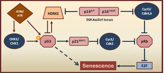
Figure 1.6- Signal transduction pathways mediating senescence.
Sequential activation of the Arf-p53-p21WAF1 and p16INK4a-pRb signaling cascades. Increased transcriptional activity of p53 is achieved by phosphorylation (P), performed by the ATM/ATR and CHK2/CHK1 proteins, or by the p14Arf product of the INK4a/Arf locus, which sequesters E3 ubiquitin-protein ligase HDM2. By its turn HDM2 regulates p53 activity by a negative feedback loop mechanism. Senescence is triggered by retinoblastoma (pRb) hypophosphorylation and consequent silencing of E2f target genes. pRb is activated either by p21WAF1, or by the p16INK4a product trough inhibition of the cyclin E/Cyclin dependent kinase 2 (CycE/Cdk2) and cyclin D/Cyclin dependent kinase 4/6 (CycD/Cdk4/6) complexes, respectively. Adapted from [2].
cyclin E/Cdk2 complexes, thereby impairing pRb phosphorylation. The accumulation of unphosphorylated pRb prevents the release of E2F transcription factors from pRb/E2F complexes, blocking cells in G1 (Figure 1.6) [97]. Primarily, p21WAF1 activates cell cycle checkpoints in G1 and in G1/S [94]. However, in response to DNA damage, p21WAF1 activates the G2/M checkpoint by degradation of cyclin B1, inducing a G2 cell cycle arrest [98].
Nevertheless, senescence is not impaired in MEFs carrying a null mutation in p21WAF1, suggesting a non-essential role for p21WAF1 or the existence of other mechanisms that compensate for its loss of function [99].
1.4.1.3 The p16INK4a-pRb pathway
p16INK4a, also encoded by the INK4a/ARF locus, is a Cdk inhibitor that forms a complex with Cdk4/6, inhibiting the interaction with cyclin D through a conformational alteration.
21
Abrogation of the activation of Cdk4/6 maintains pRb in the hypophosphorylated active form (Figure 1.6) [100].
The p16INK4a promoter is not normally expressed in differentiated tissues. However, it can be induced by stress conditions and was found to be highly expressed in senescent cells [101]. p16INK4a overexpression was first described during replicative senescence [102] and later in response to oncogenic Ras activation [57].
Stimuli that trigger a DDR can also activate the p16INK4a-pRb pathway, as a secondary response after activation of the p53 pathway [103]. As previously mentioned, the signalling pathway engaged at the onset of senescence can be different according to the stress signal (specific combination and severity), cell type and species. If, on one hand, telomere disruption activates the p53 pathway, on the other hand, other senescence-inducing stimuli, like oncogenic Ras, may act primarily through the p16INK4a-pRb pathway [67, 104].
p16INK4a KO mice are more prone to develop a variety of spontaneous long latent tumors [105]. In humans, introduction of activated Ha-ras into p16INK4a inactivated primary fibroblasts can induce neoplastic transformation [104]. All the evidences point to p16INK4a as a key player in tumor suppression and a mediator of the senescence process.
1.4.2 Senescence-associated heterochromatic foci
Senescent cells suffer a nuclear rearrangement correlated with critical gene expression alterations that will epitomize activation of senescence regulators and silencing of proliferation-associated genes.
As described before, pRb is a crucial effector of senescence and a key component in the irreversibility of the cell cycle arrest. The role of p16INK4a-pRb pathway in senescence also involves chromatin remodelling and formation of heterochromatin regions, known as senescence-associated heterochromatic foci (SAHF). At these regions heterochromatin is condensed, modified and transcriptionally silent. SAHF formation appears to be dependent on the integrity of the p16INK4a-pRb pathway, in which hypophosphorylated active pRb silences E2F target genes [106].
22
Initially, histone chaperones like histone repressor A (HIRA) and heterochromatin protein 1-γ (HP1γ) are recruited to specific subnuclear organelles, the acute promyelocytic leukaemia (PML) nuclear bodies. After HIRA’s translocation into PML bodies, chromatin condensation occurs, trimethylation of lysine 9 in histone 3 (H3K9me3) accumulates, and HP1 and histone H2A variant (a family of three related proteins, macroH2A1.1, 1.2 and 2) are recruited to SAHF [107, 108].
Proliferation-promoting genes, such as E2F target genes (e.g. cyclin A), are silenced by incorporation into the SAHF, contributing to the irreversibility of the proliferative arrest that characterizes senescence [106]. Senescent cells are, therefore, unable to re-enter the cell cycle, even in a pro-mitogenic environment. These features distinguish senescence from quiescence, a non-proliferative but reversible state [106].
Although senescence can be a stress-response mechanism to prevent proliferation, some senescent cells also become resistant to certain apoptotic signals. For example, senescent human fibroblasts do not undergo ceramide-induced apoptosis, and also become resistant to programmed cell death caused by growth factor deprivation and oxidative stress [109, 110]. In contrast, Fas death receptor activation is able to induce apoptosis in these cells [111]. Nevertheless, the molecular pathways involved in resistance to apoptosis are still poorly understood. Alterations in gene expression may contribute to inhibit, promote or establish apoptosis [112]. Furthermore, evidences show that p53 might preferentially transactivate genes that arrest proliferation, rather than those that trigger apoptosis in senescent fibroblasts [113].
1.4.3 Senescence-associated secretory phenotype
Senescent cells remain metabolically active, acquiring a designated senescence-associated secretory phenotype (SASP), with a characteristic senescence-senescence-associated secretome (SAS) composed by several pro-inflammatory cytokines, chemokines, growth factors, and proteases [114].
23
Persistent DNA damage seems to be required for induction of SASP. In fact, ectopic overexpression of p21 or p16INK4a triggers senescence without SASP, while DNA damage, dysfunctional telomeres, epigenomic disruption, mitogenic signals, oxidative stress, and other senescence-DNA damage inducing stimuli induce a SASP [115, 116].
Moreover, SASP can be different according to the inducing stimulus. Genetic alterations like loss of p53, or activation of Ras, that induces senescence by indirect DNA damage due to hipper-replication [117], lead to an accelerated acquisition of a more prominent SASP [118].
The first evidences of alterations in the cell secretome due to senescence were reported in 1991 where microarray analysis revealed a strong inflammatory secretome of fibroblasts undergoing replicative senescence [119].
Currently it is well established that normal and epithelial tumor cells have a SAS after genotoxic-induced senescence [118, 120]. In addition, it was demonstrated in vivo that DNA-damaging chemotherapy-induced senescent human prostate cancer cells have a SAS [118].
Using antibody-array technology, a large-scale characterization of SAS from normal fibroblasts, normal epithelial cells, and epithelial tumor cells after genotoxic stress, showed that SAS molecules can be divided into three major categories: soluble signaling factors (interleukins, chemokines, and growth factors), secreted proteases, and secreted insoluble proteins/extracellular matrix (ECM) components [114, 118]. Overall, these factors can induce mechanisms capable of altering cell fate of neighboring cells and modify the tissue microenvironment. Despite of a substantial overlap in the SAS composition among different cells and tissues, like interleukin (IL)-8, the SAS and its effects seem to be cell and tissue-type specific [114].
The SAS has powerful paracrine effects inducing a variety of cellular responses. With several cytokines and chemokines in SAS, it can contribute to modulate immune responses, targeting the senescent cells for elimination by the immune system, attracting and activating natural killer cells, macrophages and T cells. In addition to an eventual clearance of senescent cells, local immune reactions seem to be stimulated by the SAS, in
![Table 1.1- TNM Classification system for colorectal cancers [8].](https://thumb-eu.123doks.com/thumbv2/123dok_br/19201270.953878/32.892.127.764.84.1008/table-tnm-classification-colorectal-cancers.webp)