Ar
ti
cle
*e-mail: [email protected]
Antioxidant Properties of Plant Extracts: an EPR and DFT Comparative Study
of the Reaction with DPPH, TEMPOL and Spin Trap DMPO
Adevailton Bernardo dos Santos,*,a,d Dulce Helena Siqueira Silva,b Vanderlan da Silva Bolzani,b Luciana Ávila Santos,b Tomé Mauro Schmidtc and Oswaldo Baffad
aFaculdade de Ciências Integradas do Pontal, Universidade Federal de Uberlândia, 38302-000 Ituiutaba-MG, Brazil
bInstituto de Química, Universidade Estadual Paulista, 14800-900 Araraquara-SP, Brazil
cInstituto de Física, Universidade Federal de Uberlândia, CP 593, 38400-902 Uberlândia-MG, Brazil
dDepartamento de Física e Matemática, Faculdade de Filosoia Ciências e Letras de Ribeirão Preto,
Universidade de São Paulo, 14040-901 Ribeirão Preto-SP, Brazil
Este trabalho apresenta um estudo comparativo da atividade antioxidante dos extratos de nove espécies de plantas pertencentes à lora brasileira - Swartzia langsdorfii, Machaerium villosum, Pterogyne nitens, Pera glabrata, Aegiphyla sellowiana, Copaifera langsdorfii, Chrysophyllum inornatum, Iryanthera juruensis, Didymopanax venosum - neutralizando os radicais livres DPPH (2,2-difenil-1-picrilhidrazil) e TEMPOL (4-hidróxi-2,2,6,6-tetrametil-1-piperidiniloxi-1-oxil) utilizando ressonância paramagnética eletrônica (RPE), e agindo sobre os radicais hidroxila (OH•)
gerados por uma reação Fenton, através da técnica de spin-trapping. Os melhores resultados foram obtidos para os extratos de Iryanthera juruensis e Chrysophyllum inornatum. Os resultados dos testes foram acompanhados por cálculos computacionais dos compostos, utilizando a teoria do funcional da densidade (TFD), com aproximação local da densidade (ALD), e o código SIESTA. Os resultados indicaram que a energia liberada na reação de redução TEMPOL é inferior a DPPH. Como o DPPH é um composto de menor custo e mais disponível tem um uso mais amplo que o TEMPOL, entretanto este último deve ser considerado quando se quer estudar processos que envolvam menores energias.
This work presents a comparative study of the antioxidant activity of extracts from nine plant species belonging to the Brazilian lora - Swartzia langsdorfii, Machaerium villosum, Pterogyne nitens, Pera glabrata, Aegiphyla sellowiana, Copaifera langsdorfii, Chrysophyllum inornatum, Iryanthera juruensis, Didymopanax venosum - acting on the free-radicals DPPH (2,2-diphenyl-1-picrylhydrazyl) and TEMPOL (4-hydroxy-2,2,6,6-tetramethyl-1-piperidinyloxy-1-oxyl) by electron paramagnetic resonance (EPR), and acting on the hydroxyl radical (OH•) by the
spin-trapping technique generated by a Fenton reaction. Results showed that the extracts of Iryanthera juruensis and Chrysophyllum inornatum display the strongest antioxidant activities. The results of scavenging tests were clariied by computational calculations - density functional theory (DFT), local density approximation (LDA) with SIESTA code - indicating that the energy released in the reduction reaction of TEMPOL is less than DPPH. Due to its availability and cost DPPH is more often used in these tests than TEMPOL, however TEMPOL should be considered when studying processes dealing with smaller energies.
Keywords: EPR, TEMPOL, DPPH, DMPO, DFT
Introduction
Upon normal metabolic reaction, which includes several oxidative steps, lipids, sugars and proteins can generate aldehydes, ketones, esters and other products possessing
chemical and biological properties which may be harmful to living systems. Dietary antioxidants can prevent the oxidation of such compounds by assisting in the elimination of free radicals generated by the metabolism, mainly reactive
oxygen species (ROS).1 At the present there is a great interest
everyday diet and plants that have phytotherapeutic use such as teas beverages. Usually stable free radicals and spin traps are used in these studies to identify the reactants and
to measure reaction rates.2-6
This work used electron paramagnetic resonance (EPR), a spectroscopic technique for selective detection of free radicals. EPR has wide use such as fossils dating, identification of paramagnetic substances, ionizing
radiation dosimeters and other applications,7-8 including
antioxidant evaluation, allowing studies on the kinetics and stoichiometry of the reactions of radicals and antioxidant compounds. One way to perform these measurements is by measuring the concentration of free radicals that are in contact with the antioxidant substance by using stable free radicals as DPPH and TEMPOL (Scheme 1). DPPH and TEMPOL are known as standard substances
in EPR spectroscopy9,10 and are largely applied in studies
of the antioxidant activity. DPPH is one of the molecules frequently used for measuring antioxidant activities, in
optical spectroscopy measurements as well as in EPR.
1-3,6,11,12 TEMPOL is less used for this purpose4,13-15 once in
contact with other free radicals it can show antioxidant activity, either in its original form or in reduced form
TEMPOL-H.16
Another approach to carry out studies of antioxidant substances is to use the spin-trapping technique, consisting in generating free radicals, which normally have a short lifetime, and placing them in contact with a spin trap. The result of this reaction is the generation of a spin adduct that
has a longer lifetime2,6,17,18 allowing its monitoring by EPR
spectroscopy or other technique. So, for the study of the antioxidant activity of a compound against the hydroxyl
radical (OH•), this technique can be used: the Fenton
reaction (Fe2+ + H
2O2→ Fe3+ + OH• + HO –) generates the
hydroxyl radical, and the spin trap DMPO
(5,5-dimethyl-1-pyrroline N-oxide) traps it, leading to the formation of the spin adduct DMPO-OH (Scheme 1).
One goal in this study was to carry out a comparative analysis of the antioxidant activity of some species of the Brazilian lora. The species compared in this work are:
Swartzia langsdorfii, Machaerium villosum, Pterogyne nitens, Pera glabrata, Aegiphyla sellowiana, Copaifera
langsdorffii, Chrysophyllum inornatum, Iryanthera
juruensis, Didymopanax venosum which have been selected
due to the presence of antioxidant compounds in their ethanol extracts, evidenced by preliminary screening in a TLC test using beta-carotene solution as revealing agent. Despite the possibility of studies using extracts prepared in other solvents, this work uses only ethanol extracts for initial tests. These results could serve as a reference for future work with fractions of these extracts.
The other objective of this work is to get information on the molecular structures, energies and some electronic properties of free radicals DPPH and TEMPOL, and of spin-trap DMPO, in gas-phase, using the density functional theory (DFT) calculations, with local density approximation
(LDA)19 and use of pseudopotentials. Despite several works
with antioxidants compounds, little attention is given to the free radicals used in tests, however the knowledge on the properties of these free radicals can help to analyze the results. The results of computational calculations aided to elucidate the differences between results of antioxidant activities from the same extract, mainly that DPPH react differently than TEMPOL. The main focus of these calculations is to determine hydrogen bond dissociation energy (BDE) to estimate the energies involved in the reactions of the free radicals studied with antioxidants compounds.
The reaction with antioxidant compounds (A) and free radicals (Fr) can occur through different pathways: Hydrogen atom transfer (HAT) (reaction I) or a process involving electron transfer (ET) and proton transfer (PT). One of the processes involving ET and PT is sequential proton loss electron transfer (SPLET) (reaction II) where hydrogen bond acceptor (HBA) solvent (S) reacts by ionizing antioxidant compound for proton loss followed by electron transfer from antioxidant compound to free radical. Another process is proton coupled electron transfer (PCET) in which electron and proton transfer occurs simultaneously, similar to HAT (reaction I) but in different molecular orbital: the proton is then transferred from two bonding electron antioxidant compound to the radical’s lone pair while electron move from 2p lone pair antioxidant compound to the radical’s single occupied molecular orbital (SOMO). There is another case where irst occurs electron transfer followed by proton transfer (ETPT) (reation III), with the proton reacting with
the Fr− or with solvent, if it is good HBA.20
Fr• + A-OH → Fr-H + A-O• (I)
Fr• + A-OH --- S → Fr• + A-O− + HS+→ Fr− + A-O• + HS+ (II)
Fr• + A-OH → Fr− + A-OH+→ A-O• + H+ + Fr− (III)
Independent of the preferential reaction route followed, our calculations are focused in HAT process and the results serve for estimation of the released energy in the reduction of free radical by antioxidant compound. Thus, the main goal of the this work is to compare the anti oxidative properties of several natural extracts against DPPH, TEMPOL and the spin trapping DMPO and to give some explanation of our indings based on the density functional theory (DFT).
Experimental
Reagents and plants materials
DPPH, TEMPOL, DMPO and quercetin were purchased from Sigma-Aldrich Chemical Co and used without further puriication; ethanol (> 99.9%) and hydrogen peroxide (30%)
(H2O2) from Labsynth LTDA-Brazil; and ferrous sulphate
(FeSO4) from Vetec LTDA-Brazil. Plant materials were
collected in Mogi Guaçu State Park (Cerrado), Jureia-Itatins State Park (Atlantic Forest), Mocambo Reserve (Amazon Forest) and identiied by botanists Inês Cordeiro and Nelson Rosa. Voucher specimens were deposited at Museu Paraense Emílio Goeldi, PA, and Herbarium of Instituto de Botânica-SEMA, SP.
EPR spectrometer and data analyses
A Varian E-4 X-band spectrometer computer interfaced allowing signal averaging and fast measurements equipped with a rectangular cavity (TE-102, model E-231) was used for the EPR measurements. Operation parameters were as follows: microwave power 10 mW, center magnetic ield 338 mT, sweep width 10 mT, modulation frequency 100 kHz, modulation amplitude 0.1 mT and room temperature
24 ± 2 °C. The data and graphical analyses were carried out
using the Microcal Origin 7 software.
Plants extracts
Seeds and leaves of Iryanthera juruensis (50 g, each)
as well as leaves of Swartzia langsdorfii, Machaerium
villosum, Pterogyne nitens, Pera glabrata, Aegiphila
sellowiana, Copaifera langsdorffii, Chrysophyllum
inornatum, Didymopanax venosum (50 g, each) were dried,
ground and submitted to extraction with ethanol (cold, 3x). Each ethanol solution was evaporated under reduced pressure, affording the ethanol extracts used in this study.
DPPH scavenging test
The antioxidant activities of the plant extracts against the free radical present in DPPH were studied using ethanol solutions of the extracts at several concentrations. Aliquots
of DPPH solution in ethanol (1 mmol L-1, 40 µL) and each
extract solution (40 µL) were mixed, stirred for 10 s and then transferred to a capillary tube. The capillary tube was sealed and placed inside a standard EPR quartz tube (inner diameter 3.00 mm), which was placed in the resonant cavity. This procedure took 3 min and it was carried out at room temperature. For the reference measurement the extract solution was replaced by ethanol (40 µL). Data were reported as the average of three measurements. The procedure was repeated with quercetin.
Spectra simulation was carried out by the computer program Winsim, and the double integral of the signal was evaluated as representative of the free radical concentration. Results were plotted in a graph of the double integral intensity versus extract concentration. By graphical analysis, it was possible to determine the concentration that reduces the initial signal intensity by 50% (IC 50). The spectra are a result of 3 scans average with 30 s scan time.
Hydroxyl radical scavenging test
Extracts solutions were prepared in deionized water,
at different concentrations. H2O2 (20 µL, 100 mmol L-1),
FeSO4 (20 µL, 10 mmol L-1), DMPO (20 µL, 50 mmol L-1),
and extract solution (20 µL) in phosphate buffer solution were mixed. The resulting solution was stirred for 10 s and transferred to a capillary tube, and placed in the EPR cavity as mentioned before. This procedure took 3 min. For the measurement of a reference signal the extract solution was replaced by 20 µL deionized water. Spectra of the spin adduct DMPO-OH were recorded with 1 scan of 30 s. Data are the mean values of three measurements. Results were calculated as the sum of the intensity of the four hyperine EPR lines of the spectra, and they are then plotted in a graph of the signal intensity versus extract concentration. By graphical analysis it was possible to determine the concentration that reduces the initial signal intensity by 50% (IC 50).
TEMPOL scavenging test
Extracts solutions were prepared in both ethanol and
deionized water (20 mg mL-1). Each extract solution
(40 µL) was mixed with TEMPOL solution in deionized
water (10 µmol L-1, 40 µL). The resulting solution was
time measurement, now repeated at 1, 2 and 24 h intervals. Spectra were averaged after 5 scans with 60 s scan time. In the reference, the extract solution was substituted for 40 µL of ethanol or deionized water. Finally, the procedure was carried out with quercetin in ethanol and water solution
(2 mg mL-1) mixtures with TEMPOL in water (10 µmol L-1),
for comparative test.
The extract of Chrysophyllum inornatum, showing
good results for antioxidant activities, was tested for higher TEMPOL concentrations and longer time intervals. The comparative study with the extract in water solution and ethanol solution was performed. Mixtures of 40 µL of the
solution of TEMPOL in water (0.10 mmol L-1) and 40 µL
of the extract solution in ethanol (20 mg mL-1) or 40 µL
of the extract solution in deionized water (20 mg mL-1). In
the reference solution the extract solution was substituted for 40 µL of ethanol or water.
The results presented herein were obtained after adding up the amplitude of the 3 hyperine lines of the spectra, from which the percentage of TEMPOL neutralization was calculated. This measurement is expressed by equation 1:
P = (ref-ext)/(ref-bg) (1)
where P is the percentage of decrease in the signal intensity, ref is the intensity of the reference signal, ext is the intensity of the signal due to the mixture of the free radical with the extract solution and bg is the background signal.
Computational calculations
The molecules of DPPH, TEMPOL and DMPO were simulated in gas-phase. DFT-LDA with use of pseudopotentials provides the theoretical framework of our calculations. The pseudopotentials are generated using
the norm of Troullier-Martins.21 The computational code
used is SIESTA.22 The exchange-correlation functional
used the results of Ceperley and Alder23 as parametrized
by Perdew and Zunger.24 A double-zeta added polarized
functions (DZP) basis set was employed, and a mesh cutoff
of 140 Ry was used for the reciprocal-space expansion of the charge density. All the geometries were optimized until the remaining forces were lower than 0.01 eV/Å. The Brillouin zone was sampled by four k points.
Results and Discussions
DPPH scavenging test
The IC 50 results is presented in Table 1. The most efficient extract for DPPH scavenging was that from
Iryanthera juruensis (leaf and seed) extracts. These extracts
displayed high antioxidant activity when compared to
the one obtained for Filipendula ulmaria, a native plant
of France, in a similar experiment.3 The IC 50 values
obtained for the leaves extract (0.028 mg mL-1) and seeds
extract (0.041 mg mL-1) of Iryanthera juruensis, when
compared to quercetin (0.017 mg mL-1) indicated a great
concentration of antioxidant compounds. Copaifera
langsdorfii, Chrysophyllum inornatum and Pterogyne
nitens have also shown low IC 50 values, indicating high
antioxidant capacity. Previous phytochemical work on these species evidenced the presence of lavonoids in their leaves extracts, which may have contributed to the overall
high activity.25-27
The higher IC 50 value was obtained for the extract
from Swartzia langsdorfii, indicating that this plant has
moderate antioxidant activity. It has been reported that its extract contains isolavonoids, which might be responsible
for the observed activity.28-30
The DPPH test is easy to perform, reliable and reproducible, for this reason has been widely used in the analysis of antioxidant activities, both in EPR spectroscopy and in the bleaching technique, which measures the optical absorption intensity at 515-517 nm. The fact that the DPPH EPR spectrum has a relatively simple signal due only to the free radical is an advantage in relation to the optical spectroscopy. In the latter, besides the appearance of absorptions due to the presence of other compounds in the extract, there is also an increment at 320 nm, corresponding
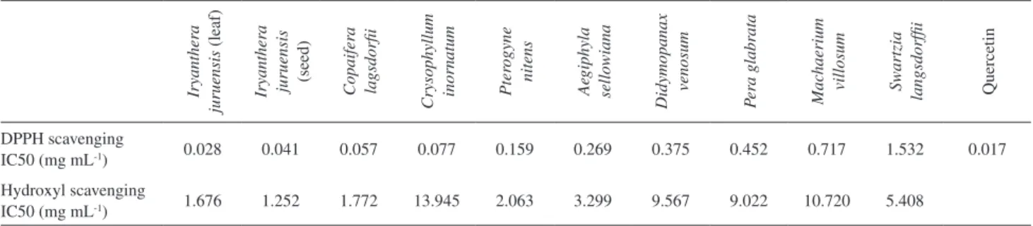
Table 1. Antioxidant activity of plant extracts against the radicals DPPH and DMPO-OH, results indicated for IC 50 (mg mL-1)
Iryanther
a
juruensis
(leaf)
Iryanther
a
juruensis (seed) Copaifer
a
la
gsdorii
Crysophyllum inornatum Pter
o
gyne
nitens
Ae
giphyla
sellowiana
Didymopanax
venosum
P
er
a glabr
ata
Mac
haerium
villosum Swartzia
langsdorfii Quercetin
DPPH scavenging
IC50 (mg mL-1) 0.028 0.041 0.057 0.077 0.159 0.269 0.375 0.452 0.717 1.532 0.017
Hydroxyl scavenging
to the increase in DPPH-H concentration due to DPPH
reduction to the respective hydrazine.9
Despite the advantage of using EPR technique for DPPH scavenging test, this result indicates only the antioxidant activity and not the mechanism of its reduction reaction. The tests were carried out in ethanol solution, polar and HBA solvent, and in this form the reaction of DPPH reduction occurs by a combination of the classical HAT mechanism and a process involving PT and ET,
mainly SPLET with ionization of antioxidant compound.20
In this case the SPLET mechanism was favored in solvents having high dielectric constant and ability to solvate anions increasing its stability similar to ethanol.
Hydroxyl radical scavenging test
Results concerning IC 50 for the hydroxyl radical scavenging test are presented in Table 1, and the small differences are in agreement with results obtained in the
other tests. As observed in DPPH test, the Iryanthera
juruensis (leaf and seed) extracts showed the strongest
antioxidant activity. The low activity of Chrysophyllum
inornatum extract contrasts with results from the other tests,
in which this extract showed a strong activity.
The spin trap technique with Fenton reaction for hydroxyl scavenging test, similar to that employed in this
work is very common,3,6,17,30 but some comments have to be
made. The EPR signal is proportional to the concentration of spin adduct in the solution after a certain time of reaction, but the form of inhibition of this signal is not indicated. It could be due to: (a) donation of a proton or electron to the radical spin adduct; (b) competition between the antioxidant species and the spin trap for the hydroxyl radical; (c) suppression of hydroxyl radical formation by reaction of the spin trap or the extract with compounds involved in the
Fenton reaction (H2O2 or Fe2+).6,31
Despite the problems in determining the reaction pathway, it is clear that in all the three forms above mentioned it is possible to measure antioxidant activity, either in relation to the radical hydroxyl or for the capacity of the extract to chelate metals. Our results are in agreement with those from previous works on plant extracts and
polyphenols.3,6,32
TEMPOL scavenging test
Despite the little use of TEMPOL scavenging test, the implementation of TEMPOL aqueous solution makes the environment more similar to that encountered in living organisms, in contrast with DPPH, which is used more
frequently in ethanol solution. We used 20 mg mL-1 of extract
for 10 µmol L-1 of TEMPOL. Despite of variations, normally
the TEMPOL reacts in different pathway, for the same antioxidant compound, when compared with DPPH, and has the capacity of reacting with free radicals in organisms,
showing antioxidant activity,4 however, its application in EPR
spectroscopy, monitoring antioxidant activities, mainly in hydrosoluble systems, is of great importance, complementing the result of the DPPH scavenging test. Kinetic data obtained from reaction of TEMPOL with TROLOX (in excess TROLOX) indicated single exponential decay of TEMPOL
concentration with rate constant 5.04 h-1, and conclusions
indicated that reaction occurs mainly via HAT due to little
dependence on the rate constant with pH variations.14 The
reaction of TEMPOL with antioxidant compounds has a second-order behavior, however in lower concentrations of nitroxide, kinetics were pseudo-irst-order with respect to
the nitroxide.15
TEMPOL scavenging test using extracts in ethanol solution
The percentage of TEMPOL neutralization, by the plant extracts and as a function of time is presented in Table 2. The most eficient extract for TEMPOL neutralization
was that from Chrysophyllum inornatum. In this case, the
TEMPOL concentration decreases from 5.0 µmol L-1 to a
value lower than 0.30 µmol L-1 in about 2 h, which is the
minimum concentration that our EPR spectrometer can detect in the present experiment.
The extracts from Machaerium villosum, Pterogyne
nitens, Pera glabrata, Iryantera juruensis (leaf), and
Copaifera langsdorffii showed similar results, with
neutralization of TEMPOL ca. 70%. The extracts of
Iryantera juruensis (leaf and seed), showed moderate
activity in this test although higher activities were observed for these extracts in the DPPH test.
By analyzing the TEMPOL concentration after its
contact with the extract from Swartzia langsdorfii, at the
beginning an increase in percentage of this radical reduction was observed, followed by a small decrease between 1 h and 2 h. It has been described that TEMPOL can be reduced to a hydroxylamine, and this substance is able to re-oxidize to
the paramagnetic nitroxide again.13 This might explain the
variation of TEMPOL concentration in this case. Similar
results were obtained with Didymopanax venosum extract.
TEMPOL scavenging test using extracts in water solution
The percentage of TEMPOL neutralization by the plant extracts as a function of time is presented in Table 2. Again, the most eficient extract is the one obtained from
Aegiphyla sellowiana and Didymopanax venosum extracts:
in ethanol solution, the antioxidant activity was not good, but in water solution, a decrease in TEMPOL concentration lower than the detection limit was observed.
The antioxidant activity of extracts in water solution is similar to extracts in ethanol solution, but it is worth noting that in water solution the kinetic is different. The initial activity, in the irst hour, is lower for seven extracts and after 24 h higher for eight extracts. Comparing with extracts dissolved in water, in general, when extracts are dissolved in ethanol, the antioxidant activity against TEMPOL is higher at irst hour, but for longer times the speed of reaction decreases, and after 24 h, the antioxidant activity is lower. This result indicates that the solvent is important for TEMPOL scavenging test. Despite the small difference, the best antioxidant performance of these extracts was obtained in water solutions.
The results of test with larger TEMPOL concentrations
and longer time intervals, using Chrysophyllum inornatum
extracts after 40 h of analysis is presented in Figure 1, with the EPR signal intensity as a function of time. The decrease of EPR signal intensity can be itted to a biexponential decay, with one fast component and another slow one (equation 2). Considering that in lower concentration of TEMPOL its reduction occurs with pseudo-irst-order kinetic and this result depends on composition of each individual extract, this result is compatible with the action of more than one antioxidant substance with different kinetic rates, and compatible with a non puriied extract.
EPR(signal) = Ae-kAt + Be-kBt (2)
Analyzing the decay rate constants (equation 2), the
following values were obtained in ethanol kA = 0.694 h-1 and
kB = 0.097 h-1 and in water k
A = 1.316 h-1 and kB = 0.093 h-1.
The main difference occurs when we compare the initial amplitude ratios: in ethanol A/B = 3.61 and water A/B = 0.68. These values are close to the ones reported
by Polovka et al.4 working with tea beverages: the formal
irst-order rate constants varying from 0.052 h-1 to 1.77 h-1,
depending on the tea sample analyzed.
It seems that in ethanol the fast component is more effective while in water it is the opposite. It can be seen
Figure 1. Above, the TEMPOL EPR signal decay, in the presence of
Crysophyllum inornatum extract solubilized in water and ethanol; and its respective it biexponential decay. Below, EPR spectra of TEMPOL with
Crysophyllum inornatum extracts after 1 h, 5 h and reference.
Table 2. Percentage of TEMPOL neutralization by the plant extracts as a function of time and solvent
Swartzia langsdorfii Iryanther
a
juruensis (leaf) Iryanther
a
ju
ru
en
si
s
(s
ee
d
)
Mac
haerium
villosum Per
a glabr
ata
Copaifer
a
la
gsdorii
Didymopanax venosum Pter
o
gyne
nitens Ae
giphyla
sellowiana Crysophyllum inornatum Quercetin
Extracts water solution
0 h 3.5 9.3 7.7 3.7 15.1 11.3 4.5 3.9 8.6 4.0 1.0
1 h 5.0 33.6 34.1 16.0 36.8 34.8 28.5 27.5 22.1 42.1 5.1
2 h 5.9 40.2 48.5 29.7 38.2 45.7 38.3 42.1 46.5 56.5 11.6
24 h 3.5 78.0 81.3 82.8 83.0 86.2 100.0 100.0 100.0 100.0 48.9
Extracts ethanol solution
0 h 20.0 12.7 13.9 20.9 8.2 8.4 17.2 17.3 4.8 24.9 1.1
1 h 22.2 43.7 30.6 32.7 29.8 45.7 15.6 43.9 16.5 71.2 6.3
2 h 20.8 49.6 40.5 53.4 33.2 48.7 27.9 53.3 20.6 100.0 17.4
that the constant A is proportional to the concentration of antioxidant compounds that react with higher decay rate
constant (kA), while the constant B is proportional to the
concentration of antioxidant compounds that react with lower
decay rate constant (kB). The result of the A/B ratio may
suggest that in ethanol solution some antioxidant compounds react more quickly than in water solution, conirming the fact that the antioxidant activity of extracts in ethanol is faster for saturation, despite the little difference in larger time intervals.
Finally, the study with quercetin indicated that this lavonoid exhibits moderate antioxidant activity towards TEMPOL, as shown in Table 2, evidencing that after 24 h about 50% of TEMPOL radical was reduced, and the reaction was faster in ethanol solution.
There is a consensus over the importance of the solvent in antioxidant reactions, mainly with phenolic compounds. The antioxidant activity is higher in non-polar organic solvents and decreases when solvent HBA (hydrogen bond acceptor)
increases.33-34 Considering that water is the best HBA solvent
and more polar than ethanol, these results agree with the expected. In addition, the low solubility of lavonoids in water contributes to explaining the lower antioxidant activity
of quercetin when compared to Chrysophyllum inornatum
extract; and also the slightly higher activity of quercetin in ethanol than in water solution.
The higher reactivity of DPPH with solution of plant extracts may be due to the energy released in its reaction of reduction being higher than compared to the TEMPOL, and justiies its broader use. On the other hand, after 24 h TEMPOL scavenging test does not show signiicant differences while being used with solution extracts in water and ethanol.
Computational calculations
The calculation of bond dissociation energy (BDE)
for phenol or p-aminophenol (A) was made through the
reaction A-OH → A-O• + H•, and the calculation of
variation BDE of p-aminophenol in relation phenol from
∆BDE = BDE(p-aminophenol) – BDE(phenol).
The molecules of DPPH, TEMPOL and DMPO were simulated and studied in gas-phase. In a second step DPPH and TEMPOL were simulated in reduced form with the hydrogen atom incorporated (DPPH-H and TEMPOL-H) and DMPO was simulated with the hydroxyl radical
incorporated (DMPO-OH•) and then with a hydrogen atom
added (DMPO-OH-H). The phenol and p-aminophenol
(Scheme 1) were also simulated as reference and parameter for the eficacy of the method. The calculation of the
released energy (∆E) with the binding of a hydrogen
atom was made through the reaction Fr• + H•→ Fr-H, in
which Fr is free radical and H is hydrogen atom, as show in equation 3,
∆E = EFr-H – EFr – EH (3)
where EFr-H is energy of free radical in reduced form, EFr is energy of free radical and EH is ground state energy of
hydrogen atom (−13.6 eV). The results are presented in
Table 3.
Phenol and p-aminophenol
Phenol was chosen as reference compound due to its known antioxidant properties and has been widely studied for
this purpose.34-36 On the other hand, p-aminophenol was the
second compound chosen due to (a) expressive ∆BDE when
compared to the BDE of phenol; (b) the easiness of simulation due to the similarity with phenol; and (c) the presence of all the atoms that constitute the studied free radicals.
The results of BDE calculations of the phenol indicated
372.3 kJ mol-1, and −47.2 kJ mol-1 (Table 3) for ∆BDE of
p-aminophenol, which are in good agreement with the
ones reported by other authors.34-38 The molecular structure
obtained for p-aminophenol has 114.3º for the H-N-H angle
and 118.0º in the respective radical. The increase in this
angle when compared to the value of 107º by NH3 molecule
indicates the action of the electron clouds from the aromatic ring approaching a planar structure. These results are in agreement with the expected chemical behavior and used to ascertain the reliability of the calculation method employed.
DPPH and DPPH-H
The rearrangement of the DPPH molecule being reduced to its respective hydrazine DPPH-H by linking a hydrogen atom to the central nitrogen atom, does not produce signiicant alterations, keeping the same structure of rings around central atoms and practically unchanged interatomic distances. The optimal structure of DPPH-H is represented in Figure 2.
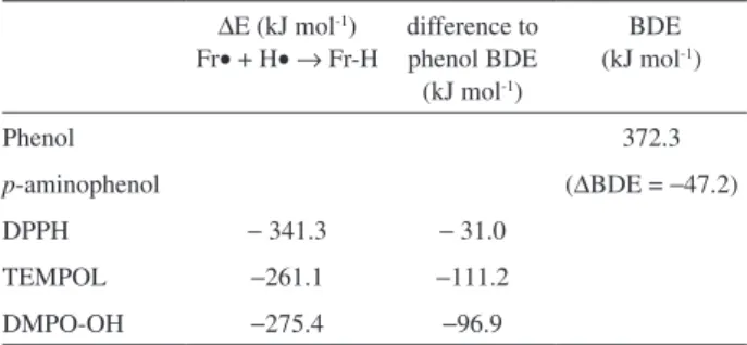
Table 3. Energies of reactions Fr· + H· → Fr-H and difference to phenol BDE
∆E (kJ mol-1)
Fr• + H•→ Fr-H
difference to phenol BDE (kJ mol-1)
BDE (kJ mol-1)
Phenol 372.3
p-aminophenol (∆BDE = −47.2)
DPPH − 341.3 − 31.0
TEMPOL −261.1 −111.2
The energy released in the reduction reaction was
−341.3 kJ mol-1 indicating the difficulty in reacting
with phenol and the relative easiness in reacting with
p-aminophenol, in accordance with experimental results.39
This result indicates that compounds with ∆BDE lower
than −31.0 kJ mol-1 can also react with DPPH. The low
IC 50 (Table 1) of quercetin, lavonoid widely studied,
agrees with this conclusion; the ∆BDE of quercetin ranges
between −122.5 kJ mol-1 and −19.1 kJ mol-1 depending on
the hydroxyl analyzed,40 and reacts quickly with DPPH.
TEMPOL and TEMPOL-H
The molecular structure of TEMPOL may be depicted in some different forms and in this study ive were simulated, changing the ring form from boat to chair and changing OH and NO positions. The optimal structure obtained with lower total energy is represented in Figure 3. The rearrangement of the TEMPOL molecule being reduced for TEMPOL-H, does not produce signiicant alterations. One alteration was the change of angles between C-N-O, having 115.5º in TEMPOL and 109.8º in its reduced form. This change may be due to the N-oxyl radical when producing a displacement in the electronic pairs, while in reduced
form it approaches NH3 form. Another important result is
the small change in the value of electric dipole, varying from 2.471 D in TEMPOL to 2.442 D in TEMPOL-H. This result indicates the possibility of the same afinity by HBA solvent, for TEMPOL and its respective hydroxylamine reduced, indicating the preferential HAT reaction pathway. Maybe this little difference is linked to the small dependence of rate constant with pH variations described
by Aliaga et al.14
The energy released in the reduction reaction of
TEMPOL is −261.1 kJ mol-1 (Table 3) indicating a greater
dificulty in reacting with antioxidant compounds than
the DPPH and agrees to its little use for this purpose.
Antioxidant compounds with ∆BDE lower than −111.2 kJ
mol-1 can react with TEMPOL considering only the
thermodynamic point of view. This value demonstrates that the study of antioxidant properties must be a different form than the DPPH scavenging test, expressing data by the use of an index different from IC 50 or in longer times. Despite these problems, the use of a different test assists a better study of antioxidant properties, especially with other solvents. In this study, the TEMPOL scavenging test was carried out in 24 h with higher concentrations of extracts and showed some different results than the DPPH scavenging test.
DMPO, DMPO-OH and DMPO-OH-H
In this study, the spin-adduct formation (DMPO-OH•)
and its neutralization for incorporation of the hydrogen atom in the structure was simulated. For the adduct formation,
the energy released in the reaction is −342.1 kJ mol-1 as
calculated from reaction DMPO + OH•→ DMPO-OH•.
Villamena et al.41 reported this value using
B3LYP/6-31+G(d,p)//B3LYP/6-31+G(d) level as −306.0 kJ mol-1,
and this difference may be due to an underestimation of the N-oxyl energy for the B3LYP method, according to the authors.
The energy released in adduct neutralization was
determined as −275.4 kJ mol-1 (Table 3), such value
indicates the necessity of ∆BDE being lower than
−96.9 kJ mol-1 so that one compound reacts to
DMPO-OH. Despite this higher value, it indicates higher reactivity than TEMPOL and lower than DPPH in agreement with calculated antioxidant activities. The reaction of adduct
neutralization with water formation (DMPO-OH• + H•
→ DMPO + H
2O) has also been tested; however the result
of −188.9 kJ mol-1 indicated that this way is less favorable.
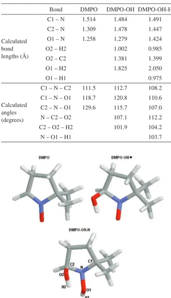
The variation of bond lengths and angles are indicated in Table 4, obtained from optimized structures of DMPO, DMPO-OH and DMPO-OH-H (Figure 4). The increase of
Figure 2. Optimized structure of DPPH-H obtained from computational
calculations.
Figure 3. Side view and top view of structural formula of TEMPOL-H
bond length of C2-N from 1.309 Å in DMPO to 1.478 Å in DMPO-OH is in agreement with the change from double to single bond. Another change is the decrease of N bond angles, which are close to the planar structure in DMPO and
close to the pyramidal NH3 structure in the DMPO-OH-H.
These results are in agreement with previous results41 and
again show the reliability of the method.
Conclusions
Ten plant extract samples from Brazilian lora were tested, in three different assays. All the extracts tested displayed signiicant antioxidant activity in at least one
test. The extracts from Iryanthera juruensis (leaf and seed)
presented the best results in DPPH and DMPO tests, while the
Chrysophyllum inornatum extract was the best in TEMPOL
Table 4. DMPO calculated bond lengths and angles
Bond DMPO DMPO-OH DMPO-OH-H
Calculated bond lengths (Å)
C1 – N 1.514 1.484 1.491 C2 – N 1.309 1.478 1.447 O1 – N 1.258 1.279 1.424
O2 – H2 1.002 0.985
O2 – C2 1.381 1.399
O1 – H2 1.825 2.050
O1 – H1 0.975
Calculated angles (degrees)
C1 – N – C2 111.5 112.7 108.2 C1 – N – O1 118.7 120.8 110.6 C2 – N – O1 129.6 115.7 107.0
N – C2 – O2 107.1 112.2
C2 – O2 – H2 101.9 104.2
N – O1 – H1 103.7
Figure 4. Structural formula of DMPO, DMPO-OH and DMPO-OH-H
obtained from computational calculations.
test. The TEMPOL scavenging test, despite the longer time of reaction, may indicate new antioxidant properties, mainly in water solution; and the differences in results indicate the importance of several tests and new studies for understanding the speciicity of each test. The differences between released energy of reduction reaction, obtained in computational calculations indicated that TEMPOL is less reactive than DPPH and justiied its restricted use. EPR spectroscopy technique proved to be an eficient and fast method for the comparative antioxidant activities, aiding in the selection of the most promising samples. Therefore, it is evident that similar studies are important in the case of a great variety of species such as for screening potential useful plants in bioprospection programs.
Acknowledgments
The Brazilian agencies FAPESP, CAPES and CNPq for partial inancial support; Carlos Brunello and Lourenço Rocha for technical support and to Angela Kinoshita for discussion about this work.
References
1. Rice-Evans, C. A.; Miller, N. J.; Paganga, G.; Free Radical Biol. Med.1996, 20, 933.
2. Calliste, C.; Trouillas, P.; Allais, D.; Simon, A.; Duroux, J.;
J. Agric. Food Chem.2001, 49, 3321.
3. Trouillas, P.; Calliste, C.; Allais, D.; Simon, A.; Marfak, A.; Delage, C.; Duroux, J.; Food Chem.2003, 80, 399.
4. McPhail, D.; Hartley, R. C.; Gardner, P. T.; Duthie, G. G.;
J. Agric. Food Chem.2003, 51, 1684.
5. Polovka, M.; Brezová, V.; Staško, A.; Biophys. Chem.2003,
106, 39.
6. Amarowicz, R.; Pegg, R. B.; Rahimi-Moghaddam, P.; Barl, B.; Weil, J. A.; Food Chem. 2004, 84, 551.
7. Kinoshita, A.; Franca, A. M.; Almeida, J. A. C.; Figueiredo, A. M.; Nicolucci, P.; Graeff, C. F. O.; Baffa, O.; Appl. Radiat. Isot.
2005, 62, 225.
8. Santos, A. B.; Rossi, A. M.; Baffa, O.; Appl. Radiat. Isot.2005,
62, 213.
9. Yordanov, N. D.; Christova, A. G.; Fresenius J. Anal. Chem.
1997, 358, 610.
10. Yordanov, N. D.; Ranguelova, K.; Spectrochim. Acta Part A
2000, 56, 373.
11. Yen, G.; Chen, H.; J. Agric. Food Chem.1995, 43, 27.
12. Valavanidis, A.; Nisiotou, C.; Papageorgiou, Y.; Kremli, I.; Satravelas, N.; Zinieris, N.; Zygalaki, H.; J. Agric. Food Chem.
2004, 52, 2358.
14. Aliaga, C.; Lissi, E. A.; Augusto, O.; Linares, E.; Free Radical Res.2003, 37, 225.
15. Kocherginsky, N. M.; Kostetski, Y. Y.; Smirnov, A. I.; J. Agric.
Food Chem.2005, 53, 1052.
16. Samuni, A.; Goldstein, S.; Russo, A.; Mitchell, J. B.; Krishna, M. C.; Neta, P.; J. Am. Chem. Soc.2002, 124, 8719.
17. Finkelstein, E.; Rosen, G. M.; Rauckman, E. J.; Arch. Biochem. Biophys.1980, 200, 1.
18. Figueiredo, M. S.; Baffa, O.; Neto, J. B.; Zago, M. A.; Res. Exp. Med. 1993, 193, 27.
19. Kohn, W.; Sham, L. J.; Phys. Rev. 1965, 140, A1133. 20. Litwinienko, G.; Ingold, K. U.; Acc. Chem. Res.2007, 40, 222.
21. Troullier, N.; Martins, J. L.; Phys. Rev. B1991, 43, 1993. 22. Ordejón, P.; Artacho, E.; Soler, J. M.; Phys. Rev. B 1996, 53,
441.
23. Ceperley, D. M.; Alder, B. J.; Phys. Rev. Lett. 1980, 45, 566.
24. Perdew, J. P.; Zunger, A.; Phys. Rev. B 1981, 23, 5048. 25. Silva, D. H. S.; Zhang Y; Santos, L. A.; Bolzani, V. S.; Nair,
M.; J. Agric. Food Chem. 2007, 55, 2569.
26. Fernandes, D. C.; Regasini, L. O.; Vellosa, J. C. R.; Bolzani, V. S.; Oliveira, O. M. M. F.; Silva, D. H. S.; Chem. Pharm. Bull.
2008, 56, 723.
27. Silva, D. H. S.; Pereira, F. C.; Zanoni, M. V. B.; Yoshida, M.;
Phytochemistry2001, 57, 437.
28. Marqui, S. R.; Lemos, R.; Santos, L. Á.; Castro-Gamboa, I.; Cavalheiro, A.; Bolzani, V. S.; Silva, D. H. S.; Scorzoni, L.; Giannini, M. J. S.; Young, M. C.; Torres, L. M.; Quim. Nova,
2008, 31, 828.
29. Magalhães, A. F.; Tozzi, A. M. G. D.; Santos, C. C; Magalhães, E. G.; J. Nat. Prod.2005, 68, 1290.
30. Burkitt, M. J.; Free Radical Res.Commun.1993, 18, 43. 31. Li, L.; Abe, Y.; Kanagawa, K.; Usui, N.; Imai, K.; Mashino, T.;
Mochizuki, M.; Miyata, N.; Anal. Chim. Acta2004, 512, 121. 32. Matthäus, B.; J. Agric. Food Chem. 2002, 50, 3444.
33. Jovanovic, S. V.; Steenken, S.; Tosic, M.; Marjanovic, B.; Simic, M. G.; J. Am. Chem. Soc.1994, 116, 4846.
34. Lucarini, M.; Mugnaini, V.; Pedulli, G. F.; Guerra, M.; J. Am. Chem. Soc. 2003, 125, 8318.
35. Brinck, T., Haeberlein, M., Jonsson, M.; J. Am. Chem. Soc.
1997, 119, 4239.
36. Lithoxoidou, A. T.; Bakalbassis E. G.; J. Phys. Chem. A 2005,
109, 366.
37. Lucarini, M.; Pedrielli, P.; Pedulli, G. F.; Cabiddu, S.; Fattuoni, C.; J. Org. Chem. 1996, 61, 9259.
38. Lind, J.; Shen, X.; Eriksen, T. E.; Merényi, G.; J. Am. Chem. Soc. 1990, 112, 479.
39. Brand-Williams, W.; Cuvelier, M. E.; Berset, C.; Food Sci. Technol. 1995, 28, 25.
40. Trouillas, P; Marsal, P.; Siri, D.; Lazzaroni, R.; Duroux, J.; Food Chem.2004, 97, 679.
41. Villamena, F. A.; Merle, J. K.; Hadad, C. M.; Zweier, J. L.;
J. Phys. Chem. A2005, 109, 6083.
Received: August 08, 2008
Web Release Date: August 28, 2009