cop
yr
ight
© ABE&M todos os direitos reser
v
ados
clinical case report
ILEANA G. S. RUBIO
ANA LUIZA GALRAO
VIVIANE PARDO
MEYER KNOBEL
ROBERTA F. POSSATO
ROSALINDA R. Y. CAMARGO
MARCELO A. FERREIRA
CRISTINA T. KANAMURA
SIMONE A. GOMES
GERALDO MEDEIROS-NETO
Thyroid Unit (LIM-25), Division of Endocrinology, University of São Paulo Medical School (IGSR, ALG, VP, MK, RFP, RRYC, GMN); Cell Biology (LIM59), Division of Pathology, University of São Paulo Medical School (MAF);
Adolfo Lutz Institute, São Paulo Public Health Service (CTK), São Paulo, SP, Brazil; Division of Endocrinology, Federal University of Sergipe Medical School (SAG), Aracajú, SE, Brazil.
Received in 2/9/2008 Accepted in 9/9/2008
ABSTRACT
Objective: To extend the molecular analysis of the IVS30+1G>T intronic thyro-globulin (TG) mutation, and to report the eleven year follow-up of the affected patients. Methods: Two siblings with severe congenital hypothyroidism with fetal and neonatal goiter, harboring the IVS30+1G>T mutation were included. Nodular and non-nodular thyroid tissue specimens were collected. Specifi c thyroid genes expression was evaluated by real-timePCR and by immunohis-tochemistry. Results: In non-nodular tissue specifi c thyroid genes mRNA were reduced when compared to normal thyroid sample. In the nodule, TPO and NIS expression was very low. Microscopic examinations showed very large follicu-lar-lumina and swollen vesicles of endoplasmatic-reticulum. Strong cytoplas-matic and low follicular-lumen TG immunostaining were detected. Intracellular NIS, membrane TPO and TSHR immunostaining had higher positivity in non-nodular sample. Both patients had a long-term adequate developmental out-come, besides one patient have been lately-treated. Conclusions: IVS30+1G>T mutation not only lead to very enlarge endoplasmatic-reticulum, but also to alterations of specifi c thyroid genes expression. The clinical evolution of pa-tients harboring these mutations strengthenthe concept of the infl uence of envi-ronment, like iodine nutrition, to determine the fi nal phenotypic appearance.
(Arq Bras Endocrinol Metab 2008; 52/8:1337-1344)
Keywords: Thyroglobulin; Congenital hypothyroidism; Gene mutations; Mo-lecular diagnosis; MoMo-lecular analysis
RESUMO
Análise Molecular e Acompanhamento a Longo Prazo de Dois Irmãos com Hipotireoidismo Congênito Portadores da Mutação Intrônica IVS30+1G>T no Gene da Tireoglobulina.
Objetivo: Aprofundar a análise molecular da mutação intrônica IVS30+1G>T do gene tireoglobulina (TG) e relatar a clínica de pacientes portadores da mutação, acompanhados por 11 anos. Métodos: Foram estudados dois irmãos com hipotireoidismo congênito grave com bócio fetal e bócio neonatal, portadores da mutação IVS30+1G>T. Foram coletadas amostras de tecido nodular e não-nodular. Avaliou-se a expressão de genes específi cos da tireóide por PCR em tempo real e imunohistoquímica. Resultados: A expressão de genes específi -cos da tireóide foi menor no tecido não-nodular que no tecido normal con-trole. Expressões de TPO e NIS foram extremamente baixas no tecido nodular. Verifi cou-se lúmen folicular aumentado com grandes vesículas de retículo en-doplasmático, e detectou-se forte marcação de TG no citoplasma e fraca no lúmen folicular. No tecido não-nodular observou-se forte positividade de NIS intracelular e, TPO e TSHR na membrana plasmática. O acompanhamento em longo prazo dos pacientes mostrou adequado desenvolvimento, apesar de um deles ter recebido tratamento tardio. Conclusões: A mutação IVS30+1G>T não só promove alterações no retículo endoplasmático, como alterações na ex-pressão de genes específi cos da tireóide. A evolução clínica destes pacientes reforça o conceito da infl uência do meio ambiente, como o aporte nutricional de iodo, no fenótipo fi nal. (Arq Bras Endocrinol Metab 200+8; 52/8:1337-1344)
cop
yr
ight
© ABE&M todos os direitos reser
v
ados
INTRODUCTION
C
ongenital hypothyroidism (CH) is the most com-mon endocrine disease in infancy, with a frequency, approximately, of 1/3000 live births (1). In 85% of pa-tients, the disorder is associated with thyroid develop-ment (dysgenesis), and 15%, harbor inborn errors in thyroid hormone synthesis (dyshormonogenesis) (2). Dyshormonogenesisis transmitted as a classical autoso-mal recessive mendelian trait and the clinical spectrum of the resulting phenotypes ranges from mild to severe goitrous hypothyroidism (3). Thyroglobulin (TG) is a large glycoprotein synthesized by the thyroid gland, and functions as a matrix for thyroid hormone synthesis (4). Thirty-eight inactivating mutations have been identifi ed, characterized in the human TG gene and as-sociated to congenital goiter and hypothyroidism (5). We have previously identifi ed the intronic IVS30+1G>T mutation in two Brazilian families with a complex his-tory of fetal goiter and congenital goiter, born to con-sanguineous parents (6-8). In this report we have extended our initial molecular and immunlogic studies of two affected goitrous siblings with defective TG syn-thesis and we describe the eleven year follow-up of the two siblings harboring this mutation.PATIENTS
We study two siblings with congenital hypothyroidism due to a defective thyroglobulin synthesis and secre-tion. They were born in Aracajú, coastal city of the nor-theast of Brazil, to a consanguineous parents (second degree cousins).
Patient 1 (AJM) was born in 1990 before the era of mandatory national neonatal screening for congenital hypothyroidism in Brazil. At birth he presented neona-tal goiter, without other typical signal of congenineona-tal hy-pothyroidism. He was referred to an endocrinologic appointment after birth but further investigations were not performed. Unfortunately early medical records were not available. His mother informed that during the fi rst years of live the patient was very calm and she suspected of a delay in development. Congenital hypo-thyroidism was fi nally diagnosed at 3 years of age. The child had a good compliance to daily thyroxin treat-ment. Clinical data and thyroid function tests are shown in Table 1. Bone age was retarded (2yrs 8m) for his chronological age of 5yrs 9m old. At puberty, bone age was similar to chronological age (Table 1). Echograph-ic studies of thyroid gland indEchograph-icated an enlarged gland at 6yrs old, and erroneously diagnosed as “chronic thy-roiditis” (hypoechogeinity). Anti-TPO antibodies were persistently negative. At 12 yrs of age intellectual and somatic development was considered as normal. Pu-berty (Tanner V) was completed at 14a 5m. During the fi rst years of elementary school he had poor school per-formance. As an young adult however, he is fi nishing the regular high school program and is considering to apply for college. During a recent medical examination he had an athletic appearance (heith 1.76m), with a very good verbal communication, talked enthusiasti-cally about his future perspectives, and gave the impres-sion of a normal 17 yrs old adolescence (Figure 1). At that age the volume of the gland was 26.1mL (goiter). Thyroidectomy was indicated due to the presence of two solid nodules (2.7 X 1.5 X 2.0 cm and 1.3 X 1.0 X 1.0 cm), respectively in the right and left lobes.
Patho-Table 1. Laboratory data, bone age and chronological age of patient 1.
Chronological age
(years, months) TSH (mU/L) (pmol/L)Free T4 (nmol/L)TotalT3
TG (ug/L)
Bone age (year, month)
Height (cm)
Weight (kg)
Newborn – – – – – 52.0 3.75
5y 9m – – – – 2y 8m – –
6y 3m 0.9 1.9 2.8 <0.5 – – –
10a 5m 0.012 2.1 – 0.1 – 144.5 39.4
12y 8m 1.12 1.5 1.61 – 11y – –
13y 8m 3.23 1.6 – – 13y 6m 161.5 53.5
14y 7m 10.08 0.93 1.38 – 13y 170.5 73.6
15y 7m 0.218 1.49 1.69 <0.2 17y – –
16a 8m 1.152 1.41 1.44 – – 175.0 65.8
17y 9m 1.74 1.22 – <0.5 18y 176.5 –
cop
yr
ight
© ABE&M todos os direitos reser
v
ados
logical diagnosis was suggested of dyshormonogenetic goiter. Both nodules were benign adenomas.
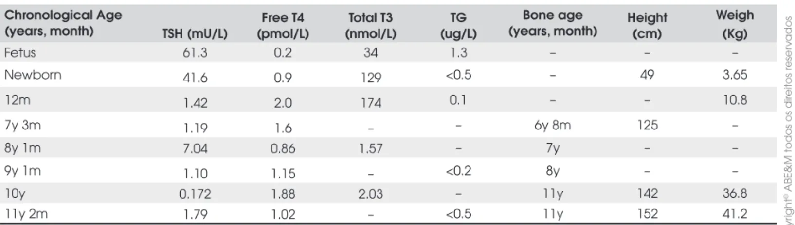
Patient 2 the younger sister of Patient 1 was born in 1997. She had a large fetal goiter (12.7mL) revealed by US of her mother at 26 weeks of gestation. Fetal hypothyroidisms was confi rmed by cordocentesis (TSH 61.3mU/L; TG 1.3mg/L) (9). After 4 weeks of a sin-gle intra-amniotic injection of 400μg L-thyroxine, a marked reduction of the goiter volume was confi rmed by ultrasound (from 12.3mL to 4.8mL). She received 12.0ug LT4/kg weight. Clinical data and thyroid func-tion tests are shown in Table 2. When she was 7 years old marked hypoechogeinity of the thyroid gland was diagnosed by ultrasonographic examination, with neg-ative anti-TPO antibodies. She had a normal somatic and intellectual development. Bone age was according with chronological age. Menarche was at 11yrs 4m of age followed by regular cycles (Figure 1).
Absence of synthesis and secretion of TG was con-fi rmed in both siblings by lack of serum TG increment 24 and 48 hours after stimulation of 0.1mg intramus-cular injection of recombinant human TSH (rhTSH). Molecular studies of the thyroglobulin gene mutations identifi ed the intronic mutation IVS30+1G>T in both patients (8). Both siblings had elevated urine iodine ex-cretion (respectively, 539 ug/L, and 492ug/L) con-fi rming a relatively high content in the diet. Both parents are euthyroid without history of thyroid disease, and harbored the heterozygous form of the intronic mutation.
METHODS
Thyroid functions tests
Serum total T4 (TT4), serum total T3 (TT3), serum TSH and serum TG levels were determined by electro-chemiluminescence immunoassays (Roche Corpora-tion, IN, USA).
Tissue samples
Nodular and non-nodular thyroid tissue specimens were collected from Patient 1 that was submitted to total thyroidectomy. One fragment of the specimen was immediately frozen in liquid nitrogen. Others were kept in formalin for immunohistochemistry and in 2.5% glutaraldehyde for electron microscopy. We also used RNA sample from a normal human thyroid tissue (con-trol tisuue).
Table 2. Laboratory data and chronological and bone age of the patient 2.
Chronological Age
(years, month) TSH (mU/L)
Free T4 (pmol/L)
Total T3 (nmol/L)
TG (ug/L)
Bone age (years, month)
Height (cm)
Weigh (Kg)
Fetus 61.3 0.2 34 1.3 – – –
Newborn 41.6 0.9 129 <0.5 – 49 3.65
12m 1.42 2.0 174 0.1 – – 10.8
7y 3m 1.19 1.6 – – 6y 8m 125 –
8y 1m 7.04 0.86 1.57 – 7y – –
9y 1m 1.10 1.15 – <0.2 8y – –
10y 0.172 1.88 2.03 – 11y 142 36.8
11y 2m 1.79 1.02 – <0.5 11y 152 41.2
–: not evaluated; references values: TSH: 0.5 – 4.0 mU/L); free T4: 11-25 (pmol/L); total T3: 1.2-3.1 nmol/L; TG: 0.5-15.0 mg/L.
cop
yr
ight
© ABE&M todos os direitos reser
v
ados
Gene expression quantifi cation by real-time PCR
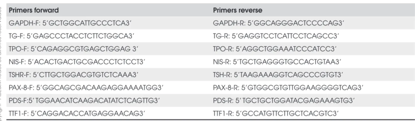
Total RNA was isolated using Trizol LS (Gibco BRL, Life Technologies, Gaithersburg, MD) and cDNA was synthesized with Super Script III RNA-H reverse tans-criptase (Invitrogen, Carlsbad, EUA). We quantifi ed gene expression of TG, Sodium-iodine symporter (NIS), thyroperoxidase (TPO), TSH receptor (TSHR), pendrin (PDS), thyroid transcription factor 1 (TTF1) and paired box transcription factor 8 (PAX-8) using Absolute QPCR SYBR®Green Mix (Abgene, Surrey, United Kingdom) in Rotor-Gene 3000 equipment (Corbett Research, Mortlake, Australia). We also quan-tifi ed the expression of GAPDH as internal control. PFAFFL (10) method was used to calculate gene ex-pression, reported as relative arbitrary units (AU). We used the expression in normal control tissue as calibra-tor sample in the PFAFFL formula, therefore expres-sion value of all the genes in normal control was 1AU. The intron spanning primers were describe in Table 3.
Immunohistochemistry analysis
Paraffi n-embedded tissue samples were stained by im-munoperoxidase (11) with primary NIS antibody (FP5A, Mayo Clinic, Rochester, MN), TSH receptor, TPO and TG antibodies (DakoCytomation, Dako, Glostrup, Denmark). Amplifi cation step was performed with Dako EnVision System, Peroxidase Kit (Dako, Glostrup, Den-mark). An immunostaining score was given according to the percentage of follicular cells with protein positive staining as follows: 0 (0%), 1+ (low, 1%-20%), 2+ (mode-rate, 21%-49%) and 3+ (high, ≥ 50%).
This work was approved by the Ethical Committee of the Hospital das Clínicas, University of São Paulo
Medical School, and was conducted according to the Helsinki Declaration. Informed consent was obtained from the parents of the patients.
RESULTS
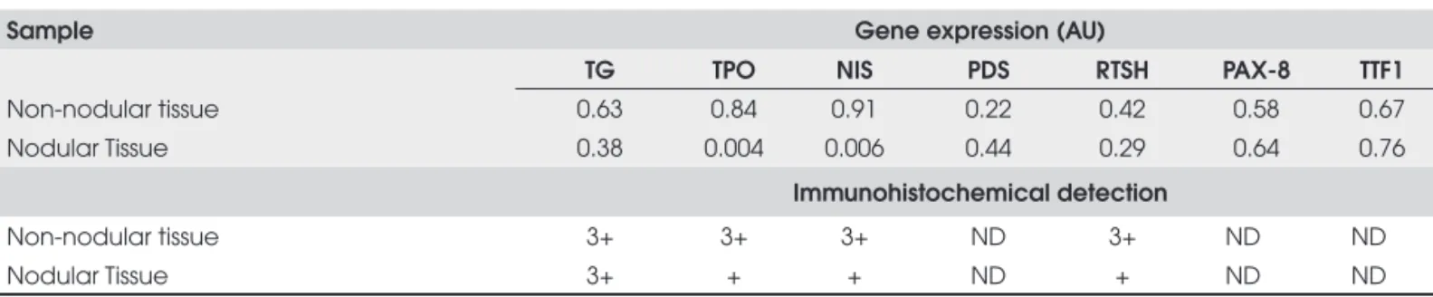
To determine whether TG and other important thyroid genes were expressed properly in the goitrous tissue of the affected patient, we performed mRNA quantifi ca-tion and immunohistochemical analysis.
Gene expression quantifi cation
We quantifi ed mRNA concentrations of TG, NIS, TPO, TSHR, PDS, TTF1 and PAX-8 genes in nodu-lar and non-nodunodu-lar thyroid samples from Patient 1 and from a normal control thyroid tissue. Messenger RNA of all genes was detected in all tissue samples (Table 4). In non-nodular tissue TG mRNA was redu-ced 37% as compared to control thyroid tissue. Similar results were observed for transcription factors PAX-8 (42%) and TTF-1 (33%), and for the TSHR (58%). NIS and TPO expression was slightly reduced when compared to control tissue (9% and 15% of reduction, respectively) and the lowest level was of PDS gene (78% of reduction).
When we compared nodular and non-nodular tis-sues TG, TSHR, TPO and NIS expression was reduced (Table 4, Figure 2). Even more, TPO and NIS expres-sion was about 150 and 230 times lower, respectively. On the other hand, higher expression of PDS (2-fold), and similar expression of PAX-8 and TTF1 genes were observed in nodular as compared to non-nodular tis-sues (Table 4, Figure 2).
Table 3. Real time PCR primers.
Primers forward Primers reverse
GAPDH-F: 5’GCTGGCATTGCCCTCA3’ GAPDH-R: 5’GGCAGGGACTCCCCAG3’
TG-F: 5’GAGCCCTACCTCTTCTGGCA3’ TG-R: 5’GAGGTCCTCATTCCTCAGCC3’
TPO-F: 5’CAGAGGCGTGAGCTGGAG 3’ TPO-R: 5’AGGCTGGAAATCCCATCC3’
NIS-F: 5’ACACTGACTGCGACCCTCTCCT3’ NIS-R: 5’TGCTGAGGGTGCCACTGTAA3’
TSHR-F: 5’CTTGCTGGACGTGTCTCAAA3’ TSH-R: 5’TAAGAAAGGTCAGCCCGTGT3’
PAX-8-F: 5’GGCAGCGACAAGAGGAAAATGG3’ PAX-8-R: 5’GTGGCGTGTTGGAAGGGGTCAG3’
PDS-F:5’ TGGAACATCAAGACATATCTCAGTTG3’ PDS-R: 5’ TGCTGCTGGATACGAGAAAGTG3’
cop
yr
ight
© ABE&M todos os direitos reser
v
ados
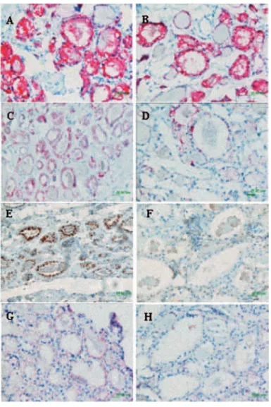
Thyroid histological, electron microscopy and immunohistochemical analysis
Light microscopic examination of nodular and non-nodular thyroid tissue stained with hematoxilin and eo-sin demonstrated that the follicle lumina were enlarged and devoid of colloid. Electron microscopy identifi ed cytoplasmatic swollen vesicles of endoplasmatic reticu-lum (ER) in both samples (Figure 3), in contrast to normally more fl attened tubular ER (12).
We performed immunohistochemical detection of TG, NIS, TSHR and TPO proteins. We detected strong (3+) cytoplasmatic TG immunostaining in non-nodular and non-nodular sample (Table 4). By contrast, very low TG protein was present in the follicular lu-men (Figure 3) as opposed to normal thyroid tissue without TG defect (12).
On the other hand, non-nodular sample NIS, TPO and TSHR proteins had higher (+3) positivity when compared with nodular (+1) (Table 4) (Figure 4).
In nodular and non-nodular samples NIS protein localization was intracellular, TPO protein was
detec-Table 4. mRNA quantifi cation of TG, TSHR, NIS, PAX-8, TTF1 and PDS and Immunohistochemical analysis of TG, TSHR, NIS proteins in nodular and non-nodular thyroid samples from patient 1.
Sample Gene expression (AU)
TG TPO NIS PDS RTSH PAX-8 TTF1
Non-nodular tissue 0.63 0.84 0.91 0.22 0.42 0.58 0.67
Nodular Tissue 0.38 0.004 0.006 0.44 0.29 0.64 0.76
Immunohistochemical detection
Non-nodular tissue 3+ 3+ 3+ ND 3+ ND ND
Nodular Tissue 3+ + + ND + ND ND
The normal control tissue was the calibrator sample in the FAFL formula, therefore expression value of all the genes in normal control was 1AU; positive protein staining:
1+ = low positive staining (1%-20%), 2+ moderate positive staining (21%-49%) and 3+ high positive staining (≥ 50%); ND: not done.
Figure 3. Electron microscopy of thyroid tissues from Patient 1, A) Non-nodular thyroid sample; B) nodular thyroid sample. Ar-rows indicate the large vesicles of endoplasmatic reticulum.
ted in the apical membrane and TSHR protein had membrane localization (Figure 4).
DISCUSSION
Defective thyroglobulin synthesis usually results in goi-trous congenital hypothyroidism. In the present study we have extended the molecular analysis of the IVS30+ 1G>T TG gene mutation and the clinical case of two siblings with congenital hypothyroidism due to thyro-globulin synthesis defect. They were born from con-sanguineous parents. The analysis of the complete coding sequence of TG gene and intro/exon borders identifi ed the intronic homozygous IVS30+1G>T mu-tation in both patients (8). The parents harbored the form of this mutations in heterozygous state.
Cases of thyroid carcinoma developing from dysor-monogenic goiters have been reported (13,14). Mo-reover, high incidence of thyroid cancer was associated with long-standing goiters with thyroglobulin muta-tions (15). Therefore, Patient 1 with the presence of two solid thyroid nodules underwent total
thyroidec-1 0,9 0,8 0,7 0,6 0,5 0,4 0,3 0,2 0,1 0
TG RTSH NIS TPO PAX-8 TTF 1 PDS Patient non nodular tissue Patient nodular tissue
Gene Espression (UA)
cop
yr
ight
© ABE&M todos os direitos reser
v
ados
tomy. The pathologic diagnosis of both nodules was benign adenoma.
Intronic mutations of the TG gene with the func-tional consequence of skipping of an entire exon are not rare in congenital hypothyroid patients (16). The IVS30+1G>T mutation is caused by guanine to thymi-ne transversion at position +1 in the donor splice site of intron 30. This mutations were previously identifi ed by our group in other two siblings from a not related fami-ly from the Northeast of Brazil (6). It was also confi r-med the compound heterozygous constellation IVS30+1G>T/A2215D in the two fi rst degree cousins
of the siblings patients of the present study (8). This intronic mutation promotes aberrant splicing and loss of 138 nucleotides of the TG mRNA, removing the entire exon 30 (6,7). Elimination of this exon does not affect the reading frame of the mRNA and potentially codifi es a shortened polypeptide. The deletion is locali-zed in the TG type III repeat domain, causing the loss of 1- putative N-linked glycosylation site (5,17). The loss of 46 aminoacids can modify the tertiary and qua-ternary structure of the protein. However, silico studies
of the mutant protein are not possible because the crys-tallographic structure of complete TG molecule is not available.
Electron microscopy confi rmed the presence of dis-tended endoplasmatic reticulum (ER) both in nodular and non-nodular thyroid samples from patient 1. These results are a consequence of the mutant TG protein re-tention inside the ER (12). Properly folded not mutant TG dimmers migrates from ER to the Golgi apparatus where glycosilation occurs (18). On the other hand, un-folded mutated protein activated the mechanism of quality control of the ER, mediated by a massive induc-tion of specifi c ER molecular chaperones including the hsp90 homolog, GRP94, and the hsp70 homolog, BiP, reducing the export of the protein to the colloid and causing thyroid ER storage disease (12). Immunohisto-chemical analysis confi rmed the defective TG traffi c both in the non-nodular tissue as well as in the nodule. In both tissues there was a marked decrease of reaction product in the follicular lumina and concomitant accu-mulation of intracellular staining. The undetectable le-vels of TG after rhTSH (8) lead us to speculate that an acute stimulation with TSH may not be enough to pass through the ER blockade, whilst that the few molecules that reach the colloid are immediately hydrolyzed after internalization into the thyroid cell.
We were able to detect the mRNA expression of the specifi c thyroid genes: TG, TPO, NIS, TSHR, TTF1, PAX-8 and PDS in the Patient 1 thyroid tissues, albeit the levels were reduced when compared with normal thyroid tissue. These data may be related to the functional state of the gland. The mRNA data of TG, TPO, NIS and TSHR were in agreement with immu-nohistochemistry evaluation of protein expression.
In nodule sample the expression levels of the va-rious genes varied widely. TG, TSHR expression was reduced as compare to non-nodular. Furthermore, the highest reduction were those of NIS and TPO expres-sion. NIS expression reduction without decrease of
cop
yr
ight
© ABE&M todos os direitos reser
v
ados
TPO expression levels in benign adenomas have been previously described (19-20). Indeed, in the present study, the expression of NIS and TPO were 150 and 230-fold lower, respectively, in the nodule when com-pared with non-nodular or normal thyroid tissue. The-se are two of the key proteins known to regulate iodide uptake and intrathyroid metabolism (20). Consequen-tly, these fi ndings may be related to defects in both the iodine-trapping ability and the iodination process in the nodule. Both physiologic functions were less pro-nounced in the non-nodular tissue.
Our results indicated that PAX-8, TTF1 and PDS gene expression were still preserved in both nodular and non-nodular samples, however the levels were re-duced as compared to normal sample. Previous report have shown that PDS gene and its product (pendrin) expression levels appeared to be similar in most hypo-functioning adenomas, whereas more than a 3-fold de-crease was observed in other samples (21,22).
Transcription factors TTF1 an PAX-8 are involved in thyroid development and in the regulation of the expression of specifi c thyroid genes (TPO, TG, NIS, TSHR, PDS) (23-29) Altogether, our fi ndings of simi-lar and signifi cant expression of PAX-8 and TTF1 in nodular and non-nodular tissue with very low expres-sion of NIS and TPO exclusively in the nodule, suggest that different mechanisms are controlling the expres-sions of these gene in these tissues.
Our immunostaining analysis indicated membrane localization of TSHR and TPO, and the intracellular localization of NIS proteins. Previous reports have sho-wn predominantly intracellular localization of NIS pro-tein in thyroid tumors (30-32).
Combined, the results of this study and the pre-viously reported molecular analysis (6,7) have proved that IVS30+1G>T TG mutation promotes a severe thyroid hormone synthesis defect with fetal or neonatal goitrous CH (elevated fetal or neonatal TSH and low TG values) (8). On the clinical side, as expected, the three years post birth of Patient 1 without L-thyroxine therapy possibly caused a mild neurological consequen-ces. This was confi rmed by the retarded bone age and early intellectual delay. Whilst after a clinical long-term follow-up (eleven years) this patient appears to be a normal adolescent with an actual normal height at 17 years old (1.76m) and presumably good school perfor-mance, due to an effective LT4 treatment. We have re-asons to believe that elevated iodine nutrition would
eventually determine this fi nal phenotypic appearance (8). In the presence of a high iodine supply, the thyroid gland would be able to overcome the severe genetic disease and induce generation of some thyroid hormo-ne, preventing more serious neurological damage (33,34). Urinary iodine excretion in the affected siblin-gs were elevated, indicating that they still are in a rela-tively high nutritional iodine environment. On the other hand, patient 2 who have received prenatal treat-ment of fetal hypothyroidism and daily LT4 replace-ment after birth, have no symptoms of CH during the clinical follow-up.
In conclusion the IVS30+1G>T not only leads to a severe intracitoplasmatic alterations, a very enlarge en-doplasmatic reticulum, but also to the alteration of mRNA expression of specifi c thyroid genes in the nod-ular and non-nodnod-ular goitrous tissues. The clinical evo-lution of patients harboring this mutations strengthen the concept of the infl uence of the environment, like iodine nutrition, to determine the fi nal phenotypic ap-pearance.
Acknowledgments: We acknowledge the fi nancial support of Fa-pesp (grant 03/02989-8) and Instituto da Tiróide, São Paulo, Brazil. No other potencial confl ict of interest relevant to this article was report.
REFERENCES
1. Loeber JG. Neonatal screening in Europe: the situation in 2004. J Inherit Metab Dis. 2007;30(4):430-8.
2. Knobel M, Medeiros-Neto G. An outline of inherited disorders of the thyroid hormone generating system. Thyroid. 2003;13(8): 771-801.
3. Caputo M, Rivolta CM, Esperante SA, Gruñeiro-Papendieck L, Chiesa A, Pellizas CG, et al. Congenital hypothyroidism with goitre caused by new mutations in the thyroglobulin gene. Clin Endocrinol. 2007;67(3):351-7.
4. Ieiri T, Cochaux P, Targovnik HM, Suzuki M, Shimoda S, Perret J, et al. A 3′ splice site mutation in the thyroglobulin gene res-ponsible for congenital goiter with hypothyroidism. J Clin In-vest. 1991;88:1901-5.
5. Rivolta CM, Targovnik HM. Molecular advances in thyroglobu-lin disorders. Cthyroglobu-lin Chim Acta. 2006;374(1-2):8-24.
6. Targovnik H, Vono J, Billerbeck AEC, Cerrone GE, Varela V, Mendive F, et al. A 138-nucleotide deletion in the thyroglobulin ribonucleic acid messenger in a congenital goiterwith defecti-ve thyroglobulin synthesis. J Clin Endocrinol Metab. 1995;80: 3356-60.
cop
yr
ight
© ABE&M todos os direitos reser
v
ados
8. Pardo V, Rubio IG, Knobel M, Aguiar-Oliveira MH, Santos MM, Gomes SA, et al. Phenotypic variation among four family members with congenital hypothyroidism caused by two dis-tinct thyroglobulin gene mutations. Thyroid. 2008;18(7):783-6. 9. Medeiros-Neto G, Bunduki V, Tomimori E, Gomes S, Knobel
M, Martin RT, et al. Clinical case seminar: Prenatal Diagnosis and Treatment of Dyshormonogenetic Fetal Goiter Due to De-fective Thyroglobulin Synthesis. J Clin Endocri Metab. 1997;82:4239-42.
10. Pfaffl MW, Tichopad A, Prgomet C, Neuvians TP. Determination of stable housekeeping genes, differentially regulated target genes and sample integrity: BestKeeper--Excel-based tool using pair-wise correlations. Biotechnol Lett. 2004;26(6):509-15. 11. Castro MR, Bergert ER, Beito TG, McIver B, Goellner JR,
Mor-ris JC. Development of monoclonal antibodies against the hu-man sodium iodide symporter: immunohistochemical charac-terization of this protein in thyroid cells. J Clin Endocrinol Metab. 1999;84:2957-62.
12. Medeiros-Neto G, Kim PS, Yoo SE, Vono J, Targovnik HM, Ca-margo R, et al. Congenital hypothyroid goiter with defi cient thyroglobulin. Identifi cation of an endoplasmic reticulum sto-rage disease with induction of molecular chaperones. J Clin Invest. 1996;98:2838-44.
13. Camargo R, Limbert E, Gillam M, Henriques MM, Fernandes C, Catarino AL, et al. Aggressive metastatic follicular thyroid car-cinoma with anaplastic transformation arising from a long-standing goiter in a patient with Pendred’s syndrome. Thyroid. 2001;11(10):981-8.
14. Alzahrani AS, Baitei EY, Zou M, Shi Y. Clinical case seminar: metastatic follicular thyroid carcinoma arising from congenital goiter as a result of a novel splice donor site mutation in the thyroglobulin gene. J Clin Endocrinol Metab. 2006;91(3):740-6. 15. Hishinuma A, Fukata S, Kakudo K, Murata Y, Ieiri T. High inci-dence of thyroid cancer in long-standing goiters with thyro-globulin mutations. Thyroid. 2005;15(9):1079-84.
16. Vono-Toniolo J, Rivolta CM, Targovnik HM, Medeiros-Neto G, Kopp P. Naturally occurring mutations in the thyroglobulin gene. Thyroid. 2005;15(9):1021-33.
17. van de Graaf SA, Ris-Stalpers C, Pauws E, Mendive FM, Targo-vnik HM, de Vijlder JJ. Up to date with human thyroglobulin. J Endocrinol. 2001;170(2):307-21.
18. Yang SX, Pollock HG, Rawitch AB. Glycosylation in human thyroglobulin: location of the N-linked oligosaccharide units and comparison with bovine thyroglobulin. Arch Biochem Biophys. 1996;327(1):61-70.
19. Bruno R, Ferretti E, Tosi E, Arturi F, Giannasio P, Mattei T, et al. Modulation of thyroid-specifi c gene expression in normal and nodular human thyroid tissues from adults: an in vivo effect of thyrotropin. J Clin Endocrinol Metab. 2005;90(10):5692-7. 20. Lazar V, Bidart JM, Caillou B, Mahé C, Lacroix L, Filetti S, et al.
Expression of the Na+/I- symporter gene in human thyroid tu-mors: a comparison study with other thyroid-specifi c genes. J Clin Endocrinol Metab. 1999;84(9):3228-34.
21. Lacroix L, Michiels S, Mian C, Arturi F, Caillou B, Filetti S, et al. HEX, PAX-8 and TTF-1 gene expression in human thyroid tis-sues: a comparative analysis with other genes involved in io-dide metabolism. Clin Endocrinol (Oxf). 2006;64(4):398-404. 22. Mian C, Lacroix L, Alzieu L, Nocera M, Talbot M, Bidart JM, et
al. Sodium iodide symporter and pendrin expression in hu-man thyroid tissues.Thyroid. 2001;11(9):825-30.
23. Ohno M, Zannini M, Levy O, Carrasco N, di Lauro R. The pai-red-domain transcription factor Pax8 binds to the upstream enhancer of the rat sodium/iodide symporter gene and partici-pates in both thyroid-specifi c and cyclic-AMP-dependent transcription. Mol Cell Biol. 1999;19(3):2051-60.
24. Dentice M, Luongo C, Elefante A, Ambrosio R, Salzano S, Zan-nini M, et al. Pendrin is a novel in vivo downstream target gene of the TTF-1/Nkx-2.1 homeodomain transcription factor in diffe-rentiated thyroid cells. Mol Cell Biol. 2005;25(22):10171-82. 25. Akagi T, Luong QT, Gui D, Said J, Selektar J, Yung A, et al.
Induc-tion of sodium iodide symporter gene and molecular characte-risation of HNF3beta/FoxA2, TTF-1 and C/EBPbeta in thyroid carcinoma cells. Br J Cancer. 2008 [Epub ahead of print]. 26. Kambe F, Seo H. Thyroid-specifi c transcription factors. Endocr
J. 1997;44(6):775-84.
27. Damante G, Tell G, Di Lauro R. A unique combination of trans-cription factors controls differentiation of thyroid cells. Prog Nucleic Acid Res Mol Biol. 2001;66:307-56.
28. Zannini M, Francis-Lang H, Plachov D, Di Lauro R. Pax-8, a paired domain-containing protein, binds to a sequence overla-pping the recognition site of a homeodomain and activates transcription from two thyroid-specifi c promoters. Mol Cell Biol. 1992;12(9):4230-41.
29. Damante G. Thyroid defects due to Pax8 gene mutations. Eur J Endocrinol. 1998;139(6):563-6.
30. Gérard AC, Daumerie C, Mestdagh C, Gohy S, De Burbure C, Costagliola S, et al. Correlation between the loss of thyroglo-bulin iodination and the expression of thyroid-specifi c pro-teins involved in iodine metabolism in thyroid carcinomas. J Clin Endocrinol Metab. 2003;88(10):4977-83.
31. Sodré AK, Rubio IG, Galrão AL, Knobel M, Tomimori EK, Alves VA, et al. Association of Low Sodium-Iodide Symposter mRNA Expression in Malignant Thyroid Nodules with Increased In-tracellular Protein Staining. J Clin Endocrinol Metab. 2008 [Epub ahead of print].
32. Dohan O, Baloch Z, Banrevi Z, Livolsi V, Carrasco N. Predomi-nant intracellular overexpression of the Na(+)/I(-) symporter (NIS) in a large sampling of thyroid cancer cases. J Clin Endo-crinol Metab. 2001;86:2697-700.
33. Vono J, Lima N, Knobel M, Medeiros-Neto G. The effect of oral administration of iodine to patients with goiter and hypo-thyroidism due to defective synthesis of thyroglobulin. Thyroid. 1996;6(1):11-5.
34. Piosik PA, van Groenigen M, van Doorn J, Baas F, de Vijlder JJ. Effects of maternal thyroid status on thyroid hormones and growth in congenitally hypothyroid goat fetuses during the second half of gestation. Endocrinology. 1997;138(1):5-11.
Correspondence to:
Ileana G S Rubio.
Thyroid Unit - LIM 25, University of São Paulo Medical School Av. Dr. Arnaldo, 455 – 4A