cop
yr
ight
© ABE&M todos os direitos reser
v
ados
Received in 25/8/2008 Accepetd in 14/10/2008
clinical case report
**FERNANDA BORCHERSCOELI
**LÚCIO FÁBIO CALDAS FERRAZ
SOFIA H. V. DE LEMOS-MARINI
SUMARA ZUANAZI PINTO RIGATTO
VERA MARIA SANTORO BELANGERO
MARICILDA PALANDIDE-MELLO
Laboratório de Genética Molecular Humana, Centro de Biologia Molecular e Engenharia Genética, Universidade Estadual de Campinas (FBC, LFCF, MPM); Centro de Investigação em Pediatria, Faculdade de Ciências Médicas,
Departamento de Pediatria, Universidade Estadual de Campinas (SHVLM, SZPR, VMSB), Campinas, SP, Brasil.
** Share fi rst authorship
ABSTRACT
The apparent mineralocorticoid excess syndrome (AME) is a rare autosomal re-cessive disorder due to the defi ciency of 11β-hydroxysteroid dehydrogenase type 2 enzyme (11beta-HSD2). The 11beta-HSD2 enzyme, encoded by HSD11B2 gene, metabolizes active cortisol in cortisone. Mutations on HSD11B2 gene affect the enzyme activity by leading to an excess of cortisol, which causes its inappro-priate access to mineralocorticoid receptor. Therefore, cortisol will bind miner-alocorticoid receptor. The human HSD11B2 gene maps to chromosome 16q22 and consists of fi ve exons encoding a protein of 405 amino acids. We present here clinical and molecular studies on a Brazilian boy who was born pre-term after an oligodramnious pregnancy. He was diagnosed as having AME at the age of 26 months. His parents are second cousins. Molecular characterization of the HSD11B2 gene revealed the homozygous mutation p.R186C. The patient
de-scribed here is the second case of HDS11B2 gene mutation reported in Brazilian patients with AME. (Arq Bras Endocrinol Metab 2008; 52/8:1277-1281)
Keywords: Apparent mineralocorticoid excess; HSD11B2 gene; Hypertension; Defi ciency of 11β-hydroxysteroid dehydrogenase type 2; Mutations
RESUMO
Síndrome de Excesso Aparente de Mineralocorticóide em um Menino Brasileiro Causada pela Mutação p.R186C em Homozigose no Gene HSD11B2.
A síndrome de excesso aparente de mineralocorticóide (AME) é uma doença autossômica recessiva rara devido à defi ciência da enzima 11β-hidroxiesteróide desidrogenase tipo 2 (11beta-HSD2). A enzima 11beta-HSD2 metaboliza o cortisol ativo a cortisona. As mutações no gene HSD11B2, que codifi ca a en-zima, afetam sua atividade levando a um excesso de cortisol, que terá acesso inapropriado ao receptor de mineralocorticóide, competindo com a ligação da aldosterona. O gene HDS11B2 humano está localizado no cromossomo 16q22 e é formado por 5 éxons que codifi cam uma proteína de 405 aminoáci-dos. Este relato apresenta os estudos clínicos e moleculares de um paciente brasileiro do sexo masculino que nasceu prematuro depois de uma gestação sob oligodrâmnio. Recebeu o diagnóstico de AME com 26 meses de idade. Seus pais são primos em segundo grau. A caracterização molecular do gene HSD11B2 revelou a mutação p.R186C em homozigose. O paciente descrito é
o segundo caso relatado de brasileiro com mutação no gene HSD11B2. (Arq
Bras Endocrinol Metab 2008; 52/8:1277-1281)
Descritores: Excesso aparente de mineralocorticóide; Gene HDS11B2; Hiper-tensão; Defi ciência da11β-hidroxiesteróide desidrogenase tipo 2; Mutações
INTRODUCTION
hyperten-cop
yr
ight
© ABE&M todos os direitos reser
v
ados
sion caused by the defi ciency of 11β-hydroxysteroid dehydrogenase type 2 (11beta-HSD2) (1-3). This en-zyme is highly expressed in mineralocorticoid target tissues and functions unidirectionally to convert corti-sol to cortisone, an inactive form (4). Although corticorti-sol is a less potent mineralocorticoid than aldosterone, the main mineralocorticoid produced in the adrenal gland, both aldosterone and cortisol bind to the mineralocor-ticoid receptor (MR) with equal affi nity (5), whereas cortisone does not. Therefore, 11beta-HSD2 enzyme protects normal subjects from cortisol intoxication by converting it into cortisone and providing a mechanism that ensures the proper occupation of MR by aldoste-rone (6). As cortisol is secreted in concentrations much higher than aldosterone, the defi ciency of 11beta-HSD2 leads to an inappropriate inactivation of cortisol, which in excess, acts as a mineralocorticoid. This corti-sol excess stimulates the MR causing intense sodium retention, hypokalemia and hypertension (1-3).
Two isoforms of 11beta-HSD have been identifi ed and characterized (7). The type 1 isoform, which is ex-pressed in several human tissues, is a NADP-dependent enzyme with reductase activity (8, 9). The type 2 iso-form, which is only 20-26% identical to type 1, is NAD-dependent and presents dehydrogenase activity (10). The human HSD11B2 gene, encoding the
11beta-HSD2 enzyme, is located on chromosome 16q22, con-sists of 5 exons spanning about 6.2 Kb, and encodes a protein of 405 amino acids (11).
AME is a result of mutations in HSD11B2 gene
(12-15). In general, the disease is present in early child-hood, with a severe phenotype including low birth weight, failure to thrive, hypokalemic metabolic alkalo-sis, and high mortality rate in untreated patients (2, 16). However, milder phenotypes have been described in adults with clinical features such as hypertension without electrolyte abnormalities (17-20). Milder phe-notypes are associated with mutations causing only par-tial inactivation of HSD11B2 (21).
In this study, we report a boy with typical clinical features of AME with p.R186C homozygous mutation in the HSD11B2 gene.
SUBJECTS AND METHODS
Blood specimens and clinical data of the patient and relatives were collected with approval by the appropria-te institutional review board; signed informed consent was obtained.
Clinical data
A Brazilian male, son of a consanguineous marriage, was born pre-term (weight = 1,700 g; length = 40 cm), Apgar score 8-9, after an oligodramnious preg-nancy. He had normal male genitalia with palpable testes. His development was normal up to the age of 5 months when he had bronchiolitis. Thereafter he presented failure to thrive. When he was 17 months old he was admitted in a hospital for pneumonia when he presented polyuria, polydipsia, hypokalemia (se-rum potassium, 1.9 mmol/L) and hypertension. He was fi rst examined by us at the age of 26 months when he was referred to our hospital for evaluation because prednisone and chlorthalidone failed to con-trol his labile hypertension. At this time his weigh (8,580 g) and height (76.8 cm) were both below the third percentile (zW = - 3.32 and zH = - 2.53). He also presented hypertension (130/90 mmHg). Labo-ratorial fi ndings indicated hypokalemia (serum potas-sium, 2.5 mmol/L), alkalosis (pH = 7.51) and normal sodium (141 mmol/L), plasma renin activity (0.48 ng/ml/h, normal 0.15 to 2.33), aldosterone (26 pg/ ml, normal 10 to 160 pg/mL), 11-deoxycortisol (2.0 ng/mL, normal < 8.0 ng/mL) and serum cortisol (9.9 µg/dL, normal 7.58 to 27 µg/dL,). On 24-hour urine he presented normal cortisol (31.9 µg/24hs, normal 20 to 90 µg/24hs) and hypercalciuria (18 mg/kg/day, normal < 4 mg/kg/day). Unfortunate-ly, neither cortisol nor cortisone urinary metabolites could be evaluated.
Renal ultrasounds revealed the presence of bilateral nephrocalcinosis (not progressive) whereas cardiac echocardiography showed mild left ventricular hyper-trophy when he was 5 years old.
cop
yr
ight
© ABE&M todos os direitos reser
v
ados
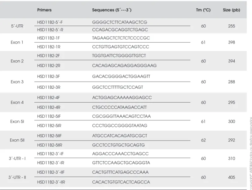
Table 1. Primers used for PCR HSD11B2 gene amplifi cation.
Primers Sequences (5´---3´) Tm (ºC) Size (pb)
5´-UTR HSD11B2-5´-F GGGGCTCTTCATAAGCTCG 60 255
HSD11B2-5´-R CCAGACGCAGGTCTGAGC
Exon 1
HSD11B2-1F TAGAAGCTCTCTCTCCCCGC
61 398
HSD11B2-1R CCTGTTGAGTGTCCAGTCCC
Exon 2
HSD11B2-2F TGGTGATTCTGGGGTTGTCT
60 394
HSD11B2-2R CACAGAGCAGAGGAGGGAAG
Exon 3
HSD11B2-3F GACACGGGGACTGGAAGTT
60 288
HSD11B2-3R GGCTCCTTTTTGCTCCAGT
Exon 4
HSD11B2-4F ACTGGAGCAAAAAGGAGCC
60 295
HSD11B2-4R CTGCCCCCATAAGACCATT
Exon 5I
HSD11B2-5IF CGCGGGTTAAACAGTCCTAA
61 300
HSD11B2-5IR CCCTGGCCGGGGTAATAG
Exon 5II
HSD11B2-5IIF ATGCCATCACAGATGCGCT
62 292
HSD11B2-5IIR GCCTCCTGTGCTGCAGTG
3´-UTR - I
HSD11B2-3´-IF AGGACCCAAACCTGAGCC
60 310
HSD11B2-3´-IR GTTCTCCAAGCTGCAGGGTA
3´-UTR - II
HSD11B2-3´-IIF CACTGTTTCATGAGCCCAAA
60 405
HSD11B2-3´-IIR CACACTGTGTCACTCAGCCA Molecular analysis
Genomic DNA was extracted from peripheral blood leukocytes by standard phenol/chloroform method (22). HSD11B2 gene was amplifi ed by PCR amplifi
ca-tion of the entire coding region including exon-intron junctions and both 5’UTR and 3’UTR regions using synthetic oligonucleotides (Invitrogen, CA, USA) as primers (table 1), which were designed using Primer 3 open access software (http://primer3.sourceforge. net). The amplifi ed fragments were directly sequenced using Big Dye TM Terminator Cycle Sequencing Kit V3.1 Ready Reaction (ABI PRISM / PE Biosystems, Foster City, CA, USA). The sequences obtained in an ABI377 Semi-Automated Sequencer (ABI PRISM / PE Biosystems) were compared to the normal sequence of the gene (NCBI # U27317).
RESULT
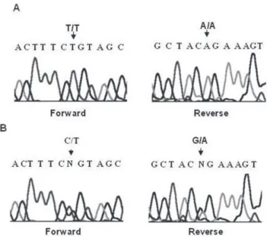
HSD11B2 sequence analysis on patient’s DNA revealed
a c.556C>T homozygous transition in codon 186 loca-ted in exon 3. This nucleotide substitution leads to the p.R186C missense mutation (Figure 1A). Further se-quence analysis on parent’s DNA samples showed hete-rozygosis for the mutation in both father and mother (Figure 1B).
DISCUSSION
featu-cop
yr
ight
© ABE&M todos os direitos reser
v
ados
are p.R186C carriers. Since this mutation was previ-ously described segregating in an African-American family (20), a founder effect should be considered. However, in the present case it is diffi cult to explain the mutation recurrence by considering a founder effect of an African-derived disease-causing allele without ana-lyzing molecular markers, which could give an estima-tive of African ancestry index for this family as defi ned by Parra and cols. (29). Therefore, we cannot discard
the possibility of this mutated allele being an African-originated allele.
Conversely, the C>T transition at codon 186 (CGT>TGT) is a typical CpG-consequence mutation.
This dinucleotide is considered to be prompt to un-dergo mutations through spontaneous deamination of cytidine to uracil (30, 31). Therefore, the occurrence of the p.R186C mutation in two unrelated families could suggest that the codon R186 is a hot spot for mutations in HSD11B2 gene, like other mutations
lo-cated in exons 3 to 5 (21, 32). However, the number of patients bearing the mutation is low, therefore there are no signifi cant evidences for this possibility unless more patients are studied.
In general there are few cases of AME reported worldwide, since this is a very rare autosomal recessive disorder (25, 27). The boy described in the present pa-per was clinically diagnosed with AME and molecular fi ndings confi rmed the phenotype. This is the second case of AME with a deleterious HDS11B2 gene
muta-tion reported in Brazilian patients (33).
Acknowledgements: We thank Maria M. Vasconcelos Rosa for assistance and technical support. We thank Conselho Nacional de Desenvolvimento Científi co e Tecnológico (CNPq) and Coorde-nação de Aperfeiçoamento de Pessoal de Nível Superior (CA-PES) for fi nancial support. The authors would also like to thank the family studied for agreeing to participate in this research.
REFERENCES
1. White PC, Mune T, Agarwal AK. 11beta-Hydroxysteroid dehydrogenase and the syndrome of apparent mineralocorti-coid excess. Endocr Rev. 1997;18(1):135-56.
2. New MI, Wilson RC. Steroid disorders in children: congenital adrenal hyperplasia and apparent mineralocorticoid excess. Proc Natl Acad Sci USA. 1999;96(22):12790-7.
3. Luft FC. Mendelian forms of human hypertension and mecha-nisms of disease. Clin Med Res. 2003;1(4):291-300.
4. Krozowski Z. The 11beta-hydroxysteroid dehydrogenases: functions and physiological effects. Mol Cell Endocrinol 1999; 151(1-2): 121-7.
res, the etiologies raised were: AME and Liddle Syn-drome. Several clinical characteristics, such as low birth weight, failure to thrive, hypercalciuria and also parent’s consanguinity favored AME diagnosis and led us to se-arch for HSD11B2 gene mutations.
Molecular investigation identifi ed the p.R186C homozygous mutation in the HSD11B2 gene. This
mutation was previously described by Wilson and cols.
(23). The aminoacid residue R186 is conserved across several species. In addition, computer analysis of the predicted protein structure revealed that p.R186C sub-stitution yields a protein with a new P-sheet from resi-dues 189 to 195 and an increased hydrophobicity from residues 180 to 191 (24). In vitro enzymatic activity
assays showed complete inactivation of 11beta-HSD2 enzyme due to p.R186C mutation (24).
To date, over 30 different mutations on HSD11B2
gene have been reported worldwide and in many ethnic groups, including Caucasians, Africans, Asians, and American Indians (25-28). Consanguinity, endogamy or a founder effect for AME have been considered in several families, especially those from ethnics in whom recurrence of certain HSD11B2 mutations is observed
(25). The Brazilian family described here is formed by a consanguineous marriage and both father and mother
Figure 1.HSD11B2 gene sequencing electropherograms
cop
yr
ight
© ABE&M todos os direitos reser
v
ados
21. Nunez BS, Rogerson FM, Mune T, Igarashi Y, Nakagawa Y, Phil-lipov G, et al. Mutants of 11beta-hydroxysteroid dehydrogenase (11-HSD2) with partial activity: improved correlations between genotype and biochemical phenotype in apparent mineralocor-ticoid excess. Hypertension. 1999;34(4 Pt 1):638-42.
22. Sambrook J, Fristsch EF, Maniatis TE. In: Molecular Cloning: A Laboratory Manual. Cold Spring Harbor Press, Cold Spring Harbor, NY, USA. 1989.
23. Wilson RC, Harbison MD, Krozowski ZS, Funder JW, Shackle-ton CH, Hanauske-Abel HM, et al. Several homozygous muta-tions in the gene for 11beta-hydroxysteroid dehydrogenase type 2 in patients with apparent mineralocorticoid excess. J Clin Endocrinol Metab. 1995;80(11):3145-50.
24. Ferrari P, Obeyesekere VR, Li K, Wilson RC, New MI, Funder JW, Krozowski ZS. Point mutations abolish 11 beta-hydroxys-teroid dehydrogenase type II activity in three families with the congenital syndrome of apparent mineralocorticoid excess. Mol Cell Endocrinol. 1996;119(1):21-4.
25. Wilson RC, Nimkarn S, New MI. Apparent mineralocorticoid excess. Trends Endocrinol Metab. 2001 12(3):104-11.
26. Quinkler M, Bappal B, Draper N, Atterbury AJ, Lavery GG, Walker EA, et al. Molecular basis for the apparent mineralo-corticoid excess syndrome in the Oman population. Mol Cell Endocrinol. 2004;217(1-2):143-9.
27. Kamide K, Kokubo Y, Hanada H, Nagura J, Yang J, Takiuchi S, et al. Genetic variations of HSD11B2 in hypertensive patients and in the general population, six rare missense/frameshift mutations. Hypertens Res. 2006;29(4):243-52.
28. Morineau G, Sulmont V, Salomon R, Fiquet-Kempf B, Jeuna-maitre X, Nicod J, Ferrari P. Apparent mineralocorticoid ex-cess: report of six new cases and extensive personal experience. J Am Soc Nephrol. 2006;17(11):3176-84.
29. Parra FC, Amado RC, Lambertucci JR, Rocha J, Antunes CM, Pena SD. Color and genomic ancestry in Brazilians. Proc Natl Acad Sci USA. 2003;100(1):177-82.
30. Ollila J, Lappalainen I, Vihinen M. Sequence specifi city in CpG mutation hotspots. FEBS Lett. 1996;396(2-3):119-22.
31. el Antri S, Mauffret O, Monnot M, Lescot E, Convert O, Fer-mandjian S. Structural deviations at CpG provide a plausible explanation for the high frequency of mutation at this site. Phosphorus nuclear magnetic resonance and circular dichro-ism studies. J Mol Biol. 1993;230(2):373-8.
32. Rogoff D, Smolenicka Z, Bergadá I, Vallejo G, Barontini M, Hei-nrich JJ, Ferrari P. The codon 213 of the 11beta-hydroxysteroid dehydrogenase type 2 gene is a hot spot for mutations in ap-parent mineralocorticoid excess. J Clin Endocrinol Metab. 1998;83(12):4391-3.
33. Li A, Li KX, Marui S, Krozowski ZS, Batista MC, Whorwood CB, et al. Apparent mineralocorticoid excess in a Brazilian kindred: hypertension in the heterozygote state. J Hypertens. 1997;15(12 Pt 1):1397-402.
Correspondence to:
Maricilda Palandi de Mello CBMEG-Unicamp
Caixa Postal 6010
13083-875 Campinas, SP, Brasil E-mail: [email protected] 5. Krozowski ZS, Funder JW. Renal mineralocorticoid receptors
and hippocampal corticosterone-binding species have identi-cal intrinsic steroid specifi city. Proc Natl Acad Sci USA. 1983;80(19): 6056-60.
6. Edwards C, Stewart P, Burt D, Brett L, McIntyre M, Sutanto W, et al. Localisation of 11b-hydroxysteroid dehydrogenase: tis-sue specifi c protector of the mineralocorticoid receptor. Lan-cet. 1988;2(8618):986-9.
7. Krozowski ZS, Provencher PH, Smith RE, Obeyesekere VR, Mercer WR, Albiston AL. Isozymes of 11 beta-hydroxysteroid dehydrogenase: which enzyme endows mineralocorticoid specifi city? Steroids. 1994;59(2):116-20.
8. Moore CC, Mellon SH, Murai J, Siiteri PK, Miller WL. Structure and function of the hepatic form of 11b-hydroxysteroid dehy-drogenase in the squirrel monkey, an animal model of gluco-corticoid resistance. Endocrinology. 1993;133(1): 368-75. 9. Stewart PM, Murry BA, Mason JI. Human kidney
11β-hydroxysteroid dehydrogenase is a high affi nity nicotin-amide adenine dinucleotide-dependent enzyme and differs from the cloned type I isoform. J Clin Endocrinol Metab. 1994;79(2):480-4.
10. Rusvai E, Naray-Fejes-Toth A. A new isoform of 11b-hydroxys-teroid dehydrogenase in aldosterone target cells. J Biol Chem. 1993;268(15):10717-20.
11. Agarwal AK, Rogerson FM, Mune T, White PC. Gene structure and chromosomal localization of the human HSD11K gene en-coding the kidney (type 2) isozyme of 11beta-hydroxysteroid dehydrogenase. Genomics. 1995;29(1):195-9.
12. Mune T, Rogerson FM, Nikkilä H, Agarwal AK, White PC. Hu-man hypertension caused by mutations in the kidney isozyme of 11beta-hydroxysteroid dehydrogenase. Nat Genet. 1995;10(4):394-9.
13. Li A, Tedde R, Krozowski ZS, Pala A, Li KX, Shackleton CH, et al. Molecular basis for hypertension in the “type II variant” of apparent mineralocorticoid excess. Am J Hum Genet. 1998;63(2):370-9.
14. New MI, Geller DS, Fallo F, Wilson RC. Monogenic low renin hypertension. Trends Endocrinol Metab. 2005;16(3):92-7. 15. Stewart PM, Krozowski ZS, Gupta A, Milford DV, Howie AJ,
Sheppard MC, Whorwood CB. Hypertension in the syndrome of apparent mineralocorticoid excess due to mutation of the 11beta-hydroxysteroid dehydrogenase type 2 gene. Lancet. 1996;347(8994):88-91.
16. Dave-Sharma S, Wilson RC, Harbison MD, Newfi eld R, Azar MR, Krozowski ZS, et al. Examination of genotype and pheno-type relationships in 14 patients with apparent mineralocorti-coid excess. J Clin Endocrin Metab. 1998;83(7):2244-54. 17. Ugrasbul F, Wiens T, Rubinstein P, New MI, Wilson RC.
Preva-lence of mild apparent mineralocorticoid excess in Menno-nites. J Clin Endocrinol Metab. 1999;84(12):4735-8.
18. Wilson RC, Dave-Sharma S, Wei JQ, Obeyesekere VR, Li K, Ferrari P, et al. A genetic defect resulting in mild low-renin hy-pertension. Proc Natl Acad Sci USA. 1998;95(17):10200-5. 19. Lavery GG, Ronconi V, Draper N, Rabbitt EH, Lyons V,
Chap-man KE, et al. Late-onset apparent mineralocorticoid excess caused by novel compound heterozygous mutations in the HSD11B2 gene. Hypertension. 2003;42(2):123-9.