cop
yr
ight
© ABE&M todos os direitos reser
v
ados
clinical case report
PATRICIA B. MORY
FELIPE CRISPIM
TERESA KASAMATSU
MONICA A. L. GABBAY
SERGIO A. DIB
REGINA S. MOISÉS
Disciplina de Endocrinologia, Escola Paulista de Medicina, Universidade Federal de São Paulo, São Paulo, SP, Brasil.
Received in 25/8/2008 Accepted in 17/10/2008
ABSTRACT
Lipodystrophies are a group of heterogeneous disorders characterized by the loss of adipose tissue and metabolic complications. The main familial forms of lipodystrophy are Congenital Generalized Lipodystrophy and Fa-milial Partial Lipodystrophy (FPLD). FPLD may result from mutations in the
LMNA gene. Besides FPLD, mutations in LMNA have been shown to be re-sponsible for other inherited diseases called laminopathies. Here we de-scribe the case of a 15-year-old girl who was referred to our service due to diabetes mellitus and severe hypertriglyceridemia. Physical examination re-vealed generalized loss of subcutaneous fat, confi rmed by DEXA (total body fat 8.6%). As the patient presented with pubertal-onset of generalized lip-odystrophy and insulin resistance, molecular analysis of the LMNA gene
was performed. We identifi ed a heterozygous substitution in exon 1 (c.29C>T) predicting a p.T10I mutation. In summary, we describe an atypical pheno-type of lipodistrophy associated with a de novo appearance of the p.T10I
mu-tation in LMNA gene. (Arq Bras Endocrinol Metab 2008; 52/8:1252-1256) Keywords: Lamin A/C; Lipodystrophy; Insulin resistance; LMNA gene
RESUMO
Lipoatrofi a Generalizada Atípica e Resistência Insulínica Grave Devi-do à Mutação p.T10I em Heterozigose no Gene LMNA.
As lipodistrofi as são um grupo heterogêneo de doenças caracterizadas por perda de tecido adiposo e complicações metabólicas. As formas heredi-tárias mais importantes de lipodistrofi as são: lipodistrofi a congênita gene-ralizada e lipodistrofi a parcial familiar (LDPF). LDPF resulta de mutações no gene LMNA que codifi cam as lâminas tipo A. Além da LDPF, mutações no
gene LMNA são responsáveis por outras doenças hereditárias,
denomina-das laminopatias. Descrevemos o caso de uma paciente de 15 anos de idade encaminhada por diabetes melito e hipertrigliceridemia grave. Ao exame físico, apresentava perda generalizada de gordura subcutânea que foi con-fi rmada por DEXA (gordura corporal total 8,6%). Como a paciente apresen-tava perda de gordura de início na puberdade e resistência insulínica, foi realizada análise molecular do gene LMNA. Identifi camos uma substituição
em heterozigose no éxon 1 (c.29C>T), resultando na mutação p.T10I. Em sumário, um caso de fenótipo atípico de lipodistrofi a generalizada devido à mutação de novo p.T10I no gene LMNA é descrito. (Arq Bras Endocrinol
Metab 2008; 52/8:1252-1256)
cop
yr
ight
© ABE&M todos os direitos reser
v
ados
SUBJECTS AND METHODS
We studied a 15-year old girl who was referred to our hospital due to diabetes mellitus and hypertriglyceride-mia. The patient and her parents were informed about the aims of the study and gave their written consent.
Molecular analysis of LMNA gene
DNA was extracted from peripheral blood leukocytes using a commercial kit (Puregene DNA Isolation Kit, Gentra System, Minneapolis, MN, USA). Exons 1-12 and the intron-exon boundaries of LMNA gene were
ampli-fi ed by PCR. The PCR products were directly sequenced with the use of Big Dye Terminator Cycle Sequencing Reaction Kit version 3.1 and analyzed on an ABI Prism 3100 Genetic Analyzer (Applied Biosystems, CA, USA).
Analysis of total and segmental body fat Dual-energy-x-ray absorptiometry (DEXA) was used to evaluate whole-body and regional fat with the Holo-gic QDR-4500A equipment.
Biochemical measurements
Plasma glucose was determined by the glucose-oxidase method. Cholesterol contents of lipoproteins fractions and triglycerides were measured enzymatically. Plasma adiponectin concentrations were measured by radioim-munoassay (Linco Research, St Charles, MI, USA).
RESULTS
Case Report
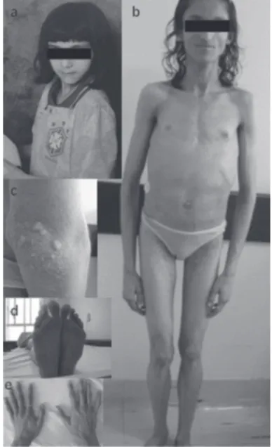
In 2001, a 15-year-old girl was referred to our hospital due to diabetes mellitus and hypertriglyceridemia. Both conditions were diagnosed at age of 14 yr during inves-tigation for delayed puberty. She was using NPH Insu-lin 14U/day, Metformin 1 g/day and Bezafi brate 400 mg/day. Physical examination revealed generalized loss of subcutaneous fat, including palmar aspects of hand and plantar aspects of feet. Prominent superfi cial veins and xantomas were seen on limbs (Figure 1). The pa-tient referred that her subcutaneous fat gradually disa-ppeared from the age of 11. Acanthosis nigricans was present on axilla and neck. Her weight was 33.9 kg, height 1.51 m and BMI 15 kg/m2. Blood pressure was
150x100 mmHg. She had pubic hair at Tanner stage 4,
L
ipodystrophies are a group of clinicallyheteroge-neous disorders characterized by the loss of adipo-se tissue. Metabolic complications such as insulin resistance, impaired glucose tolerance, dyslipidemia and hepatic steatosis are generally present in affected patients and their severity is determined by the extent of fat loss (1,2). Lipodystrophies are classifi ed accor-ding to their origin as familial (or genetic) and acquired types; and according to their clinical pattern of fat loss as generalized or partial.
The main familial forms of lipodystrophy are Con-genital Generalized Lipodystrophy (CGL) or Berar-dinelli-Seip Syndrome (OMIM 269700) and Familial Partial Lipodystrophy (FPLD), Dunnigan variety (OMIM 51660). CGL is a rare autosomal recessive disorder characterized by a nearly complete absence of adipose tissue since birth or early infancy. Familial Partial Lipodystrophy, Dunnigan variety is an auto-somal dominant disease characterized by gradual loss of adipose tissue from the extremities and trunk start-ing at the puberty, and subsequent fat accumulation on the face and neck. Two distinct genes were found to be responsible for the majority of CGL cases: the gene encoding the enzyme 1-acyl-glycerol phosphate acyltransferase 2 (AGPAT2) and the BSCL2 gene
which encodes a protein called seipin of unknown function (3,4). FPDL may result from heterozygous mutations in the LMNA gene (5,6). This gene is
lo-cated on chromosome 1q 21-22 and contains 12 ex-ons. LMNA gene, through alternative splice, encodes
lamin A and lamin C (7). Lamins are structural com-ponents of nuclear lamina and belong to the interme-diate fi lament family of proteins. Besides FPLD, mutations in LMNA have been shown to be
respon-sible for other inherited diseases such as Emery-Dreifuss muscular dystrophy type 2, dilated cardiomyopathy and conduction system disease, Hutchinson-Gilford Progeria Syndrome, Charcot-Marie-Tooth disorder and others (8-11). The term laminopathies is used to collectively call these diseases. Furthermore, LMNA
mutations were found in atypical progeroid syndromes and in a pubertal-onset generalized lipodystrophy (12-14).
cop
yr
ight
© ABE&M todos os direitos reser
v
ados
however no breast development (Tanner stage 1). The liver was palpable at 6 cm below the right costal margin and spleen at 1 cm below the left costal margin. She presented thinning of scalp hair and sparse grey hair; no other progeroid characteristics were noted.
Laboratory measurements showed fasting plasma glucose of 143 mg/dl, HbA1C of 6.5%, triglycerides of 877 mg/dL, total cholesterol of 170 mg/dL, HDL-cholesterol of 20 mg/dL. Liver function tests showed alanine aminotransferase of 42 U/L (normal <31 U/L), aspartate aminotransferase of 36 U/L (normal <32 U/L), alkaline phosphatase of 562 U/L (normal
<460 U/L) and γ glutamyltranspeptidase of 56 U/L (normal <32 U/L). Plasma adiponectin concentration of 0.6 ng/mL (reference range: 5-30 ng/mL). Blood electrolytes, renal function and thyroid hormone level were normal.
Abdominal ultrasonography showed hepatospleno-megaly and liver steatosis. Doppler echocardiography showed small atrial septum defect, ostium secundum
type (4 mm), without any other abnormality. Dual en-ergy x-ray absorptiometry was performed to evaluate whole-body and regional fat. The percentage of total body fat was 8.6%, corresponding to 2,924 g (reference range: 30.3 %± 1.5), legs 6.3% (reference range: 33.1%±1.5), arms 8.5% (reference range: 30.2%±1.8), trunk 7.8% (reference range: 29.0%±1.6). Therefore, a generalized loss of body fat was present. The reference values were reported by Mazess and cols. (15).
During the follow-up period a worsening on her metabolic state was observed coincident with a decrease in body fat (percentage of total body fat in 2004: 4.0%). The most recent evaluation showed an HbA1C of 13.5% despite high doses of insulin (1200 U/day) and triglycerides of 9,838 mg/dL despite the use of statin, fi brate and nicotinic acid.
Molecular analysis of LMNA gene
As the patient presented with pubertal-onset of genera-lized lipodystrophy and insulin resistance, molecular analysis of the LMNA gene was performed. We
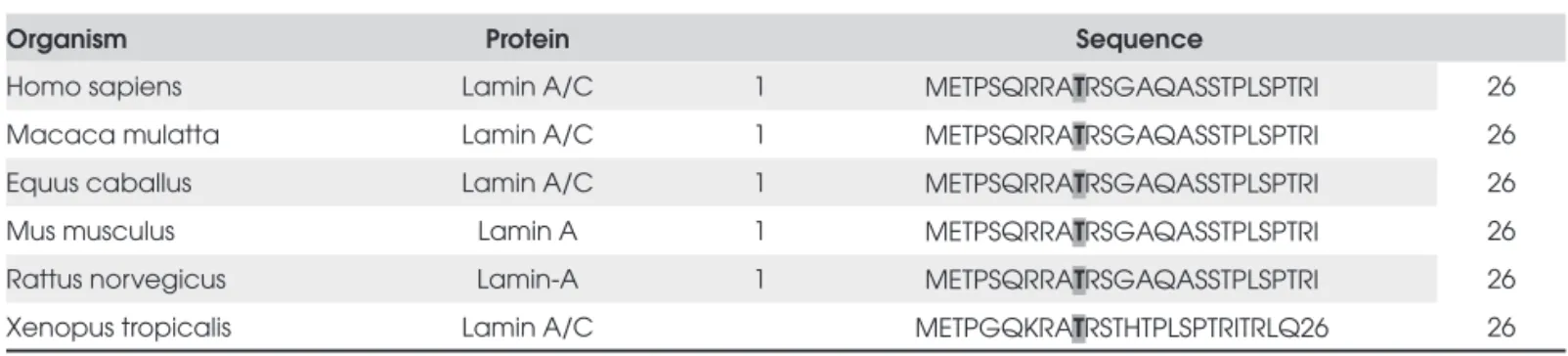
identi-fi ed a heterozygous substitution in exon 1 (c.29C> T) predicting the substitution of threonine, a polar hydro-philic amino acid, to isoleucine, a non polar hydropho-bic amino acid, at codon 10 (p.T10I mutation) in the N-terminal domain of the protein (Figure 2). The ami-no acid threonine at this position in LMNA gene is
hi-Figure 1. Photography of the patient at approximately 8 years of age showing the normal appearance (A) and at current age showing generalized lipoatrophy, sparse body hair, protu-berant abdomen, and no breast development (B). C, detail of xantomas in the elbow. D,E, close-up of her feet and hands. Note loss of fat and the presence of callosities in her feet.
cop
yr
ight
© ABE&M todos os direitos reser
v
ados
ghly conserved across different species (Table 1), suggesting an important role in protein function. We did not observe this mutation in both of her parents, indicating that it is a de novo mutation.
DISCUSSION
Here we report the case of a patient with pubertal-on-set generalized lipoatrophy, severe insulin resistance and hepatic steatosis harboring the p.T10I mutation in
LMNA gene. The switch from polar hydrophilic
threo-nine to non polar hydrophobic isoleucine in the lamin A/C N-terminal head may have functional consequen-ces. Accordingly, Csoka et al showed in cultured fi bro-blasts that the p.T10I mutation causes severe consequences for nuclear morphology such as lobula-tions of the nuclear membrane, and some of these lo-bulations contained no chromatin, indicating that the lamina had detached from the chromatin (14).
Laminopathies are characterized by a complex gen-otype/phenotype relationship and LMNA mutations
and redistribution of adipose tissue are more frequently observed in Familial Partial Lipodystrophy (Dunnigan variety) (6,16,17). Also, lipodystrophy and metabolic complications associated with insulin resistance have been reported in Mandibuloacral Dysplasia (18), and some affected patients were found to carry homozy-gous missense mutations in LMNA gene (19-21). The
clinical phenotype of the patient reported herewith does not resemble any of these two conditions.
Recently, Caux et al described a male patient har-boring the heterozygous R133L LMNA mutation that
has some features similar to those found in our patient: pubertal-onset generalized lipodystrophy, insulin-resis-tant diabetes, hypertriglyceridemia and liver steatosis (13). However, hypertrophic cardiomyopathy and
dis-semined leucomelanodermic papules also described by Caux et al were not observed in our patient.
The p.T10I mutation was previously described by Csoka et al in a patient who was originally diagnosed with atypical progeria, but was later reclassifi ed as Seip Syndrome based on generalized lipoatrophy, hypergly-cemia and hypertriglyceridemia (14). However, no fur-ther information is provided such as the age of onset of these abnormalities, making diffi cult the comparison with this patient. Metabolic alterations such as insulin resistance, hypertriglyceridemia and liver steatosis are characteristics of lipodystrophies. Recent studies de-scribe new phenotypes of metabolic laminopathies, even in the absence of obvious clinical lipoatrophy (22) or associated with premature ageing process (14).
Mutations associated with FPLD are frequently clus-tered in the LMNA region encoding the carboxi-terminal
domain (2). The present observation illustrates a general-ized lipoatrophy with a very severe insulin resistance due to a mutation in LMNA region encoding the N-terminal
head. Previous reports of generalized lipoatrophy as a manifestation of laminopathy were also associated with mutations which affect the amino-terminal head domain or the α-helical rod domain of the protein (13,14,23). These observations pointed out that the heterogeneity of lipodystrophies as a manifestation of laminopathy.
In summary, here we describe an atypical phenotype of lipodystrophy associated with a de novo appearance of
the p.T10I mutation in LMNA gene. The identifi cation
of mutation in this gene implies in a careful clinical and laboratory evaluation and follow-up of the affected pa-tient. Also, family members should be screened for an early detection and adequate treatment.
Acknowledgment: This work was supported by a grant from Fundação de Amparo à Pesquisa do Estado de São Paulo (Fa-pesp). No potencial confl ict of interest relevant to this article was reported.
Table 1. Amino acid sequence alignment for lamin A/C from various species. The residue affected by the mutation described (LMNA T10I) is shown in bold.
Organism Protein Sequence
Homo sapiens Lamin A/C 1 METPSQRRATRSGAQASSTPLSPTRI 26
Macaca mulatta Lamin A/C 1 METPSQRRATRSGAQASSTPLSPTRI 26
Equus caballus Lamin A/C 1 METPSQRRATRSGAQASSTPLSPTRI 26
Mus musculus Lamin A 1 METPSQRRATRSGAQASSTPLSPTRI 26
Rattus norvegicus Lamin-A 1 METPSQRRATRSGAQASSTPLSPTRI 26
cop
yr
ight
© ABE&M todos os direitos reser
v
ados
REFERENCES
1. Garg A. Acquired and Inherited Lipodystrophies. New Engl J Med. 2004;350(18):1220-34.
2. Agarwal AK, Garg A. Genetic basis of lipodystrophies and ma-nagement of metabolic complications. Annu Rev Med. 2006;57:297-311.
3. Agarwal AK, Arioglu E, de Almeida S, Akkoc N, Taylor SI, Bo-wcock AM, et al. AGPAT2 is mutated in congenital generalized lipodystrophy linked to chromosome 9q34. Nat Genet. 2002;31(1):21-3.
4. Magre J, Delépine M, Khallouf E, Gedde-Dahl Jr T, Van Mal-dergem L, Sobel E, et al. Identifi cation of the gene altered in Berardinelli-Seip congenital lipodystrophy on chromosome 11q13. Nat Genet. 2001; 28(4):365-70.
5. Peters JM, Barnes R, Bennett L, Gitomer WM, Bowcock AM, Garg A. Localization for the gene for familial partial lipodystro-phy (Dunnigan variety) to chromosome 1q21-22. Nat Genet. 1998;18(3):292-5.
6. Cao H, Hegele RA. Nuclear lamin A/C R482Q mutation in Cana-dian kindreds with Dunnigan-type familial partial lipodystro-phy. Hum Mol Genet. 2000;9(1):109-12.
7. Lin F, Worman HJ. Structural organization of the human gene encoding nuclear lamin A and nuclear lamin C. J Biol Chem. 1993;268(22):16321-6.
8. Di Barletta MR, Ricci E, Galluzzi G, Tonali P, Mora M, Morandi L, et al. Different mutations in the LMNA gene cause autoso-mal dominant and autosoautoso-mal recessive Emery-Dreifuss mus-cular dystrophy. Am J Hum Genet. 2000;66(4):1407-12. 9. Fatkin D, MacRae C, Sasaki T, Wolff MR, Porcu M, Frenneaux
M, et al. Missense mutations in the rod domain of the lamin A/C gene as causes of dilated cardiomyopathy and conduc-tion-system disease. N Engl J Med. 1999;341(23):1715-24. 10. Cao H, Hegele RA. LMNA is mutated in Hutchinson-Gilford
pro-geria (MIM 176670) but not in Wiedemann-Rautenstrauch proge-roid syndrome (MIM 264090). J Hum Genet. 2003;48(5):271-4. 11. De Sandre-Giovannoli A, Chaouch M, Kozlov S, Vallat JM, Tazir
M, Kassouri N, et al. Homozygous defects in LMNA, encoding lamin A/C nuclear-envelope proteins, cause autosomal recessi-ve axonal neuropathy in human (Charcot-Marie-Tooth disorder type 2) and mouse. Am J Hum Genet. 2002;70(3):726-36. 12. Chen L, Lee L, Kudlow BA, Dos Santos HG, Sletvold O,
Shafe-ghati Y, et al. LMNA mutations in atypical Werner’s syndrome. Lancet. 2003;362(9382):440-5.
13. Caux F, Dubosclard E, Lascols O, Buendia B, Chazouillères O, Cohen A, et al. A New Clinical Condition Linked to a Novel Mutation in Lamins A and C with Generalized Lipoatrophy, Insulin-Resistant Diabetes, Disseminated Leukomelanodermic Papules, Liver Steatosis, and Cardiomyopathy J Clin Endo-crinol Metab.2003; 88(3):1006-13.
14. Csoka AB, Cao H, Sammak PJ, Constantinescu D, Schatten GP, Hegele RA. Novel lamin A/C gene (LMNA) mutations in atypi-cal progeroid syndromes. J Med Genet. 2004;41(4):304-8. 15. Mazess RB, Barden HS, Bisek JP, Hanson J. Dual-energy x-ray
absorptiometry for total-body and regional bone-mineral and soft-tissue composition. Am J Clin Nutr. 1990;51(6):1106-12. 16. Shackleton S, Lloyd DJ, Jackson SN, Evans R, Niermeijer MF,
Singh BM, et al. LMNA, encoding lamin A/C, is mutated in par-tial lipodystrophy. Nat Genet. 2000;24(2):153-6.
17. Speckman RA, Garg A, Du F, Bennett L, Veile R, Arioglu E, et al. Mutational and haplotype analyses of families with familial partial lipodystrophy (Dunnigan variety) reveal recurrent mis-sense mutations in the globular C-terminal domain of lamin A/C. Am J Hum Genet. 2000;66(4):1192-8.
18. Cutler DL, Kaufmann S, Freidenberg GR. Insulin-resistant dia-betes mellitus and hypermetabolism in mandibuloacral dys-plasia: a newly recognized form of partial lipodystrophy. J Clin Endocrinol Metab. 1991;73(5):1056-61.
19. Novelli G, Muchir A, Sangiuolo F, Helbling-Leclerc A, D’Apice MR, Massart C, et al. Mandibuloacral dysplasia is caused by a mutation in LMNA-encoding lamin A/C. Am J Hum Genet. 2002;71(2):426-31.
20. Garg A, Cogulu O, Ozkinay F, Onay H, Agarwal AK. A novel homozygous Ala529Val LMNA mutation in Turkish patients with mandibuloacral dysplasia. J Clin Endocrinol Metab. 2005;90(9):5259-64.
21. Simha V, Agarwal AK, Oral EA, Fryns JP, Garg A. Genetic and phenotypic heterogeneity in patients with mandibuloacral dysplasia-associated lipodystrophy. J Clin Endocrinol Metab. 2003;88(6):2821-4.
22. Decaudain A, Vantyghem MC, Guerci B, Hécart AC, Auclair M, Narbonne YRH, et al. New Metabolic Phenotypes in Laminop-athies: LMNA Mutations in Patients with Severe Metabolic Syndrome. J Clin Endocrinol Metab. 2007;92(12):4835-44 23. Jacob KN, Baptista F, Santos HG, Oshima J, Agarwal AK, Garg
A. Phenotypic heterogeneity in body fat distribution in pa-tients with atypical Werner´s Syndrome due to heterozygous Arg133Leu laminin A/C mutation. J Clin Endocrinol Metab. 2005;90(12):6699-06.
Correspondence to:
Regina S. Moisés
Universidade Federal de São Paulo, Escola Paulista de Medicina, Disciplina de Endocrinologia,