Cop
yright
© ABE&M t
odos os dir
eit
os r
eser
vados
.
Congenital hypothyroidism:
recommendations of the Thyroid
Department of the Brazilian Society
of Endocrinology and Metabolism
Hipotireoidismo congênito: recomendações do Departamento de Tireoide da Sociedade Brasileira de Endocrinologia e Metabologia
Léa Maria Zanini Maciel1, Edna Teruko Kimura2, Célia Regina Nogueira3, Glaucia Maria F. Silva Mazeto3, Patrícia Künzle Ribeiro Magalhães1, Marilza Leal Nascimento4, Suzana Nesi-França5, Sandra E. Vieira6
ABSTRACT
Congenital hypothyroidism (CH) is the most common congenital endocrine disorder, with an incidence of 1:2,000 to 1:4,000 live births and it is a leading preventable mental retardation. Neonatal Screening Programs allow early identiication of the disease and the adequate treat-ment of affected children can avoid the complications related to deprivation of the hormone. Most cases of primary congenital hypothyroidism (85%) are due to thyroid dysgenesis (ectopia, hypoplasia or agenesis) while the remaining result from defects in hormone synthesis. Affected children (> 95%) usually have no symptoms suggesting the disease at birth. The most frequent symptoms and signs are prolonged neonatal jaundice, hoarse cry, lethargy, slow movements, constipation, macroglossia, umbilical hernia, large fontanelle, hypotonia and dry skin. Around the world, various strategies are used for the screening of the CH. In Brazil, screening for CH is mandatory by law and usually done by serum TSH in dried blood collected from the heel. The recommended age for performing this test is after 48 hours of life until the 4th day. Diag-nostic conirmation is required dosing TSH and free T4 or total T4 in serum. Arq Bras Endocrinol Metab. 2013;57(3):184-92
Keywords
Congenital hypothyroidism; neonatal screening
RESUMO
O hipotireoidismo congênito (HC) é o distúrbio endócrino congênito mais frequente, com in-cidência variando de 1:2.000 a 1:4.000 crianças nascidas vivas e uma das principais causas de retardo mental que pode ser prevenida. Os Programas de Triagem Neonatal para a doença permitem a identiicação precoce dos afetados e seu tratamento de modo a evitar as complica-ções da falta do hormônio. A maioria dos casos de hipotireoidismo congênito é decorrente de disgenesias tireoidianas (85%), entre elas a ectopia, hipoplasia ou agenesia tireoidianas, e os demais resultam de defeitos de síntese hormonal. As crianças afetadas (> 95%) geralmente não apresentam sintomas sugestivos da doença ao nascimento. Os sintomas e sinais mais comuns são: icterícia neonatal prolongada, choro rouco, letargia, movimentos lentos, constipação, ma-croglossia, hérnia umbilical, fontanelas amplas, hipotonia e pele seca. Várias estratégias são utilizadas para a triagem do HC. No Brasil, esta é obrigatória por lei e geralmente é feita com a dosagem de TSH em sangue seco coletado do calcanhar. A idade recomendada para sua reali-zação é após as 48 horas de vida até o quarto dia. A conirmação diagnóstica é obrigatória com as dosagens de TSH e T4 livre ou T4 total. Arq Bras Endocrinol Metab. 2013;57(3):184-92
Descritores
Hipotireoidismo congênito; triagem neonatal
Correspondence to:
Léa Maria Zanini Maciel Av. Bandeirantes, 3900 Departamento de Clínica Médica 14049-900 – Ribeirão Preto, SP, Brasil [email protected]
Received on Mar/19/2013 Accepted on Mar/19/2013
1 Endocrinology and Metabolism
Division, Faculdade de Medicina de Ribeirão Preto, Universidade de São Paulo (FMRP-USP), Ribeirão Preto, SP, Brazil
2 Institute of Biomedical Sciences,
USP, São Paulo, SP, Brazil
3 Endocrinology and Metabolism
Division, Department of Clinical Medicine, Faculdade de Medicina de Botucatu, Universidade Estadual de São Paulo (Unesp), Botucatu, SP, Brazil
4 Universidade Federal de
Santa Catarina (UFSC), Florianopólis, SC, Brazil
5 Pediatric Endocrinology Unit,
Department of Pediatrics, Universidade Federal do Paraná (UFPR), Curitiba, PR, Brazil
6 Department of Pediatrics,
Cop
yright
© ABE&M t
odos os dir
eit
os r
eser
vados
.
INTRODUCTION
C
ongenital hypothyroidism (CH) is the most common congenital endocrine disorders, with an incidence of 1:2,000 to 1:4,000 live births in io-dine-suficient countries (1,2) (B). In Brazil, the in-cidence of CH is close to these values, ranging from 1:2,595 to 1:4,795 (3,4)(B). However, recent stu-dies indicate a higher incidence of CH in the United States, from 1:4,094 in 1987 to 1:2,372 in 2002 (5) (B). This higher incidence may be due to improved means of detecting subclinical cases of the disease as a consequence of a lower cutoff point for thyroid sti-mulating hormone (TSH) levels and the inclusion of transient hypothyroidism in the screening process (6-9) (D).The prevalence of CH varies among ethnic groups and is signiicantly less prevalent among African Ameri-cans compared to Hispanics (1:10,000 vs. 1:2,700). Regarding gender, CH is more prevalent in females (2:1). In addition, children with Down syndrome have a 35-fold increased risk of CH compared to the general population (10) (B).
In the absence of early diagnosis and proper treat-ment, most children develop varying degrees of neuro-logical, motor and growth deicits, including irreversi-ble mental retardation.
METHODS
Active searches were conducted in the primary databa-ses Medline and SciELO using the following keywords (MeSH Terms): congenital hypothyroidism and neo-natal screening.
Grade of recommendation and strength of evi-dence
The strength of evidence was evaluated according to the Oxford classiication system and established ba-sed on the experimental design uba-sed, considering the best available evidence for each question and the Bra-zilian experience.
A: Most consistent experimental and/or observa-tional studies.
B: Less consistent experimental and/or observatio-nal studies.
C: Case reports.
D: Opinion without critical evaluation based on consensus, physiological studies or animal models.
1. WHAT ARE THE CAUSES OF CH?
The most frequent cause of permanent CH is thyroid dysgenesis, which results from defects in glandular for-mation during embryogenesis, and represents 85% of the cases (Table 1). This group encompasses thyroid ectopy, agenesis and hypoplasia, which account for 30%-45%, 35%-45% and 5% of cases, respectively (11) (D). The precise reasons for these alterations remain unclear, although mutations in transcriptional fac-tors that regulate thyroid gland development, such as thyroid transcription factor 2 (TTF-2), NKX2.1 (also known as TTF-1) and paired box gene 8 (PAX-8) have been reported to be involved. However, only 2% of dys-genesis cases exhibit these genetic mutations (12) (B). These transcriptional factors are present in other tissues and are associated with CH syndrome. Thus, mutations in NKX2.1, a gene that participates in lung and brain development, cause neonatal respiratory distress and choreoathetosis in the newborn (NB). Patients with mutations in TTF-2 present with spiky hair, cleft palate, anal atresia and thyroid agenesis (11) (D).
Other etiologies of permanent CH are defects in hormone production called dyshormonogenesis that represent approximately 15% of cases. The defects are autosomal recessive and include mutations in genes en-coding the sodium-iodide symporter (NIS) (SLC5A5
gene), thyroperoxidase (TPO), hydrogen peroxide generation factors [thyroid oxidase and dual oxidase maturation factors (DUOXA1 and DUOX2 genes)], thyroglobulin (Tg) and iodothyronine deiodinases (13).
Uncommon causes of CH include defects in thyroid hormone (TH) transport, such as mutations in the mo-nocarboxylase transporter 8 (MCT8) gene (14) (C); re-sistance to TH (syndrome of rere-sistance to thyroid hor- (syndrome of resistance to thyroid hor-mone) (15) (D), resistance to TSH (16) (C) and central hypothyroidism (17,18) (B).
Central hypothyroidism can be due to isolated TSH deiciency or, more commonly, hypopituitarism, whi-ch causes deiciency in several adenohypophysis hor-mones. Mutations in many genes involved in pituitary development or function have been implicated, inclu-ding HESX1, LHX4, PIT-1 and PROP1. Resistance to thyrotropin-releasing hormone (TRH) due to a muta-tion in the gene encoding the TRH receptor may also cause central hypothyroidism (18).
Cop
yright
© ABE&M t
odos os dir
eit
os r
eser
vados
.
Resistance to TSH is deined as elevated serum TSH concentrations (hyperthyrotropinemia) in the absence of goiter. Affected individuals have normal or hypoplastic thyroid glands and their serum T4 and T3 values are normal or low.
Defects in TH transport caused by mutations in the
MCT8 gene, which is located on the X chromosome, impairs T3 transport and leads to mental retardation. The syndrome is characterized by high serum T3, low serum T4 and high serum TSH concentrations.
Recommendation 1
The most frequent cause of permanent CH is thyroid dysgenesis, which includes thyroid agenesis, ectopy and hypoplasia (B). Dyshormonogenesis is the second most common cause (B). Rare causes of CH include central hypothyroidism (B), syndrome of resistan-ce to thyroid hormone (D), TSH resistance syndrome (C) and MCT8 mutations (C).
2. CAN CH BE TRANSIENT?
CH can be transient and can result from several causes:
• Excessive (or deicient) iodine intake by the mother.
• Maternal anti-thyroid drugs intake (mothers with hyperthyroidism).
• Transplacental passage of maternal antibodies that block the TSH receptor. This diagnosis should be considered when the mother reports the occurrence of more than one child with transient hypothyroidism detected by neonatal screening. It usually lasts 1-3 months, i.e., until the antibodies disappear from circulation.
• Heterozygous mutations in enzymes DUOX1
(DUOXA1 gene) or DUOX2/THOX (DUOX2
gene).
• Large liver hemangiomas (increased deiodinase type 3 activity) (7,8) (D).
Patients classiied as having HC due to synthesis defects were followed for 3 years and, at re-evalua-tion, only 47% had permanent hypothyroidism. Thus, it is recommended to evaluate all children with topic thyroid at age of 3 to deine the presence or absence of the disease (19) (B).
Recommendation 2
Neonatal hypothyroidism may be permanent or tran-sient. It is recommended that children be re-evaluated at 3 years of age; for patients with unclear hypothyroi-dism etiology, levothyroxine (L-T4) treatment should be discontinued (B).
3. CLINICAL MANIFESTATIONS OF CH
Most children with CH (> 95%) have little or no clini-cal manifestation of the disease at birth (20) (B) due to the transplacental passage of maternal T4 (21) (B) and because most affected children have some functioning thyroid tissue. As TH has a half-life of 7 days, the ma-ternal hormone is metabolized and excreted approxi-mately 3-4 weeks after birth.
Affected children typically present normal weight and height. One of the irst signs is prolonged neonatal jaundice (22,23) (B). Over time, undiagnosed children appear lethargic, with slow movements, hoarse cry, fe-eding dificulties, constipation, macroglossia, umbilical hernia, large anterior or posterior fontanels, hypotonia, dry skin, thinning hair and typical facies with saddle nose. Some NBs with dyshormonogenesis present with a palpable goiter at birth, but this condition may also appear later, even with treatment (23). An x-ray of knee epiphyses may reveal delayed ossiication, which relects fetal hypothyroidism severity. Table 2 lists symptoms or
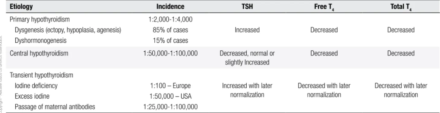
Table 1. Main etiologies of CH and hormonal changes
Etiology Incidence TSH Free T4 Total T4
Primary hypothyroidism
Dysgenesis (ectopy, hypoplasia, agenesis) Dyshormonogenesis
1:2,000-1:4,000 85% of cases 15% of cases
Increased Decreased Decreased
Central hypothyroidism 1:50,000-1:100,000 Decreased, normal or slightly Increased
Decreased Decreased
Transient hypothyroidism Iodine deiciency Excess iodine
Passage of maternal antibodies
1:100 – Europe 1:50,000 – USA 1:25,000-1:100,000
Increased with later normalization
Decreased with later normalization
Cop
yright
© ABE&M t
odos os dir
eit
os r
eser
vados
.
signs exhibited in affected children according to dise-ase severity (24) (B). Screening of Brazilian newborns with CH was associated with umbilical hernia (48.9%), saddle nose (46.6%), prolonged jaundice beyond 7 days (44.4%) and 20% of cases had no clinical manifestation (25) (A).
When the etiologic diagnosis of CH is hypopituita-rism, the child will be predisposed to hypoglycemia due to growth hormone and adrenocorticotropic hormone (ACTH)/cortisol deiciency, and males will exhibit mi-cropenis. These children are at risk of death if the disea-se is not detected early, and it is usually not detected by NB screening utilizing TSH measurement.
Children with CH and cleft palate may have mu-tations in TTF-2 (FOXE-1 gene) (28) (C), and those with persistent neurological symptoms, including ata-xia, may have mutations in the NKX2.1 gene (29) (C). Hearing problems occur in approximately 20% of children with CH, and all affected children should un-dergo a hearing screening test (30,31) (B).
There is no consensus for conducting screening tests for congenital anomalies. However, a careful phy-sical examination is important, and the child should be referred for evaluation if any alteration is detected.
The early detection of other malformations in tients with CH may modify the prognosis of these pa-tients (32) (B).
Recommendation 4
Hearing screening and a careful physical examination are recommended to search for other congenital ab-normalities in children with congenital hypothyroidism (B).
5. IS NB SCREENING EFFECTIVE FOR TRACKING
EARLY HYPOTHYROIDISM?
The main purpose of NB screening for CH is to avoid sequelae, especially hypothyroidism-induced mental re-tardation, which can be achieved initiating therapy wi-thin the irst 2 weeks of life (33) (B).
Neonatal screening for CH is routine in the Uni-ted States, Canada, Europe, Israel, Japan, Australia and New Zealand, and it is in development in Eastern Eu-rope, South America, Asia and Africa.
The high sensitivity of NB screening tests for CH makes them an effective way to identify disease. Po-pulation studies conducted in Europe and the United States have reported sensitivities of 97-100% and speci-icities of 98%-100% (34,35) (A).
Recommendation 5
Neonatal screening is recommended to track CH (A).
6. WHEN SHOULD NB SCREENING TESTS FOR
HYPOTHYROIDISM BE PERFORMED?
For the NB screening test, blood is collected from the heel and placed on ilter paper, which is added to a card that lists the child’s data (date of birth, gestational age, sex, weight, whether there was a blood transfusion etc.)
Table 2. Occurrence of symptoms and signs of CH at the time of diagnosis according to disease severity
Total T4 < 2.5 µg/dL
n = 215 (%)
Total T4 > 2.5 µg/dL
n = 232 (%)
Prolonged jaundice 128 (59) 77 (33)**
Feeding dificulty 75 (35) 36 (16)**
Lethargy 73 (34) 32 (14)**
Umbilical hernia 68 (32) 42 (18)*
Macroglossia 53 (25) 28 (12)*
Constipation 38 (18) 24 (10)
Cold skin 39 (18) 24 (10)
Hoarse cry 16 (7) 15 (6)
Hypothyroid appearance 12 (6) 6 (2)
Hypothermia 6 (3) 7 (3)
Hypotonia 6 (3) 7 (3)
No symptoms 34 (16) 78 (33)**
* p < 0.01; ** p < 0.001.
Modiied from Grant and cols., 1992 (24) (B).
Recommendation 3
Despite the possibility of the absence of clinical symp-toms in infants with congenital hypothyroidism, the signs and symptoms described in table 2 should serve as a warning (B).
4. CAN CH OCCUR IN ASSOCIATION WITH
CONGENITAL ABNORMALITIES?
Cop
yright
© ABE&M t
odos os dir
eit
os r
eser
vados
.
and contact information. It is recommended that blood collection be performed after 48 hours of birth to 4 days of life (36), when the physiological postnatal TSH peak has decreased. Ideally, the blood samples should be collected prior to hospital discharge; however, blood collection performed at early discharge (< 48 hours) may result in a false-positive result. In critically ill or preterm children, blood collection should be perfor-med at 7 days of life; however, it is important to note that it may be too late for children with congenital adrenal hyperplasia or metabolic disease when blood samples are collected after 4 days of life (36). Due to the immaturity of the hypothalamic-pituitary-thyroid axis in preterm infants, some authors recommend re-peating their screening test within 2 to 4 weeks of age. When there is need for whole blood transfusion, heel blood should be collected before the child is trans-fused, regardless of age (37) (D).
Recommendation 6
Blood should be collected from NBs for screening after 48 hours of birth to 4 days of life or before the NB leaves the hospital and always before blood transfusion (D).
7. WHICH TESTS ARE PERFORMED IN BRAZIL TO
SCREEN FOR CH?
There are several strategies to screen for CH: 1. TSH measurement.
2. Simultaneous measurement of TSH and T4. 3. Initial measurement of T4 followed by TSH if T4 is below a certain limit (usually below the 10th
percen-tile).
In Brazil, the public system performs TSH scree-ning (TSHneo), with cutoff TSHneo values ranging from 5-20 μU/ml. Children with high TSHneo values are called for evaluation and conirmation.
The dosage of TSH has greater speciicity than an isolated T4 dosage. The simultaneous dosage of TSH and T4 has higher sensitivity in all protocols, but also leads to a higher number of false positives (38) (B).
In some neonatal screening centers, children with TSHneo values between 10-20 μU/ml are recalled for a second collection onto ilter paper, and if TSH is above 10 μU/ml, the results need to be conirmed in serum.
When only TSH is evaluated, children with cen-tral hypothyroidism or delayed TSH elevation may not be identiied. Late TSH elevations are parti-cularly common in children with low birth weight (< 2,500 g) and in preterm births (39) (B).
Screening with initial measurement of T4 follo-wed by TSH can detect cases of primary hypothyroi-dism, as well as central hypothyroidism and children with deiciency of thyroid hormone carrier protein (TBG). The latter group exhibits low total T4 (TT4) and normal free T4 (FT4) and will not require CH treatment. However, this approach will not detect chil-dren with CH that have normal T4 due to less severe thyroid dysfunction. A comparison between the two approaches that involve TSH and T4 measurements in different sequences showed that 1 in 93,000 screened children would not be diagnosed with the initial ap-proach with T4, which would not occur if TSH was evaluated irst (40) (B).
Recommendation 7
In Brazil, NB screening for CH is performed by TSH determination on ilter paper, followed by total T4 and/ or free T4 measurement in serum, when necessary. This strategy is effective and has also been adopted in other countries (A).
8. SHOULD ABNORMAL SCREENING TEST
RESULTS BE CONFIRMED?
NB screening tests for CH are not diagnostic, and ab-normal results should be conirmed by quantitative me-thods to measure serum TSH and TT4 or FT4 (41) (B). Most conirmatory tests should be performed between the irst and second weeks of life, when the upper le-vel of the TSH normal range drops to 10 μU/ml. It should be noted that the normal range is different from that observed in adults. Between 4 to 30 days of life, the normal ranges of TT4 and FT4 are 7-16 μg/dL and 0.8-2.3 ng/dL, respectively (42).
TSH levels above 10 μU/mL and low FT4 or TT4 values conirm the diagnosis of primary hypothyroi-dism, and these children should undergo appropriate treatment (36).
Cop
yright
© ABE&M t
odos os dir
eit
os r
eser
vados
.
irst month of life, even with T4 in the normal range, some researchers suggest treatment and re-evaluation at 3 years of age (19,43) (B,D).
Preterm infants and infants that are ill for any reason (euthyroid sick syndrome) may have low TT4/FT4 with normal TSH levels, and treatment with L-T4 is not re-commended unless there is evidence of hypothalamic or pituitary disease (44).
Recommendation 8
Screening tests for CH that yield abnormal results should be conirmed by quantitative measurement of venous TSH and total T4/free T4 (B).
9. IDENTIFICATION OF HYPOTHYROIDISM
ETIOLOGY
If the diagnosis of hypothyroidism is conirmed, further studies are necessary to determine the disease etiology; however, the decision to treat the disease is based on hormone levels, and additional tests are optional and should not delay the beginning of treatment.
Tests that may be required to elucidate CH etiology are as follows:
• Cervical ultrasonography – the main initial examination. Once the thyroid is located, the most common etiologies of agenesis and ectopy would be ruled out. However, ultrasonography is less sensitive than thyroid scintigraphy for de-tection of ectopic gland, even though Doppler ultrasound can successfully identify 90% of ec-topic glands (45). The ultrasound has a sensiti-vity and speciicity of 90.5% and 47.8%, respec-tively, for the diagnosis of agenesis and 100% and 80.4%, respectively, for hypoplasia, but has low sensitivity for the diagnosis of ectopy (only 10%) (46) (B). The advantages are to avoid ex-posure to radiation and lower cost (47) (B).
• Mapping with 99mTc – indicated when
ultrasou-nd does not detect the ectopic glaultrasou-nd. Scintigra-phy can be performed with technetium (99mTc)
pertechnetate or iodine (123I) instead of 131I due
to lower irradiation. Goiter can be observed when there is an enzyme defect. For detection of ectopic gland, it has a sensitivity and speci-icity of 92% and 97.1% respectively (46) (B).
• Thyroglobulin measurement – there is a large overlap of Tg values for different CH etiologies;
therefore, Tg values are used only in special si-tuations. The association between Tg levels and ultrasonography can distinguish between
athyreosis and ectopic glandular tissue. Some functional ectopic tissue is present if no thyroid tissue is visualized in the normal location but T4 and Tg levels are measurable (36,42).
• Measurement of antithyroid antibodies [an-ti-peroxidase (TPO) antibody and antibody that blocks TSH receptor (TRAb)] – this test may be useful to justify the presence of elevated TSH in the infants of mothers that have Hashi-moto’s thyroiditis (transient hypothyroidism) or Graves’ disease (36).
• Urinary iodine – can conirm the lack or excess of iodine in suspected cases, and treatment with L-T4 should be established for several months until be gradually reduced.
Recommendation 9
Complementary investigations are necessary to deter-mine the etiology of congenital hypothyroidism (B), but should never delay the start of treatment.
10. WHEN SHOULD TREAMENT BEGIN?
The age of treatment onset, the L-T4 dose administe-red and appropriate monitoring are essential for brain development of CH patients. There is an inverse rela-tionship between the age of diagnosis/treatment and intelligence quotient (IQ). Children that are identiied in NB screening programs and treated in the irst weeks of life usually have a normal IQ, although some stu-dies have shown that they also have cognitive deicits (48,49).
Recommendation 10
The beginning of the treatment should be as early as possible, preferably within the irst 2 weeks of life (B).
11. DOES SODIUM L-T4 THERAPY NORMALIZE
HORMONE LEVELS OF CHILDREN WITH CH?
Cop
yright
© ABE&M t
odos os dir
eit
os r
eser
vados
.
(D). Studies show that with these doses, FT4 or TT4 and TSH concentrations normalize in 3 days and 2-4 weeks, respectively (50) (A).
L-T4 tablets should be used because the liquid form of the hormone is not approved for clinical use. The ta-blet should be crushed and dissolved in a small amount of water and administered in the morning, ideally while fasting. Food should be avoided for 30 minutes. In case of immediate vomiting, the same dose should be repea-ted. With good oral absorption and a half-life of appro-ximately 7 days, L-T4 is administered daily (36) (D). Although it is recommended that L-T4 be given on an empty stomach and food should be avoided for 30-60 minutes, this is not practical in infants. Thus, L-T4 may be administered between feedings, and doses should be adjusted based on serum hormone levels. L-T4 cannot be used with other substances that interfere with its ab-sorption, such as soybeans, iron or calcium.
Recommendation 11
CH treatment should be initiated as soon as possible, preferably within the irst 15 days of life. Oral L-T4 is recommended at the initial dose of 10-15 µg/kg/day (A).
12. HOW SHOULD TREATMENT BE MONITORED?
Brain development is highly dependent on thyroid hormone levels for the irst 2-3 years of life. There are studies showing that persistently low serum T4 concen-trations (TT4 below 10 μU/ml) in the irst year of life are associated with IQ approximately 18 points lower than the average IQ (51). The recommendations of the American Academy of Pediatrics (36) (D) regarding
the treatment and monitoring of children are listed in table 3 (43).
More frequent laboratory testing may be necessa-ry when there is poor compliance with the treatment, abnormal values are obtained or the dosage has been changed.
The goal of treatment is to ensure that the children have adequate growth and psychomotor development as close as possible to their genetic potential.
Care must be taken to avoid excessive treatment for prolonged periods, which may lead to craniosynostosis and changes in the child’s temperament (51).
Disclosure: no potential conlict of interest relevant to this article was reported.
REFERENCES
1. Waller DK, Anderson JL, Lorey F, Cunningham GC. Risk factors for congenital hypothyroidism: an investigation of infant’s birth weight, ethnicity, and gender in California, 1990-1998. Teratology. 2000;62(1):36-41.
2. Corbetta C, Weber G, Cortinovis F, Calebiro D, Passoni A, Vigone MC, et al. A 7-year experience with low blood TSH cutoff levels for neonatal screening reveals an unsuspected frequency of congenital hypothyroidism (CH). Clin Endocrinol (Oxf). 2009;71(5):739-45.
3. Ramos HE, Nesi-França S, Maciel RM. New aspects of genetics and molecular mechanisms on thyroid morphogenesis for the understanding of thyroid dysgenesia. Arq Bras Endocrinol Metabol. 2008;52(9):1403-15.
4. Magalhães PK, Turcato M de F, Angulo Ide L, Maciel LM. Neonatal screening program at the university hospital of the Ribeirão Preto School of Medicine, São Paulo University, Brazil. Cad Saude Publica. 2009;25(2):445-54.
5. Harris KB, Pass KA. Increase in congenital hypothyroidism in New York State and in the United States. Mol Genet Metab. 2007;91(3):268-77.
6. Shapira SK, Lloyd-Puryear MA, Boyle C. Future research directions to identify causes of the increasing incidence rate of congenital hypothyroidism in the United States. Pediatrics. 2010;125 Suppl 2:S64-8.
7. Parks JS, Lin M, Grosse SD, Hinton CF, Drummond-Borg M, Borgfeld L, et al. The impact of transient hypothyroidism on the increasing rate of congenital hypothyroidism in the United States. Pediatrics. 2010;125 Suppl 2:S54-63.
8. Olney RS, Grosse SD, Vogt RF Jr. Prevalence of congenital hypothyroidism--current trends and future directions: workshop summary. Pediatrics. 2010;125 Suppl 2:S31-6.
9. Hertzberg V, Mei J, Therrell BL. Effect of laboratory practices on the incidence rate of congenital hypothyroidism. Pediatrics. 2010;125 Suppl 2:S48-53.
10. Roberts HE, Moore CA, Fernhoff PM, Brown AL, Khoury MJ. Population study of congenital hypothyroidism and associated birth defects, Atlanta, 1979-1992. Am J Med Genet. 1997;71(1):29-32.
11. Park SM, Chatterjee VK. Genetics of congenital hypothyroidism. J Med Genet. 2005;42(5):379-89.
Table 3. CH treatment, monitoring and target hormone concentrations
Starting L-T4 dose 10-15 μg/kg/day
Monitoring free T4 or total T4 and TSH
At 2 and 4 weeks after indication of L-T4 treatment
Every 1-2 months during the irst months Every 2-3 months from 6-36 months Every 6-12 months until growth is complete
Target values for thyroid hormones
T4: upper reference range for age
Example: Free T4: reference range: 0.8-2.3 ng/dL,
aim for 1.4-2.3 ng/dL
Total T4: 10-16 μg/dL in the irsts 2 years of life;thereafter, upper half of the age-speciic reference range
Cop
yright
© ABE&M t
odos os dir
eit
os r
eser
vados
.
12. Al Taji E, Biebermann H, Límanová Z, Hníková O, Zikmund J, Dame C, et al. Screening for mutations in transcription factors in a Czech cohort of 170 patients with congenital and early-onset hypothyroidism: identiication of a novel PAX8 mutation in dominantly inherited early-onset non-autoimmune hypothyroidism. Eur J Endocrinol. 2007;156(5):521-9.
13. Cangul H, Aycan Z, Olivera-Nappa A, Saglam H, Schoenmakers NA, Boelaert K, et al. Thyroid dyshormonogenesis is mainly caused by TPO mutations in consanguineous community. Clin Endocrinol (Oxf). 2012 Dec 13. doi: 10.1111/cen.12127. [Epub ahead of print].
14. Friesema EC, Grueters A, Biebermann H, Krude H, von Moers A, Reeser M, et al. Association between mutations in a thyroid hormone transporter and severe X-linked psychomotor retardation. Lancet. 2004;364(9443):1435-7.
15. Refetoff S, Weiss RE, Usala SJ. The syndromes of resistance to thyroid hormone. Endocr Rev. 1993;14(3):348-99.
16. Alberti L, Proverbio MC, Costagliola S, Romoli R, Boldrighini B, Vigone MC, et al. Germline mutations of TSH receptor gene as cause of nonautoimmune subclinical hypothyroidism. J Clin Endocrinol Metab. 2002;87(6):2549-55.
17. Hanna CE, Krainz PL, Skeels MR, Miyahira RS, Sesser DE, LaFranchi SH. Detection of congenital hypopituitary hypothyroidism: ten-year experience in the Northwest Regional Screening Program. J Pediatr. 1986;109(6):959-64.
18. Persani L. Clinical review: central hypothyroidism: pathogenic, diagnostic, and therapeutic challenges. J Clin Endocrinol Metab. 2012;97(9):3068-78.
19. Korzeniewski SJ, Grigorescu V, Kleyn M, Young WI, Birbeck G, Todem D, et al. Transient hypothyroidism at 3-year follow-up among cases of congenital hypothyroidism detected by newborn screening. J Pediatr. 2013;162(1):177-82.
20. Alm J, Hagenfeldt L, Larsson A, Lundberg K. Incidence of congenital hypothyroidism: retrospective study of neonatal laboratory screening versus clinical symptoms as indicators leading to diagnosis. Br Med J (Clin Res Ed). 1984;289(6453):1171-5.
21. Vulsma T, Gons MH, de Vijlder JJ. Maternal-fetal transfer of thyroxine in congenital hypothyroidism due to a total organiication defect or thyroid agenesis. N Engl J Med. 1989;321(1):13-6.
22. LaFranchi SH, Murphey WH, Foley TP Jr, Larsen PR, Buist NR. Neonatal hypothyroidism detected by the Northwest Regional Screening Program. Pediatrics. 1979;63(2):180-91.
23. Ramos JC, Lacerda Filho Ld, DeMartini Ade A, Silveira RB, Pereira RM, Sandrini Neto R, et al. Clinical and laboratory features of children and adolescents with congenital hypothyroidism due to dyshormonogenesis in southern Brazil. Arq Bras Endocrinol Metabol. 2012;56(3):201-8.
24. Grant DB, Smith I, Fuggle PW, Tokar S, Chapple J. Congenital hypothyroidism detected by neonatal screening: relationship between biochemical severity and early clinical features. Arch Dis Child. 1992;67(1):87-90.
25. Nascimento ML, Rabello FH, Ohira M, Simoni G, Cechinel E, Linhares RM, da Silva PC. [Newborn Screening Program for congenital hypothyroidism of the State of Santa Catarina, Brazil: etiological investigation in the irst visit]. Arq Bras Endocrinol Metabol. 2012;56(9):627-32.
26. Olivieri A, Stazi MA, Mastroiacovo P, Fazzini C, Medda E, Spagnolo A, et al.; Study Group for Congenital Hypothyroidism. A population-based study on the frequency of additional congenital malformations in infants with congenital hypothyroidism: data from the Italian Registry for Congenital Hypothyroidism (1991-1998). J Clin Endocrinol Metab. 2002;87(2):557-62.
27. Kumar J, Gordillo R, Kaskel FJ, Druschel CM, Woroniecki RP. Increased prevalence of renal and urinary tract anomalies in children with congenital hypothyroidism. J Pediatr. 2009;154(2):263-6.
28. Castanet M, Park SM, Smith A, Bost M, Léger J, Lyonnet S, et al. A novel loss-of-function mutation in TTF-2 is associated with congenital hypothyroidism, thyroid agenesis and cleft palate. Hum Mol Genet. 2002;11(17):2051-9.
29. Doyle DA, Gonzalez I, Thomas B, Scavina M. Autosomal dominant transmission of congenital hypothyroidism, neonatal respiratory distress, and ataxia caused by a mutation of NKX2-1. J Pediatr. 2004;145(2):190-3.
30. Pharoah PO, Buttield IH, Hetzel BS. Neurological damage to the fetus resulting from severe iodine deiciency during pregnancy. Lancet. 1971;1(7694):308-10.
31. Léger J, Ecosse E, Roussey M, Lanoë JL, Larroque B; French Congenital Hypothyroidism Study Group. Subtle health impairment and socioeducational attainment in young adult patients with congenital hypothyroidism diagnosed by neonatal screening: a longitudinal population-based cohort study. J Clin Endocrinol Metab. 2011;96(6):1771-82.
32. Azar-Kolakez A, Ecosse E, Dos Santos S, Léger J. All-cause and disease-speciic mortality and morbidity in patients with congenital hypothyroidism treated since the neonatal period: a national population-based study. J Clin Endocrinol Metab. 2013;98(2):785-93.
33. Bongers-Schokking JJ, Koot HM, Wiersma D, Verkerk PH, de Muinck Keizer-Schrama SM. Inluence of timing and dose of thyroid hormone replacement on development in infants with congenital hypothyroidism. J Pediatr. 2000;136(3):292-7.
34. Pharoah PO, Madden MP. Audit of screening for congenital hypothyroidism. Arch Dis Child. 1992;67(9):1073-6.
35. Kwon C, Farrell PM. The magnitude and challenge of false-positive newborn screening test results. Arch Pediatr Adolesc Med. 2000;154(7):714-8.
36. American Academy of Pediatrics, Rose SR; Section on Endocrinology and Committee on Genetics, American Thyroid Association, Brown RS; Public Health Committee, Lawson Wilkins Pediatric Endocrine Society, Foley T, Kaplowitz PB, Kaye CI, Sundararajan S, Varma SK. Update of newborn screening and therapy for congenital hypothyroidism. Pediatrics. 2006;117(6):2290-303.
37. Kaye CI; Committee on Genetics, Accurso F, La Franchi S, Lane PA, Northrup H, et al. Introduction to the newborn screening fact sheets. Pediatrics. 2006;118(3):1304-12.
38. Korzeniewski SJ, Grigorescu V, Kleyn M, Young W, Birbeck GL, Todem D, et al. Performance metrics after changes in screening protocol for congenital hypothyroidism. Pediatrics. 2012;130(5):e1252-60.
39. Tylek-Lemańska D, Kumorowicz-Kopiec M, Starzyk J. Screening
for congenital hypothyroidism: the value of retesting after four weeks in neonates with low and very low birth weight. J Med Screen. 2005;12(4):166-9.
40. Dussault JH, Morissette J. Higher sensitivity of primary thyrotropin in screening for congenital hypothyroidism: a myth? J Clin Endocrinol Metab. 1983;56(4):849-52.
41. Zilka LJ, Lott JA, Baker LC, Linard SM. Finding blunders in thyroid testing: experience in newborns. J Clin Lab Anal. 2008;22(4):254-6.
Cop
yright
© ABE&M t
odos os dir
eit
os r
eser
vados
.
43. LaFranchi SH. Approach to the diagnosis and treatment of neonatal hypothyroidism. J Clin Endocrinol Metab. 2011;96(10):2959-67. 44. Larson C, Hermos R, Delaney A, Daley D, Mitchell M. Risk factors
associated with delayed thyrotropin elevations in congenital hypothyroidism. J Pediatr. 2003;143(5):587-91.
45. Ohnishi H, Sato H, Noda H, Inomata H, Sasaki N. Color Doppler ultrasonography: diagnosis of ectopic thyroid gland in patients with congenital hypothyroidism caused by thyroid dysgenesis. J Clin Endocrinol Metab. 2003;88(11):5145-9.
46. Karakoc-Aydiner E, Turan S, Akpinar I, Dede F, Isguven P, Adal E, et al. Pitfalls in the diagnosis of thyroid dysgenesis by thyroid ultrasonography and scintigraphy. Eur J Endocrinol. 2012;166(1):43-8.
47. Supakul N, Delaney LR, Siddiqui AR, Jennings SG, Eugster EA, Karmazyn B. Ultrasound for primary imaging of congenital hypothyroidism. AJR Am J Roentgenol. 2012;199(3):W360-6. 48. LaFranchi SH, Austin J. How should we be treating children
with congenital hypothyroidism? J Pediatr Endocrinol Metab. 2007;20(5):559-78.
49. Huo K, Zhang Z, Zhao D, Li H, Wang J, Wang X, et al. Risk factors for neurodevelopmental deicits in congenital hypothyroidism after early substitution treatment. Endocr J. 2011;58(5):355-61. 50. Selva KA, Mandel SH, Rien L, Sesser D, Miyahira R, Skeels
M, et al. Initial treatment dose of L-thyroxine in congenital hypothyroidism. J Pediatr. 2002;141(6):786-92.