Cop
yright
© ABE&M t
odos os dir
eit
os r
eser
vados
.
Absence of mutations in
PAX8
,
NKX2.5
, and
TSH
receptor genes in
patients with thyroid dysgenesis
Ausência de mutações nos genes PAX-8, NKX2.5 e receptor
de TSH em pacientes com disgenesia tireoidiana
Ester S. Brust1, Cristine B. Beltrao2, Maria C. Chammas3, Tomoco Watanabe3, Marcelo T. Sapienza3, Suemi Marui1
ABSTRACT
Objectives: To precisely classify the various forms of TD, and then to screen for mutations in transcription factor genes active in thyroid development. Subjects and methods: Patients under-went ultrasound, thyroid scan, and serum thyroglobulin measurement to accurately diagnose the form of TD. DNA was extracted from peripheral leukocytes. The PAX8, and NKX2.5 genes were evaluated in all patients, and TSH receptor(TSHR) gene in those with hypoplasia. Results: In 27 nonconsanguineous patients with TD, 13 were diagnosed with ectopia, 11 with hypoplasia, and 3 with athyreosis. No mutations were detected in any of the genes studied. Conclusion: Sporadic cases of TD are likely to be caused by epigenetic factors, rather than mutations in thyroid transcription factors or genes involved in thyroid development. Arq Bras Endocrinol Metab. 2012;56(3):173-7
Keywords
Thyroid dysgenesis; PAX8; NKX2.5; TSHR; congenital hypothyroidism; mutation
RESUMO
Objetivos: Classiicar corretamente as várias formas de DT e depois rastrear por mutações em genes que participam no desenvolvimento da tireoide. Sujeitos e métodos: Os pacientes realizaram ultrassonograia, cintilograia e tireoglobulina sérica para o diagnóstico preciso de DT. DNA foi extraído de leucócitos periféricos. Os genes PAX8 e NKX2.5 foram estudados em todos os pacientes e o gene do receptor do TSH (TSHR) foi estudado na hipoplasia. Resultados: Avaliaram-se 27 pacientes sem consanguinidade com DT, dos quais 13 foram diagnosticados com ectopia, 11 com hipoplasia e 3 com atireose. Nenhuma mutação foi detectada nos genes estudados. Conclusão: Casos esporádicos de DT são provavelmente causados mais por fatores epigenéticos do que por mutações em fatores de transcrição ou genes envolvidos no desenvol-vimento tireoidiano. Arq Bras Endocrinol Metab. 2012;56(3):173-7
Descritores
Disgenesia tireoidiana; PAX-8; NKX2.5; TSHR; hipotireoidismo congênito; mutação
1 Unidade de Tireoide, Laboratório
de Endocrinologia Celular e Molecular (LIM-25), Disciplina de Endocrinologia, Hospital das Clínicas, Faculdade de Medicina da Universidade de São Paulo (HCFMUSP), São Paulo, SP, Brazil
2 Associação de Pais e Amigos
dos Excepcionais (APAE) de São Caetano do Sul; Unidade de Tireoide, Laboratório de Endocrinologia Celular e Molecular (LIM-25), Disciplina de Endocrinologia, HCFMUSP, São Paulo, SP, Brazil
3 Instituto de Radiologia (InRad),
FMUSP, São Paulo, SP, Brazil
Correspondence to:
Suemi Marui Faculdade de Medicina, Universidade de São Paulo Av. Dr. Arnaldo, 455, sala 4305, 4º andar
01243-903 – São Paulo, SP, Brazil [email protected]
Received on 3/Aug/2011 Accepted on 13/Mar/2012
INTRODUCTION
T
hyroid dysgenesis (TD) is the major cause of con genital and permanent hypothyroidism. Most ca ses are sporadic, affecting more females, and frequently associated to heart defects (1). Clinical presentation of TD includes athyreosis, the absence of thyroid tissue, hemiagenesis, the presence of only one thyroid lobe, hypoplasia, and ectopia (2,3). Transcription factorsPAX8 and NKX2.5 are active in thyroid development (4), and are possible candidates for TD. Mutations in the TSH receptor (TSHR) cause hypothyroidism of variable severity via thyroid hypoplasia (5).
Cop
yright
© ABE&M t
odos os dir
eit
os r
eser
vados
.
Absence of mutation in thyroid dysgenesis
Few patients with congenital hypothyroidism caused by ectopia or athyreosis have PAX8 mutations inherited in an
autosomal dominant fashion (79).
NKX2.5 is a homeoboxcontaining transcription fac tor essential to heart morphogenesis. In mouse embryos,
Nkx2.5 transcripts were observed in thyroid precursor cells
in the pharyngeal loor. In later stages of development,
Nkx2.5 expression is limited to the thyroid primordium
area. Mutations in NKX2.5 have been described not only in
patients with heart defects, but also in patients with thyroid ectopia or athyreosis without heart involvement (1,10).
Inactivating mutations in TSHR have been described
in patients with congenital hypothyroidism and thyroid hypoplasia. Some of these patients had been diagnosed with athyreosis, but serum thyroglobulin was detectable, denoting thyroid tissue that was not visible by conventio nal imaging methods (11). The deinition of hypoplasia is extremely dificult due to many variables that determi ne thyroid size in childhood, such as gender, age, height, body surface area, puberty, and iodine suficiency. In prior studies, we used ultrasound in combination with thyroid scan and serum thyroglobulin levels to more precisely de ine primary congenital hypothyroidism (12).
In addition to deining the conditions of ectopia, athyreosis, and thyroid hypoplasia in patients with perma nent and primary hypothyroidism using a combination of ultrasound, thyroid scan, and serum thyroglobulin levels, the aim of this study was to deine candidate genes and sear ch for mutations related to various clinical presentations of TD. Since PAX8 and NKX2.5 genes are involved in all
steps of thyroid development (formation, migration, di fferentiation, and proliferation), these transcription factors were studied in all patients with TD. The TSHR gene was
studied only in patients with thyroid hypoplasia, since this gene is involved only in the proliferation of thyroid cells.
SUBJECTS AND METHODS
Twentyseven patients aged 319 years, diagnosed with primary congenital hypothyroidism were recruited in the outpatient clinic of the Association for Parents and Friends of Disabled Individuals (APAE), São Caetano, and referred to the Governmental Neonatal Screening Service to be studied at the Hospital das Clinicas – FMUSP (12). Informed consent was obtained from all parents, and the protocol was approved by the Ethics Committee of the Institution. Patients underwent co lor Doppler ultrasound (CDUS), combined serum thyroglobulin (TG) measurement, and thyroid scan
with uptake of 99Tc Pertechnetate (99mTc) and radio
active iodine (131I).
CDUS was performed using a Phillips scanner with a 7.512 MHZ transducer focusing on the thyroid gland and cervical region, from the mandible bone to the manu brium. Total thyroid volume was calculated as described elsewhere (13,14), and compared according to height, sex, age, and body surface area.
After a fourweek washout with no levothyroxine treat ment, total T3 and T4, free T4 (FT4), TSH, thyroglobulin (TG) and antiTG antibody were measured in all patients by immunoluorometric assays (Autodelia®, Wallac Oy,
Turku, Finland). Patients with antiTG antibodies were ex cluded from the study. A radionuclide scan was performed after the four week levothyroxine washout and two weeks on a low iodine diet. Uptake of 131I (5 μCi) was measu
red at 2 and 24 h after oral administration. Similar uptake measurements were carried out on the following day after intravenous injection of 99Tc Pertechnetate (10 mCi).
Athyreosis diagnosis was determined when no thyroid was visualized by any of the imaging techniques used (CD US and scan). Thyroid hypoplasia was diagnosed when to tal thyroid volume measured by CDUS was calculated to be less than 2 SD from the normal value for height, gender, chronological age, and body surface area. Ectopia was diag nosed when thyroid tissue was observed outside the normal bed. After patients were diagnosed and classiied according to the various clinical presentations of TD, the TSHR gene
was studied only in patients with thyroid hypoplasia. The
PAX8 and NKX2.5 genes were studied in all patients.
Mutation screening by DNA sequencing
DNA was extracted from peripheral leukocytes (15). Coding regions and exonintron boundaries of the candidate genes were ampliied by PCR and sequenced using the ABI Prism 3130 xl (Applied Biosystems, Fos
ter City, CA, USA), as previously described (1,7). DNA from 50 adults with normal thyroid function was used for comparison in gene sequencing.
RESULTS
Cop
yright
© ABE&M t
odos os dir
eit
os r
eser
vados
.
Table 1. Clinical and serum thyroglobulin levels from patients with thyroid dysgenesis
Diagnosis Ectopia Athyreosis Hypoplasia
F/M* 12/1 2/1 5/6
Thyroglobulin (ng/mL)†
(NR: 1.7-35)
4.5-123 < 1.0 4.0-65.2
PAX8 No mutation No mutation No mutation
NKX2.5 No mutation No mutation No mutation
TSHR - No mutation No mutation
* F: female; M: male; † Minimal and maximal serum thyroglobulin levels; NR: normal reference
values.
Twelve patients were diagnosed with ectopia, having thyroid tissue in the submandibular region observed on thyroid scan. One additional patient presented ectopia as sociated with left lobe hemiagenesia. Thyroglobulin levels of the 13 patients with ectopia ranged from 4.5 to 123 ng/mL (mean and SD = 46.2 ± 37.9 ng/mL, median 28.4 ng/mL) (Figure 1).
Eight patients were diagnosed with thyroid hypopla sia, having less than 2 SD of the normal thyroid volume, as assessed by CDUS. Thyroglobulin levels of these patients ranged from 6.8 to 65.2 ng/mL (mean and SD = 34.7 ± 18.6 ng/mL, median 35.2 ng/mL) (Figure 1).
Six patients were diagnosed with athyreosis using CD US and thyroid scan. However, 3 of these 6 patients had measurable thyroglobulin levels of 4.0, 5.4, and 9.1 ng/mL. Therefore they were reclassiied as having thyroid hypo plasia.
The coding regions and exonintron boundaries of the
PAX8 and NKX2.5 genes were fully sequenced in all 27
patients with TD. We identiied no mutations. The single nucleotide polymorphism (SNP) rs2277923 (www.ncbi.
nlm.nih.gov/snp) within the NKX2.5 gene was detected
in 93% of patients, but the differences in the allele and genotype frequencies between patients and controls were not statistically signiicant (p > 0.05).
The coding regions and exonintron boundaries of the
TSHR gene were fully sequenced in the 8 patients with
thyroid hypoplasia and in the 3 patients with athyreosis who were reclassiied as having thyroid hypoplasia due to detectable serum thyroglobulin. Similarly, no mutations were identiied.
DISCUSSION
Thyroid dysgenesis (TD) comprises a broad spectrum of clinical presentations, including athyreosis, hemiagenesia, hypoplasia, and ectopia. All cause deinitive hypothyroi dism. It is possible that each subgroup of TD could be caused by a speciic genetic modiication (1). We therefore decided to better deine each clinical presentation in order to more eficiently search for the involvement of candidate genes.
Kreisner and cols. suggested an initial approach with ultrasound to deine congenital hypothyroidism (16). If the thyroid gland was absent, TD is the most probable diagnosis, with athyreosis or ectopia. If the thyroid gland was present at ultrasound, more exams are necessary to establish the etiology. Combined use of ultrasound and serum thyroglobulin measurement allows the differentia tion between patients with ectopia, athyreosis, and thyroid hypoplasia. For example, we were able to reclassify three patients who had been misdiagnosed with athyreosis using only imaging methods. The presence of serum thyroglo bulin in these patients revealed that their correct diagnosis was actually thyroid hypoplasia. Since thyroid scan is more dificult to perform, serum thyroglobulin measurement is an important tool in the differential diagnosis of TD, and should be routinely performed in cases of primary con genital hypothyroidism. Patients with undetectable serum thyroglobulin, in the absence of antithyroglobulin anti bodies, certainly present athyreosis or thyroglobulin dei ciency (dyshormonogenesis). In thyroglobulin deiciency, the presence of the thyroid, palpable or visible on the ul trasound, enables dyshormonogenesis diagnosis (12).
In this study, all 27 patients with TD were screened for mutations in the PAX8 and NKX2.5 genes, since these are
transcription factors involved in thyroid embryogenesis. Mutations in these genes have been previously described in cases of athyreosis, ectopia, and hypoplasia (7,8,10,17 20). We found no mutations in the PAX8 or NKX2.5 ge
120
100
80
60
40
20
0
Serum thyroglobulin (ng/dL)
Athyreosis Ectopia Hypoplasia
Cop
yright
© ABE&M t
odos os dir
eit
os r
eser
vados
.
nes in our cohort. The only variation we observed was SNP rs2277923 within the NKX2.5 gene, which is a sy
nonymous variation, and does not cause an amino acid change. However, we do not believe this SNP to be rela ted to TD since the frequency of the SNP within patients and controls was not signiicantly different. In the patients with thyroid hypoplasia, we looked for mutations in the
TSHR gene, but found none.
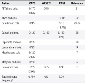
Our negative indings demonstrate the rare genetic etiology in sporadic cases of TD, at least in genes known to be involved in the formation and migration of folli cular cells. Several previously studies in different ethnic populations with TD did not ind mutations, either, par ticularly in sporadic cases (1,7,9,2127) (Table 2). It is likely that other epigenetic factors determine TD, such as differential gene expressions or methylation (28,29). Considering the candidate genes for TD described above in a large number of negative patients with different gene tic backgrounds studied so far, including those from our group, we can conclude that TD should be weakly corre lated with inherited genetic defects. This inding make it necessary to carry out further molecular analyses, as the genes that are known to cause the disorder account for only very few cases.
Financial support: Fundação de Amparo à Pesquisa do Estado de São Paulo (Fapesp) 06/058001 and 08/047860.
Disclosure: no potential conlict of interest relevant to this article was reported.
REFERENCES
1. Ramos HE, Nesi-Franca S, Boldarine VT, Pereira RM, Chiamolera
MI, Camacho CP, et al. Clinical and molecular analysis of thyroid hypoplasia: a population-based approach in southern Brazil. Thyroid. 2009;19(1):61-8.
2. Larsen PR, Davies TF, Schlumberger M, Hayan I. Thyroid
physiolo-gy and diagnostic evaluation of patients with thyroid disorders. In: Larsen K, Melmed P, editors. Williams Textbook of Endocrino-logy. New York: Saunders; 2002. p. 331-73.
3. Dias VM, Campos AP, Chagas AJ, Silva RM. Congenital
hypothyroi-dism: etiology. J Pediatr Endocrinol Metab. 2010;23(8):815-26. 4. Kopp P. Perspective: genetic defects in the etiology of congenital
hypothyroidism. Endocrinology. 2002;143(6):2019-24.
5. De Felice M, Di Lauro R. Thyroid development and its disorders:
ge-netics and molecular mechanisms. Endocr Rev. 2004;25(5):722-46. 6. Trueba SS, Auge J, Mattei G, Etchevers H, Martinovic J,
Czerni-chow P, et al. PAX8, TITF1, and FOXE1 gene expression patterns during human development: new insights into human thyroid development and thyroid dysgenesis-associated malformations. J Clin Endocrinol Metab. 2005;90(1):455-62.
7. Macchia PE, Lapi P, Krude H, Pirro MT, Missero C, Chiovato L, et
al. PAX8 mutations associated with congenital hypothyroidism caused by thyroid dysgenesis. Nat Genet. 1998;19(1):83-6. 8. Vilain C, Rydlewski C, Duprez L, Heinrichs C, Abramowicz M,
Malvaux P, et al. Autosomal dominant transmission of congeni-tal thyroid hypoplasia due to loss-of-function mutation of PAX8. J Clin Endocrinol Metab. 2001;86(1):234-8.
9. Lanzerath K, Bettendorf M, Haag C, Kneppo C, Schulze E, Gru-lich-Henn J. Screening for Pax8 mutations in patients with con-genital hypothyroidism in South-West Germany. Horm Res. 2006;66(2):96-100.
10. Dentice M, Cordeddu V, Rosica A, Ferrara AM, Santarpia L, Salva-tore D, et al. Missense mutation in the transcription factor NKX2-5: a novel molecular event in the pathogenesis of thyroid dysgene-sis. J Clin Endocrinol Metab. 2006;91(4):1428-33.
11. Gagne N, Parma J, Deal C, Vassart G, Van Vliet G. Apparent con-genital athyreosis contrasting with normal plasma thyroglobulin levels and associated with inactivating mutations in the thyro-tropin receptor gene: are athyreosis and ectopic thyroid distinct entities? J Clin Endocrinol Metab. 1998;83(5):1771-5.
12. Beltrao CB, Juliano AG, Chammas MC, Watanabe T, Sapienza MT, Marui S. Etiology of congenital hypothyroidism using thyroglo-bulin and ultrasound combination. Endocr J. 2010;57(7):587-93. 13. Ueda D. Normal volume of the thyroid gland in children. J Clin
Ultrasound. 1990;18(6):455-62.
14. Duarte GC, Tomimori EK, de Camargo RY, Catarino RM, Ferreira JE, Knobel M, et al. Excessive iodine intake and ultrasonographic thyroid abnormalities in schoolchildren. J Pediatr Endocrinol Me-tab. 2009;22(4):327-34.
15. Abrao MG, Billerbeck AE, Nishi MY, Marui S, Mendonca BB. [Standardization of DNA extraction with NaCl from oral mucosa cells: application in PROP1 gene study]. Arq Bras Endocrinol Me-tabol. 2005;49(6):978-82.
16. Kreisner E, Camargo-Neto E, Maia CR, Gross JL. Accuracy of ul-trasonography to establish the diagnosis and aetiology of perma-nent primary congenital hypothyroidism. Clin Endocrinol (Oxf). 2003;59(3):361-5.
Table 2. Frequency of described mutations in PAX8, NKX2.5 and TSHR
genes in thyroid dysgenesis cohorts
Author PAX8 NKX2.5 TSHR Reference
Al Taji and cols. 1/170 (0.6%)
0/15 21
Alves and cols. 0/90# 22
Camilot and cols. 0/15 3/16 (18.7%)
23-24
Cangul and cols. 0/120 0/120 6/120* (5%)
25
Esperante and cols. 0/60 26
Lanzerath and cols. 0/95 9
Macchia and cols. 3/120 (2.5%)
7
Mahjoubi and cols. 0/50 27
Ramos and cols. 1/35 (2.9%)
0/35 0/35 1
Total estimated frequency&
0.75% 0% 3.9%
# Only exon 10 TSHR was studied. * Only familial thyroid dysgenesis. & Based on total cohort
from cited authors.
Acknowledgments: the authors would like to thank the patients and their parents, as well as the staff at Associação de Pais e Ami gos do Excepcional (APAE), São Caetano do Sul.
Cop
yright
© ABE&M t
odos os dir
eit
os r
eser
vados
.
17. Bereket A, Liao XH, Turoglu T, Aribal E, Refetoff S. Analysis of the PAX8 gene in congenital hypothyroidism caused by different for-ms of thyroid dysgenesis in a father and daughter. J Pediatr En-docrinol Metab. 2004;17(7):1021-9.
18. Congdon T, Nguyen LQ, Nogueira CR, Habiby RL, Medeiros-Neto G, Kopp P. A novel mutation (Q40P) in PAX8 associated with con-genital hypothyroidism and thyroid hypoplasia: evidence for phe-notypic variability in mother and child. J Clin Endocrinol Metab. 2001;86(8):3962-7.
19. Meeus L, Gilbert B, Rydlewski C, Parma J, Roussie AL, Abramo-wicz M, et al. Characterization of a novel loss of function mu-tation of PAX8 in a familial case of congenital hypothyroidism with in-place, normal-sized thyroid. J Clin Endocrinol Metab. 2004;89(9):4285-91.
20. Tonacchera M, Banco ME, Montanelli L, Di Cosmo C, Agretti P, De Marco G, et al. Genetic analysis of the PAX8 gene in children with congenital hypothyroidism and dysgenetic or eutopic thyroid glands: identiication of a novel sequence variant. Clin Endocrinol (Oxf). 2007;67(1):34-40.
21. Al Taji E, Biebermann H, Limanova Z, Hnikova O, Zikmund J, Dame C, et al. Screening for mutations in transcription factors in a Czech cohort of 170 patients with congenital and early-onset hypothyroidism: identiication of a novel PAX8 mutation in do-minantly inherited early-onset non-autoimmune hypothyroidism. Eur J Endocrinol. 2007;156(5):521-9.
22. Alves EA, Cruz CM, Pimentel CP, Ribeiro RC, Santos AK, Caldato MC, et al. High frequency of D727E polymorphisms in exon 10 of the TSHR gene in Brazilian patients with congenital hypothyroi-dism. J Pediatr Endocrinol Metab. 2010;23(12):1321-8.
23. Camilot M, Teofoli F, Gandini A, Franceschi R, Rapa A, Corrias A, et al. Thyrotropin receptor gene mutations and TSH resistance: variable expressivity in the heterozygotes. Clin Endocrinol (Oxf). 2005;63(2):146-51.
24. Camilot M, Teofoli F, Vincenzi M, Federici F, Perlini S, Tatò L. Im-plementation of a congenital hypothyroidism newborn screening procedure with mutation detection on genomic DNA extracted from blood spots: the experience of the Italian northeastern refe-rence center. Genet Test. 2007;11(4):387-90.
25. Cangul H, Morgan NV, Forman JR, Saglam H, Aycan Z, Yakut T, et al. Novel TSHR mutations in consanguineous families with congenital nongoitrous hypothyroidism. Clin Endocrinol (Oxf). 2010;73(5):671-7.
26. Esperante SA, Rivolta CM, Miravalle L, Herzovich V, Iorcansky S, Baralle M, et al. Identiication and characterization of four PAX8 rare sequence variants (p.T225M, p.L233L, p.G336S and p.A439A) in patients with congenital hypothyroidism and dysgenetic thyroid glands. Clin Endocrinol (Oxf). 2008;68(5):828-35.
27. Mahjoubi F, Mohammadi MM, Montazeri M, Aminii M, Hashe-mipour M. Mutations in the gene encoding paired box domain (PAX8) are not a frequent cause of congenital hypothyroidism (CH) in Iranian patients with thyroid dysgenesis. Arq Bras Endo-crinol Metabol. 2010;54(6):555-9.
28. Vassart G, Dumont JE. Thyroid dysgenesis: multigenic or epige-netic ... or both? Endocrinology. 2005;146(12):5035-7.